Flesh fly myiasis (Diptera, Sarcophagidae) in
Peruvian poison frogs genus Epipedobates (Anura,
Dendrobatidae)
Mattias Hagman1, Thomas Pape2 and Rainer Schulte3
1 Biological Sciences, Building AO8, The University of Sydney, NSW 2006, Australia. E-mail: mattias.hagman@bio.usyd.edu.au.
2 Zoological Museum, University of Copenhagen, Universitetsparken 15, DK 2100, Copenhagen, Denmark.
E-mail: tpape@snm.ku.dk.
3 Inibico, Jr Ramirez Hurtado 608, Tarapoto, San Martin, Peru. E-mail: inibico@terra.com.pe.
Received 14 February 2005. Accepted 29 September 2005. Distributed October 2005.
Myiasis – the invasion of fly larvae in vertebrate tissues – is a phenomenon well documented in many animals, predominantly mammals and birds, with the most frequent forms being wound and dermal myiasis (Zumpt 1965, Guimarães et al. 1983, Hall and Wall 1995, Hall and Farkas 2000). Amphibians world wide are also parasitized by larvae of numerous fly species, however this is a poorly studied area of amphibian biology. In Europe, North America and India, amphibians are commonly attacked by larvae of several species of blow flies (Callipho-ridae), flesh flies (Sarcophagidae) and muscid flies (Muscidae) (James and Maslin 1947, Das-gupta 1962, Roy and DasDas-gupta 1977, Strijbosch 1980, Bolek and Coggins 2002, Bolek and Janovy 2004) while in Australia amphibians are infected with the larvae of grass flies (Chloropidae) in the genus Batrachomyia (Schell and Burgin 2001). In the Neotropics, dermal myiasis of amphibians
Keywords: Anura, Dendrobatidae, Epipedobates, myiasis, Sarcodexia lambens, flesh fly, Diptera, Sarcophagidae, Peru.
by flesh flies has been reported from harlequin frogs (Atelopus spp.) in Costa Rica (Crump and Pounds 1985, Pounds and Crump 1987), from the granular toad (Bufo granulosus) in Venezuela (Lopes and Vogelsang 1953), from the bullfrog (Rana catesbeiana) in Brazil (Souza et al. 1990), and from leptodactylid frogs (Eleutherodactylus
sp., Proceratophrys sp.) in Panama and Brazil (Dodge 1968, Lopes 1981). When fly identifi-cation was possible most of these cases have been attributed to Notochaeta bufonivora
(Lopes and Vogelsang 1953).
myiasis in poison frogs collected in various locations in Peru during 1982-2005 and present evidence that larger and medium-sized poison frogs (Epipedobates) are infected with sarco-phagid fly larvae. We hope that our observa-tions will stimulate awareness of myiasis in poison frogs and stimulate more research on myiasis in amphibians.
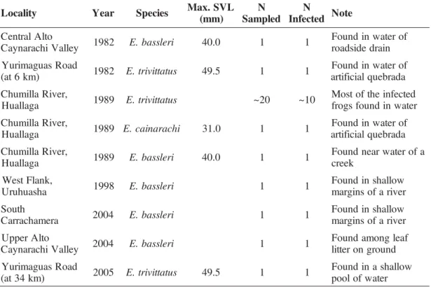
One of us (RS) first observed myiasis in Peruvian poison frogs on two occasions in 1982 (Schulte 1984) and on three occasions during fieldwork with a Kansas University team headed by W. E. Duellman in 1989. Subsequent observa-tions were made in 1998 and again in 2004 and 2005. These observations are summarized in Table 1. With the purpose of studying the prevalence of myiasis we sampled individuals of
three species of poison frogs – Epipedobates trivittatus (n=111), Epipedobates bassleri (n=5) and Epipedobates cainarachi (n=1) – at six localities (the same localities as given in Table 1) from 27th February to 28th March of 2004. Only
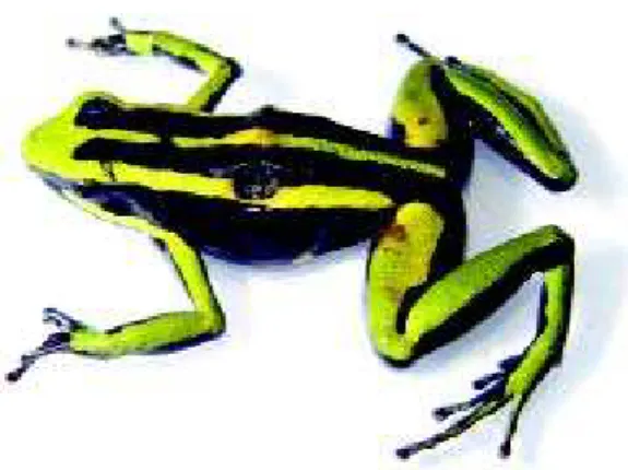
one individual (E. cainarachi) of the 117 frogs collected was infected. Eight maggots were retrieved after preservation. Maggots were preserved in 80% alcohol and identified as a species of flesh fly (Sarcophagidae, subfamily Sarcophaginae) due to the large size and characteristically recessed posterior spiracles. Species-level identification of flesh fly larvae, however, is still not possible. In June 2005 an infected E. trivittatus (Figure 1) was found near San Jose village (J. L. Brown, pers. comm.; Table 1). The frog, which died within hours after it was
Table 1 - Number of myiasis observations in poison frogs genus Epipedobates collected in various locations in Peru during 1982–2005. Data on maximum snout-vent length (SVL) include both sexes and were obtained from the literature (Schulte 1989, Haddad and Martins 1994, Walls 1994).
y t i l a c o
L Year Species Max.SVL ) m m ( N d e l p m a S N d e t c e f n
I Note
o t l A l a r t n e C y e l l a V i h c a r a n y a
C 1982 E.bassleri 40.0 1 1
f o r e t a w n i d n u o F n i a r d e d i s d a o r d a o R s a u g a m i r u Y ) m k 6 t a
( 1982 E.trivittatus 49.5 1 1
f o r e t a w n i d n u o F a d a r b e u q l a i c i f i t r a , r e v i R a l l i m u h C a g a l l a u
H 1989 E.trivittatus ~20 ~10
d e t c e f n i e h t f o t s o M r e t a w n i d n u o f s g o r f , r e v i R a l l i m u h C a g a l l a u
H 1989 E.cainarachi 31.0 1 1
f o r e t a w n i d n u o F a d a r b e u q l a i c i f i t r a , r e v i R a l l i m u h C a g a l l a u
H 1989 E.bassleri 40.0 1 1
a f o r e t a w r a e n d n u o F k e e r c , k n a l F t s e W a h s a u h u r
U 1998 E.bassleri 1 1
w o l l a h s n i d n u o F r e v i r a f o s n i g r a m h t u o S a r e m a h c a r r a
C 2004 E.bassleri 1 1
w o l l a h s n i d n u o F r e v i r a f o s n i g r a m o t l A r e p p U y e l l a V i h c a r a n y a
C 2004 E.bassleri 1 1
f a e l g n o m a d n u o F d n u o r g n o r e t t i l d a o R s a u g a m i r u Y ) m k 4 3 t a
( 2005 E.trivittatus 49.5 1 1
Figure l -Epipedobates trivittatus parasitized by larvae of Sarcodexia lambens (Sarcophagi-dae). This specimen (SVL > 45mm) was found in a small water pool near San Jose village, Peru, in June 2005.
collected, was placed in a ventilated container. Within a week thirty larvae had emerged from the dead frog, and about three weeks later three adults (two males, one female, all = Sarcodexia lambens (Wiedemann, 1830) and deposited in the Zoological Museum, Copenhagen) hatched from their puparia. Unfortunately, wasps of an unknown species had parasitized the remaining larvae.
S. lambens is widely distributed in the New World, from the south-eastern US to northern Argentina and Paraguay (Pape 1996). This species has been bred from dead bird nestlings, snails, dead and living (but probably often injured or otherwise weakened) insects and scorpions, and vertebrate carrion, and it is known from cases of human myiasis (Townsend 1893, Almeida 1933, Callan 1946, Parker et al.
1953, Harrison 1963, Stegmaier 1972, Cornaby 1974, Jones 1988, Fessl et al. 2001, Janzen and Hallwachs 2005). With such diversity of breeding habits, a record of amphibian myiasis would seem at first to be no surprise. Still, it is noteworthy that no records exist of S. lambens
involved in amphibian myiasis outside Peru, and the pattern of infection in the poison frogs is
here considered as indicative of a specialised predator. Precautions were taken to avoid contamination of the infected frog after being brought to the laboratory, but clearly the small parasitic wasps (1.5-2.0 mm) could penetrate the fine nylon mesh covering the breeding con-tainer. Some flesh flies will larviposit on the mesh without direct contact with the bait (Bänziger and Pape 2004), but with only one breeding event giving adult flies, and thereby a reliable identification, data does not allow a clearcut answer.
The present records represent the first evi-dence of myiasis on frogs from Peru as well as the first evidence of fly larvae parasitizing poison frogs. Most of the infected frogs that have been found are the larger species Epipedo-bates trivittatus, and a few are from the medium-sized species E. bassleri and E. cainarachi (see Table 1 for data on body size). No maggot infections have been recorded from any of the smaller sympatric species E. hahneli (maximum SVL = 23.0 mm; Haddad and Martins 1994) and
E. pongoensis (max. SVL = 26.0 mm; Schulte 1999). It is unclear if these flies can infect small
Epipedobates species. Other studies on fly larvae that cause myiasis in amphibians clearly show that smaller frogs under 30 mm die quicker and get consumed within one to three days of infection (Bolek and Coggins 2002, Bolek and Janovy 2004), suggesting that small frogs also may be infected but that they are less likely to be found by researchers due to the rapid death and consumption by fly maggots.
observed larvae of Notochaeta bufonivora
parasitizing Atelopus varius in Costa Rica, and they reported that all hosts died within four days after they were found. Maggots that crawled out from the dead frogs pupated within 48 hours and eclosion occurred in 17 to 30 days.
Notochaeta bufonivora is the only fly species known to parasite diurnal, toxic frogs in the Neotropics (A.varius; Crump and Pounds 1985). Although not closely related, harlequin frogs (Atelopus) and poison frogs (Dendrobatidae) have similar life-history traits and are often found living sympatrically. Like poison frogs, harlequin frogs are small, diurnal and have noxious toxins. Crump and Pounds (1985) reported a SVL range of 25.0 – 29.5 mm for male
A. varius and 30.0 – 41.0 mm for females. Interestingly, male A. varius were more common in their study location, although females were more commonly infected (Crump and Pounds 1985).
Note: the area where we made our observa-tions is under heavy pressure of illegal collecting. We have therefore omitted latitude and longitude coordinates for our study sites. We will provide them to researchers on request.
Acknowledgments
The study was supported financially by a grant from SIDA (Swedish International Deve-lopment Cooperation Agency) to the senior author. We thank J. L. Brown for photographs and R. Shine for helpful comments on an earlier draft of the manuscript and P. Venegas from the Cordillera Oriental Park RAP (Rapid Assessment Procedures) team for sharing his observations.
References
Almeida, J. L. 1933. “Otomyase” em bezerro por Sarcophaga sternodontes (Towsend [sic], 1892). Archivos da Escola Superior de Agricultura e Medicina Veterinaria 10: 171–172.
Bänziger, H. and T. Pape. 2004. Flowers, faeces and cadavers: natural feeding and laying habits of flesh
flies in Thailand (Diptera: Sarcophagidae, Sarco-phaga spp.). Journal of Natural History 38: 1681– 1 6 9 8 .
Bolek, M. G. and J. R. Coggins. 2002. Observations on myiasis by the Calliphorid, Bufolucilia silvarum in the Eastern American toad (Bufo americanus americanus) from Southeastern Wisconsin. Journal of Wildlife Disease 38: 598–603.
Bolek, M. G. and J. Janovy. 2004. Observations on myiasis by the Calliphorids, Bufolucilia silvarum and Bufolucilia elongata, in the wood frogs, Rana sylvatica from Southeastern Wisconsin. Journal of Parasitology 90: 1169–1171.
Callan, E. McC. 1946. A note on Sarcophaga lambens (Wied.), a parasite of the South American Bollworm, Sacadodes pyralis Dyar. Revista de Entomologia 17: 474–475.
Cornaby, B. W. 1974. Carrion reduction by animals in contrasting tropical habitats. Biotropica 6: 51–63.
Crump, M. L. and J.A. Pounds. 1985. Lethal parasi-tism of an aposematic anuran (Atelopus varius) by Notochaeta bufonivora (Diptera: Sarcophagidae). Journal of Parasitology 71: 588–591.
Daly, J. 1978. A dangerously toxic frog (Phyllobates) used by Embera Indians of Western Colombia, with discussion of blowgun fabrication and dart poisoning. Bulletin of the American Museum of Natural History 161: 311–365.
Daly, J. 1995. The chemistry of poisons in amphibian skin. Proceedings of the National Academy of Sciences 92: 9–13.
Daly, J. and C. Myers. 1983. Dart-poison frogs. Scientific American 248: 120–133.
Dasgupta, B. 1962. On myiasis of Indian toad Bufo melanostictus. Parasitology 52: 63–66.
Dodge, H. R. 1968. The Sarcophagidae of Barro Colorado Island, Panama (Diptera). Annals of the Entomological Society of America 61: 421–450.
Fessl, B., M. S. Couri and S. Tebbich. 2001. Philornis downsi Dodge & Aitken, new to the Galapagos Islands (Diptera, Muscidae). Studia Dipterologica 8: 317–322.
Guimarães, J. H., N. Papavero and A. Prado. 1983. As miíases na região Neotropical (identificação, bio-logia, bibliografia). Revista Brasileira de Zooloica 1: 239–416.
Haddad, C. F. B. and M. Martins. 1994. Four species of Brazilian poison frogs related to Epipedobates pictus (Dendrobatidae): taxonomy and natural history observations. Herpetologica 50: 282–295.
Hall, M. J. R. and R. Farkas. 2000. Traumatic myiasis of humans and animals. Pp. 751–768 in L. Papp et al. (eds), Contributions to a Manual of Palae-arctic Diptera. Vol. 1, General and Applied Dipterology 1. Budapest. Science Herald.
Hall, M. J. R. and R. Wall. 1995. Myiasis of humans and domestic animals. Advances in Parasitology 35: 257–334.
Harrison, J. D. 1963. On the biology of three banana pests in Costa Rica (Lepidoptera: Limacodidae, Nymphalidae). Annals of the Entomological Society of America 56: 87–94.
Heselhaus, R. 1992. Poison Arrow Frogs. UK. Cassel Plc.
James, T. M. and T. P. Maslin. 1947. Notes on myiasis of the toad, Bufo boreas (Baird and Girard). Journal of the Washington Academy of Sciences 37: 366–368.
Janzen, D. H. and W. Hallwachs. 2005. Dynamic database for an inventory of the macrocaterpillar fauna, and its food plants and parasitoids, of Area de Conservacion Guanacaste (ACG), northwestern Costa Rica (nn-SRNP-nnnnn voucher codes). http:/ /janzen.sas.upenn.edu/caterpillars/database.lasso . [Accessed 26 September 2005.]
Jones, W. A., Jr. 1988. World review of the parasitoids of the southern green stink bug, Nezara viridula (L.) (Heteroptera: Pentatomidae). Annals of the Entomological Society of America 81: 262–273.
Lopes, H. S. 1981. Notes on American Sarcophagidae (Diptera). Revista Brasileira de Biologia 41: 149– 1 5 2 .
Lopes, H. S. and E. G. Vogelsang. 1953. Notochaeta bufonivora n. sp., paràsita de Bufo granulosus Spix em Venezuela (Diptera Sarcophagidae). Anais da Academia Brasileira de Ciências 25: 139–143.
Pape, T. 1996. Catalogue of the Sarcophagidae of the world (Insecta: Diptera). Memoirs of Entomology, International 8: 1–558.
Parker, H. L., P. A. Berry and A. Silveira. 1953. Host-parasite and Host-parasite-host lists of insects reared in the South American Parasite Laboratory during the period 1940-46. Revista de la Asociacion Ingenie-ría Agronómica 92: 1–101.
Pounds, J. A. and M. L. Crump. 1987. Harlequin frogs along a tropical montane stream: aggregation and
the risk of predation by frog eating flies. Biotro-pica 19: 306–309.
Roy, P. and B. Dasgupta. 1977. Sarcophaga ruficornis fabr (Sarcophagidae – Diptera) as a parasite of common Indian toad. Proceedings of the Indian Academy of Science 86: 207–209.
Schell, C. B. and S. Burgin. 2001. Batrachomyia strigapes (Diptera) parasitism of Uperoleia laevi-gata (Anura). Journal of Parasitology 87: 1215– 1 2 1 6 .
Schulte, R. 1984. Frösche und Kröten. Hohenheim. Ulmer Verlag Stuttgart. 240 pp.
Schulte, R. 1989. Nueva especie de rana venenosa del género Epipedobates registrada en la Cordillera Oriental, departamento de San Martin. Boletin Lima 63: 41–46.
Schulte, R. 1999. Pfeilgiftfrösche- Artenteil Peru. Vol. 2. Waiblingen, Germany. INIBICO. 292 pp.
Souza, F. L. S., Jr., C. W. O. Souza, M. Hipolito, L. Baldassi, and M. L. Martins. 1990. Cases of buccal myiasis in the bullfrog (Rana catesbeiana Shaw, 1802), with larvae of Notochaeta sp. Aldrich, 1916 (Diptera: Sarcophagidae) in São Paulo, Brazil. Memórias do Instituto Oswaldo Cruz 84: 517–518.
Stegmaier, C. E., Jr. 1972. Notes on some Sarcopha-gidae (Diptera) reared from snails (Mollusca) in Florida. Florida Entomologist 55: 237–242.
Strijbosch, H. 1980. Mortality in a population of Bufo bufo resulting from the fly Lucilia bufonivora. Oecologia 45: 285–286.
Summers, K. and M. E. Clough. 2001. The evolution of coloration and toxicity in the poison frog family (Dendrobatidae). Proceedings of the National Academy of Sciences 98: 6227–6232.
Townsend, C. H. T. 1893. A scorpion parasite. Journal of the Institute of Jamaica 1: 221.
Walls, J. G. 1994. Jewels of the Rainforest. USA T. F. H. Publications Inc.
Wiedemann, C. R. W. 1830. Aussereuropäische zweiflü-gelige Insekten. Als Fortsetzung des Meigenschen Werkes. Vol. 2. Schulz, Hamm; xii + 684 pp.

