Mitochondrial Physiology in the Major
Arbovirus Vector
Aedes aegypti: Substrate
Preferences and Sexual Differences Define
Respiratory Capacity and Superoxide
Production
Juliana B. R. Correa Soares1,2, Alessandro Gaviraghi1,2, Marcus F. Oliveira1,2*
1Laboratório de Bioquímica de Resposta ao Estresse, Instituto de Bioquímica Médica Leopoldo de Meis, Universidade Federal do Rio de Janeiro, Cidade Universitária, Rio de Janeiro, Brazil,2Laboratório de Inflamação e Metabolismo, Instituto Nacional de Ciência e Tecnologia de Biologia Estrutural e Bioimagem (INBEB), Universidade Federal do Rio de Janeiro, Rio de Janeiro, RJ, Brazil
Abstract
Adult females ofAedes aegyptiare facultative blood sucking insects and vectors of Dengue and yellow fever viruses. Insect dispersal plays a central role in disease transmission and the extremely high energy demand posed by flight is accomplished by a very efficient oxida-tive phosphorylation process, which take place within flight muscle mitochondria. These or-ganelles play a central role in energy metabolism, interconnecting nutrient oxidation to ATP synthesis, but also represent an important site of cellular superoxide production. Given the importance of mitochondria to cell physiology, and the potential contributions of this organ-elle forA.aegyptibiology and vectorial capacity, here, we conducted a systematic assess-ment of mitochondrial physiology in flight muscle of young adultA.aegyptifed exclusively with sugar. This was carried out by determining the activities of mitochondrial enzymes, the substrate preferences to sustain respiration, the mitochondrial bioenergetic efficiency and capacity, in both mitochondria-enriched preparations and mechanically permeabilized flight muscle in both sexes. We also determined the substrates preferences to promote mitochon-drial superoxide generation and the main sites where it is produced within this organelle. We observed that respiration inA.aegyptimitochondria was essentially driven by complex I and glycerol 3 phosphate dehydrogenase substrates, which promoted distinct mitochondri-al bioenergetic capacities, but with preserved efficiencies. Respiration mediated by proline oxidation in female mitochondria was strikingly higher than in males. Mitochondrial superox-ide production was essentially mediated through proline and glycerol 3 phosphate oxida-tion, which took place at sites other than complex I. Finally, differences in mitochondrial superoxide production among sexes were only observed in male oxidizing glycerol 3 phos-phate, exhibiting higher rates than in female. Together, these data represent a significant step towards the understanding of fundamental mitochondrial processes inA.aegypti, with potential implications for its physiology and vectorial capacity.
OPEN ACCESS
Citation:Soares JBRC, Gaviraghi A, Oliveira MF
(2015) Mitochondrial Physiology in the Major Arbovirus VectorAedes aegypti: Substrate Preferences and Sexual Differences Define Respiratory Capacity and Superoxide Production. PLoS ONE 10(3): e0120600. doi:10.1371/journal. pone.0120600
Academic Editor:Marc Liesa, Boston University
School of Medicine, UNITED STATES
Received:October 16, 2014
Accepted:January 24, 2015
Published:March 24, 2015
Copyright:© 2015 Soares et al. This is an open
access article distributed under the terms of the
Creative Commons Attribution License, which permits unrestricted use, distribution, and reproduction in any medium, provided the original author and source are credited.
Data Availability Statement:All relevant data are
within the paper and its Supporting Information files.
Funding:This work was supported by grants from
Introduction
Aedes aegyptifemale mosquitoes are facultative blood sucking insects and this unusual dietary source is absolutely essential not only to trigger the oogenesis process, but also to transmit pathogens, such as the viruses that cause Dengue and yellow fever. Collectively, these diseases afflict nearly 400 million individuals each year globally and increasing concern on public health has prompted policies directing vectors control in endemic areas [1]. The adult individuals of A.aegyptiexhibit remarkable sexual dimorphism, where males can clearly be distinguished from the females by a number of morphological and behavioral features [2]. These sexual dif-ferences can also be extended to other aspects ofA.aegyptibiology, such as gene expression [3,
4], hemocyte counts, and pathogen-induced immune response [5].
Dispersal of adultA.aegyptiindividuals plays a central ecological and medical role and field studies pointed out that a wide range of distances are covered by this insect [6,7]. Insect flight is accomplished by the contractile activity of flight muscle, which has an extremely high energy demand, and represents one of the most metabolic active tissues found in nature. The metabol-ic provision to power flight is diverse and the substrates required for this purpose depend on the species, flight duration, diet and aging [8–16]. For example, in honeybees carbohydrates seems to be the sole energy source to sustain flight activity [15], whereas in locusts, carbohy-drates are oxidized only at the initial periods of flight, being further replaced by lipids [16]. Be-yond these substrates, proline was also reported to play a key energetic role during flight activity in many insect species [11,13,17–24]. Interestingly, the high rates of proline oxidation observed in blood sucking insects would represent an important adaptation to blood feeding habit [25]. Also, hemolymph levels of proline are quite high in many blood feeding insect vec-tors of neglected tropical diseases (NTD), representing the most abundant aminoacid found in that fluid [13,25–28]. Another important energetic substrate in flight muscle is glycerol 3 phosphate (G3P), which connects glycolysis and the ubiquinone reduction, channeling elec-trons from cytosol directly into the mitochondrial electron transport system [9,12,17,29–34]. G3P oxidation is not only directly involved on the re-oxidation of cytosolic NADH produced during glycolysis in insect flight muscle [35], but also on the generation of mitochondrial hy-drogen peroxide (H2O2) [29,31,34].
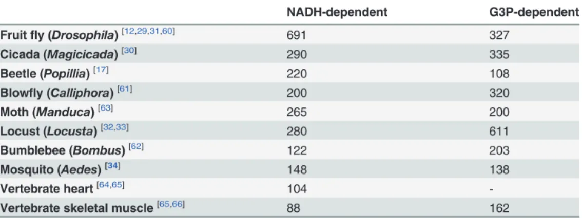
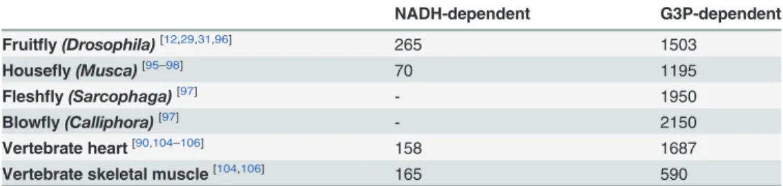
Since the so-called "sarcosomes" were defined as mitochondria in insect flight muscle [36], a number of studies have demonstrated the structural and functional properties of these organ-elles in that tissue [12,18,23–25,29–37]. However, despite the dominance of insects as a major taxonomic group in number of species [38,39], studies devoted to explore mitochondrial phys-iology in this important group remain largely underrepresented in the literature (only about 1.8% of all mitochondrial studies found to date in Pubmed). In this sense, a brief survey over the literature searching the respiratory activities of flight muscle mitochondria in different in-sect species was compiled inTable 1. We can observe that mitochondrial respiratory rates dur-ing phosphorylatdur-ing conditions varies greatly among insect species and substrates, in such a way that complex I substrates contribute more importantly to oxygen consumption in some species (Drosophila,Popillia,Manduca), while G3P oxidation plays a prominent role in respi-ration in others (Magicicada,Calliphora,LocustaandBombus). With exception of the bumble-bee, all NADH-dependent respiratory rates were expressively higher in insects when compared to vertebrate heart and skeletal muscle. Similarly, G3P-mediated respiration in insect flight muscle mitochondria were in general higher than in vertebrate muscle mitochondria, with re-spiratory rates ofManducaandPopilliasimilar to vertebrate skeletal muscle, with the highest respiration observed inLocusta(5.6 times higher than inPopillia). Thus, high respiratory rates of insect flight muscle mitochondria agrees with the concept of intense oxidative metabolism in flight muscle which is essential to provide the huge energy demand posed by flight. In fact,
(Produtividade em Pesquisa—PQ 301571/2009-0).
JBRCS is a PAPD-FAPERJ fellow (Apoio à Pós-Doutorado no Estado do Rio de Janeiro, 45879) and AG is a CsF-BJT-CNPq fellow (BJT-Ciências sem Fronteiras, 402409/2012-4). The funders had no role in study design, data collection and analysis, decision to publish, or preparation of the manuscript.
Competing Interests:The authors have declared
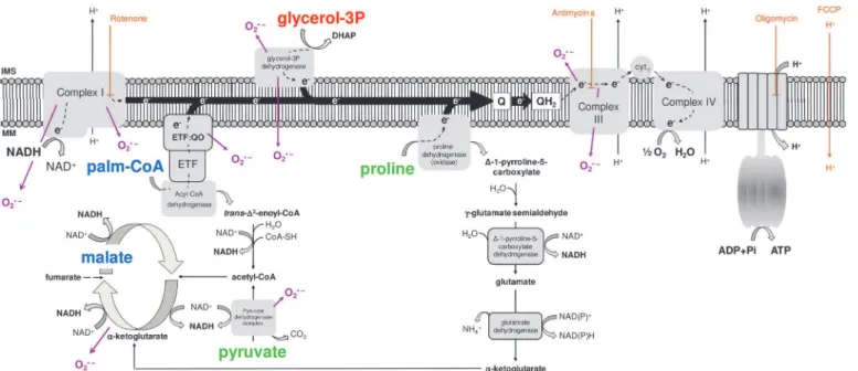
most of the ATP required to sustain muscle contraction and flight activity are essentially pro-vided by a highly efficient oxidative phosphorylation (OXPHOS) process within flight muscle mitochondria. These organelles play a central role in energy and redox metabolism, intercon-necting the energy transduction, by means of carbohydrates, lipids and amino acids oxidation, to ATP synthesis. The electron flow at the inner membrane is channeled to ubiquinone by dis-tinct sites, which are further transferred to complex III, cytochromec, cytochromecoxidase and finally to molecular oxygen by the electron transport system (Fig. 1). The energy released by electron flow is partially conserved as a proton gradient across the inner mitochondrial membrane, known as the protonmotive force (pmf), which is utilized to allow ATP generation by the F1FoATP synthase complex activity. Mitochondrial function is also directly involved on cellular redox balance, representing an important source of the so-called reactive oxygen spe-cies (ROS), which contributes to a number of redox-dependent signaling cascades [40]. The mechanisms involved on mitochondrial oxygen consumption and ROS generation are complex and highly regulated, and the contribution of mitochondrial physiology to basic aspects of in-sect biology has previously been appraised [12,24,25,29,34,41–46]. Interestingly, recent evi-dence pointed out that proline oxidation promotes mitochondrial ROS formation in
Drosophilaflight muscle, which occurs essentially at levels of complex I and II [37]. Particularly relevant in the context of NTD insect vectors, recent evidence demonstrate the involvement of mitochondrial processes mediating the innate immune response against pathogens [44–46].
Previous investigations of mitochondrial physiology inA.aegyptiflight muscle revealed that both respiration and ADP phosphorylation were induced by tricarboxylic acid cycle intermedi-ates, in DDT-sensitive reactions [47,48]. Our group has shown that in females ofA.aegypti, flight muscle mitochondrial oxygen consumption and hydrogen peroxide generation were transiently reduced along the blood digestion cycle [34]. Interestingly, these effects were paral-lel to reduced cytochrome levels and activation of mitochondrial fusion. Therefore, in an effort to understand fundamental aspects of mitochondrial physiology in this important NTD insect
Table 1. Mitochondrial oxygen consumption of insect flight muscle compared to vertebrate muscle induced by two distinct electron transport system sites.
NADH-dependent G3P-dependent
Fruitfly (Drosophila)[12,29,31,60] 691 327
Cicada (Magicicada)[30] 290 335
Beetle (Popillia)[17] 220 108
Blowfly (Calliphora)[61] 200 320
Moth (Manduca)[63] 265 200
Locust (Locusta)[32,33] 280 611
Bumblebee (Bombus)[62] 122 203
Mosquito (Aedes)[34] 148 138
Vertebrate heart[64,65] 104
-Vertebrate skeletal muscle[65,66] 88 162
Values of mitochondrial oxygen consumption rates during phosphorylating conditions were expressed as nmol of O2/min/mg protein. Data originally expressed in other units were accordingly converted to nmol of O2/min/mg protein, based onhttp://bioblast.at/images/5/5d/MiPNet12.15_RespiratoryStates.pdf. In
Drosophila,Locustaand vertebrate heart and skeletal muscle, where multiple articles were utilized for calculations, an average value was obtained. NADH-dependent substrates comprise pyruvate, proline, malate and other electron donors for complex I. Superscript letters within the brackets indicate the references where data were collected.
vector, we carried out here a systematic functional assessment of this organelle in flight muscle of young adultA.aegyptifed exclusively with sugar. This was accomplished by examining the activities of tricarboxylic acid cycle and electron transport system enzymes, the contribution of different substrates on respiration and H2O2generation, the mitochondrial bioenergetic effi-ciency and capacity, the sites of H2O2generation, as well as the differences observed in all these parameters between the sexes. The data presented here indicate thatA.aegyptimitochondria utilize preferentially complex I and glycerol 3-phosphate dehydrogenase (G3PDH) substrates to sustain respiration, presenting distinct mitochondrial bioenergetic capacities among these substrates, but with preserved efficiency. In addition, the contribution of proline oxidation to respiration in female mitochondria was strikingly higher than in males. Mitochondrial super-oxide (O2•) production is essentially mediated through proline and G3P oxidation, which takes place at distinct electron transport system points other than complex I site IF. Finally, sexual differences on H2O2generation were only observed when using G3P as substrate, with male mitochondria exhibiting higher rates of O2•formation than females. Therefore, the set of data described here represent a significant step towards the understanding of mitochondrial func-tional processes in this important insect NTD vector, with potential implications for dispersal, reproduction, survival, aging, insecticide resistance and pathogen transmission.
Materials and Methods
Insects
Aedes aegypti(Red eyes strain) were maintained at 28°C, 70–80% relative humidity with a pho-toperiod of 12h light/dark (L:D, 12:12h) during all life cycle. Larvae were reared on a diet con-sisting of commercial dog chow. Insects utilized in all experiments were adult individuals 5–7 days after the emergence. Usually about 200 insects were placed in 5 L plastic cages in a 1:1 sex-ratio and allowed to feedad libitumon cotton pads soaked with 10% (w/v)
sucrose solution.
Fig 1. Schematic representation of the electron transport system, showing the sites of action of oxidative phosphorylation (OXPHOS) modulators (brown), the different substrates utilized throughout this study (pyruvate and proline, green; glycerol 3 phosphate, red; palmitoylcarnitine and malate, blue) and the known sites of superoxide (O2•) production (purple).
Mitochondria
Mitochondria isolation fromA.aegyptiflight muscle was carried out by using a method previ-ously established by our group with minor modifications [34]. About 120 mosquitoes were im-mobilized by chilling on ice, dissected to obtain the thoraxes, and then gently homogenized in a 15 mL Potter-Elvehjem tissue grinder in a Teflon pestle with 10 mL of ice-cold isolation me-dium (250 mM sucrose, 5 mM Tris-HCl, 2 mM EGTA, 1% (w/v) fatty acid free bovine serum albumin, pH 7.4). The preparation was maintained at 4°C throughout the subsequent washing and centrifugation procedures. The liquid was centrifuged at 300 xgfor 5 min in an Eppendorf centrifuge model 5810-R with a rotor F34-6-38. The supernatant was collected and further centrifuged at 10,000 xgfor 10 min. The brown pellet was carefully re-suspended in approxi-mately 0.1 mL of "respiration buffer" (120 mM KCl, 5 mM KH2PO4, 3 mM Hepes, 1 mM EGTA, 1.5 mM MgCl2, and 0.2% fatty acid free bovine serum albumin, pH 7.2) to give a prepa-ration with about 20 mg of protein/mL. Protein concentprepa-ration was determined by the Lowry method, using bovine serum albumin as standard [49]. Usually this method yields about 16 mg mitochondrial protein/mL/120 thoraces.
Enzymatic activities
Enzyme activities were determined in mitochondrial preparations following methods described in the literature [50] with modifications by incubating 1.7 mg of mitochondrial preparation in 0.1 mL of hypotonic buffer (25 mM potassium phosphate and 5 mM MgCl2, pH 7.2) and sub-sequently subjected to three freeze-thawing cycles. The enzyme activities were measured at room temperature, in 1 mL of 100 mM potassium phosphate, pH 7.4 (complex I-III) or hypo-tonic buffer (complex IV), using a Shimadzu spectrophotometer model visible 2450 (Shimadzu Scientific Instruments, Tokyo, Japan). All enzyme activities in this study were determined using samples corresponding to about 80μg of mitochondrial protein and at least three
sepa-rate cohorts of insects. NADH:Cytcoxido-reductase activity was measured as the increase in absorbance at 550 nm due to the ferricytochromecreduction. The reaction was initiated by the addition of 50μM cytochromec, 200μM NADH, 1 mM KCN and followed by a sample of
freeze-thawed mitochondria and the absorbance was monitored at 550 nm for about 10 min-utes. Rotenone (0.5μM) was added to inhibit complex I activity, which was considered as the
rotenone-sensitive rate of cytochromecreduction (ε= 18.7 mM−1
cm−1). Proline:Cyt
c oxido-reductase was measured in the same conditions described for NADH:Cytcoxido-reductase ac-tivity, but using 25 mM of proline as substrate and 2.5μg/mL antimycin A to inhibit complex
III, instead of rotenone. G3P:Cytcoxido-reductase was measured in the same conditions de-scribed for NADH:Cytcoxido-reductase activity, but using 20 mM ofsn-glycerol 3 phosphate as substrate and 2.5μg/mL antimycin A to inhibit complex III, instead of rotenone.
Palmitoyl-carnitine (PC):Cytcoxido-reductase was measured in the same conditions described for NADH:Cytcoxido-reductase activity, but using 10μM of palmitoylcarnitine as substrate and
2.5μg/mL antimycin A to inhibit complex III, instead of rotenone. Cytochromecoxidase
activ-ity was measured by following the decrease in absorbance due to the oxidation of ferrocyto-chromec(ε= 18.7 mM−1
cm−1). The reaction was initiated by the addition of freeze-thawed
mitochondria and the reduction in absorbance was monitored at 550 nm. KCN (1 mM) was added to inhibit cytochromecoxidase activity, which was considered as the cyanide-sensitive rate of cytochrome c oxidation. Citrate synthase (CS) activity was determined spectrophoto-metrically [51] by incubating a sample corresponding to 17μg of protein from freeze-thawed
5,50-dithiobis (2-nitrobenzoic acid) (DTNB), which has an absorption maximum at 412 nm.
The reaction was started by the addition of 0.5 mM oxaloacetate.
Respirometry analyses on isolated mitochondria
The respiratory activity ofA.aegyptiflight muscle mitochondria was analyzed in a two-channel titration injection respirometer (Oxygraph-2k, Oroboros Instruments, Innsbruck, Austria) at 27.5°C. Aliquots corresponding about 200μg of protein from freshly isolated mitochondria
were transferred to the respirometer chambers containing the "respiration buffer" in a final vol-ume of 2.2 mL and allowed to equilibrate for about 15 minutes, with continuous stirring set up at 750 rpm. Then, the oxygen concentrations and the rates of oxygen consumption were simul-taneously recorded in real time in both respirometer chambers by using the DatLab 4.0 soft-ware (Oroboros Inc., Austria). The routine of electron transport system activities inA.aegypti mitochondria was carried out by following the high resolution respirometry (HRR) analyses coupled to substrate-uncoupler-inhibitor titration (SUIT) protocols established in the litera-ture [52]. HRR-SUIT analyses were conducted using three distinct oxidizable substrate combi-nations as following: pyruvate + proline (Pyr+pro),sn-glycerol 3-phosphate (G3P), and palmitoylcarnitine + malate (PC+Mal) as indicated in different colors fonts inFig. 1. The rou-tine was started by the addition of substrates to final concentrations of 10 mM Pyr + 10 mM pro, 20 mM G3P, or 10μM PC + 10 mM Mal. When using G3P, 0.5μM rotenone was added
before substrates addition in order to avoid electron backflow from glycerol 3 phosphate dehy-drogenase (G3PDH) to complex I. ATP synthesis coupled to oxygen consumption through the oxidative phosphorylation (OXPHOS) was promoted by the addition of 1 mM ADP followed by a second shot reaching a final ADP concentration of 2 mM. The substrates concentration utilized in this work to induce ADP-stimulated respiration were established based on previous experiments showing that, in these conditions, oxygen consumption was maximum (data not shown). Then, 10μM cytochromecwere added to each respirometer chamber and the absence
of a significant stimulatory effect on respiration was used as a quality control test for integrity of the outer mitochondrial membrane [53]. The maximum non-coupled respiration was in-duced by stepwise titration of carbonyl cyanide p-(trifluoromethoxy) phenylhydrazone (FCCP) from 0.5 to 3μM. The contribution of complexes I and III on electron flow were
deter-mined by the addition of 0.5μM rotenone and 2.5μg/mL antimycin A, respectively. All the
Respirometry analyses on permeabilized flight muscle
The general concept of permeabilized muscle fiber was applied to determine respiratory capaci-ties in permeabilized flight muscle fromA.aegypti[52]. Briefly, adult mosquitoes (5–7 days old), were immobilized by chilling on ice onto a pre-cooled Petri dish and then dissected to ob-tain the thoraces. To determine the respiratory capacities, the flight muscle were accessible by cracking the thoraces using fine forceps and then a single thorax was immediately transferred into the O2k chamber containing 2.2 mL of the "respiration buffer" (120 mM KCl, 5 mM KH2PO4, 3 mM Hepes, 1 mM EGTA, 1.5 mM MgCl2, and 0.2% fatty acid free bovine serum al-bumin, pH 7.2) supplemented with 280 U/mL of catalase. Respirometry analyses were per-formed at 27.5° C, using DatLab 4.0 software (Oroboros Inc., Austria), with continuous stirring at 750 rpm and all experiments started by registering the endogenous substrate supported res-piration, following the HRR-SUIT protocols established in the literature [52]. In order to pre-vent limitations in oxygen diffusion and artificial hypoxic conditions, we carried out all experiments with whole flight muscle in an environment enriched with oxygen within the O2k chamber (O2concentrations maintained between 400 and 520 nmol/mL), as recommended by the literature [52]. Experiments started by injecting a suitable amount of oxygen-enriched gas-eous mixture (70% O2and 30% N2mol/mol) into the O2k-chamber to reach an oxygen con-centration of about 500 nmol/mL. To avoid substantial drops in oxygen levels and the risk of bubble formation within the oxygraph chamber during the experiments, we injected small volumes (μL) of a H2O2stock solution (200 mM) into the O2k-chamber. The routine of elec-tron transport system activities in flight muscle tissue was the same utilized for isolated mito-chondria and was carried out using either 10 mM Pyr+pro or 20 mM G3P as substrates. The concentrations of substrates and OXPHOS modulators were established based on the data ob-tained for isolated mitochondria. Typical traces of oxygen consumption by whole thorax are shown inS2andS3Figs. To determine the contribution of the electron transport system dehydrogenases to respiration, we utilized the same approach as described above for isolated mitochondria.
Hydrogen peroxide (H
2O
2) release
Mitochondrial H2O2production was assessed by monitoring resorufin fluorescence due to the oxidation of Amplex red (Invitrogen, USA) as described previously [34]. Briefly, 0.17 mg of mi-tochondrial preparations were incubated in the presence of 1.0 unit/mL horseradish peroxidase (Sigma Co., USA) on“respiration buffer”and the same substrates concentrations described for respirometry above. The rate of amplex red oxidation was recorded at room temperature using a Cary Eclipse spectrofluorimeter (Varian, USA) adapted with a continuous stirring device, op-erating at excitation and emission wavelengths of 530 nm and 590 nm, respectively. Typical amplex red fluorescence traces are shown inS4 Fig.Standard curves of reagent-grade H2O2 (Merck, Germany) were performed in the presence of each pharmacological OXPHOS mod-ulator as well as of 0.17 mg of mitochondrial protein. To assess the topology of H2O2 genera-tion sites, we utilized classical electron transport system inhibitors aiming the manipulagenera-tion of the redox states of specific sites within mitochondria. Then, mitochondria were incubated with the four different substrates investigated for respirometry plus ADP, oligomycin and FCCP (5μM) and the effects of sequential addition of rotenone and antimycin A on the amplex red
fluorescence rates were registered. Specific contribution of site IFof complex I to H2O2 produc-tion using pyr+pro as substrates was assessed and calculated by subtracting the rates induced by 0.5μM rotenone from their respective rates of FCCP. Although this rotenone concentration
respiration driven by pyr + pro or PC+Mal, it is possible that the contribution of site IFof com-plex I to H2O2production would be affected by higher levels of rotenone. The remaining sites of H2O2formation promoted by pyr+pro comprised ProDH and other dehydrogenases [37,
54] that were not assigned in this work, and calculated by subtracting the rates of amplex red fluorescence induced by antimycin a by their respective rates obtained by rotenone. The contri-bution of G3PDH and other dehydrogenases to H2O2generation was calculated as the amplex red fluorescence rates induced by antimycin A and then subtracted by their respective FCCP rates using G3P as substrate. The contribution of ETF:QOR and other dehydrogenases to H2O2 generation was determined by subtracting the rates of amplex red fluorescence induced by anti-mycin A by their respective rates obtained by rotenone, using PC+Mal as substrates. Finally, to assess the specific contribution of site IFof complex I to H2O2production using PC+Mal as substrates, the rates in the presence of rotenone were subtracted by their respective rates with FCCP.
Data and statistics
Data in graphs were presented as bars with mean ± SD values for each condition. D´Agostino and Pearson normality tests were done for all values to assess their Gaussian distribution. Com-parisons between groups were done by one-way ANOVA anda posterioriTukey’s test for pair-wise comparisons. When appropriate, unpaired Student’s t-tests or Mann-Whitney´s test were employed. Differences ofp<0.05 were considered to be significant. Correlation and linear
re-gression analyses of data were conducted, obtaining Spearman´s or Pearson´s correlation coef-ficients for comparative analyses among variables. Statistical comparisons among linear regression slopes were conducted by using analysis of covariance (ANCOVA). When Gaussian distribution was achieved, outlier values were excluded by performing the Grubbs' test using the online tool available athttp://graphpad.com/quickcalcs/Grubbs1.cfm. All graphs and anal-yses were carried out by using the GraphPad Prism software version 5.00 for Windows (Graph-Pad Software, USA).
Results and Discussion
a) Sexual size dimorphism and flight muscle mitochondrial protein
recovery
Adult males and females ofA.aegyptiwere chilly-anesthetized, their total body weight deter-mined, as well as mitochondrial protein yields from flight muscle preparations.S1 Tableshows that total body weight was significantly higher in females compared to males (p<0.0001) in
ac-cordance with previous studies [2]. Data of wings length and area were obtained from the liter-ature [55,56] and shows that both parameters were significantly higher in females (p<0.0001
andp<0.007, respectively), despite the mitochondrial protein yield were roughly the same,
from about 120 individuals of each sex. The sexual size dimorphism observed here is in agree-ment with the general trend observed in many organisms, including most insects species [57]. These data demonstrate very similar mitochondrial protein recovery from flight muscle of both sexes.
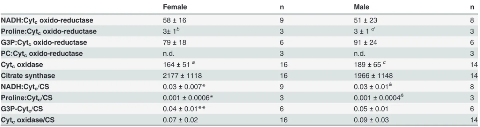
b) Cytochrome c is essentially reduced by means of NADH and G3P
oxidation
as the common intermediate electron acceptor in many organisms (see the substrates depicted in different colors onFig. 1and ref [58]), we assessed the activities of four different electron transport system branches coupled to cytochromecreduction using NADH, G3P, proline, and PC as substrates, as well as the cytochromecoxidase activity.Table 2shows the absolute and CS-normalized electron transport system complex activities found inA.aegyptimitochondria from both sexes. We observed that cytochromecoxidase activity exhibited the highest levels compared to all other electron transport system enzymes, whereas PC oxidation was unable to promote cytochromecreduction. This strongly indicates that lipids are not suitable substrates to support respiration inA.aegyptiflight muscle, as previously suggested [47]. CS activities were high and compatible with values obtained for other insects [59]. The electron transport system enzymatic profile normalized by CS followed a pattern very close to the absolute activity values, with G3P and NADH as the main substrates to allow cytochromecreduction and cyto-chromecoxidase exhibiting the highest activity. These data demonstrate thatA.aegyptiflight muscle mitochondria utilize complex I and G3P dehydrogenase (G3PDH) as the main electron fueling sites to reduce cytochromec. Interestingly, all enzymatic profiles shown inTable 2were roughly the same in both sexes, following the trend observed in mitochondrial protein recovery
(S1 Table) and independent of mitochondrial mass.
c) Pyuvate+proline and G3P are the main substrates to fuel flight muscle
mitochondrial oxygen consumption
Table 1shows the compiled published data on respiration induced by complex I
(NADH-de-pendent) and G3PDH (G3P-de(NADH-de-pendent) from flight muscle of seven different insect species, and vertebrate heart and skeletal muscle during phosphorylating conditions (ADP). A closer
Table 2. Enzyme activities ofA.aegyptiflight muscle mitochondria.
Female n Male n
NADH:Cytcoxido-reductase 58±16 9 51±23 8
Proline:Cytcoxido-reductase 3±1b 3 3±1d 3
G3P:Cytcoxido-reductase 79±18 6 91±24 6
PC:Cytcoxido-reductase n.d. 3 n.d. 3
Cytcoxidase 164±51a 16 189±65c 14
Citrate synthase 2177±1118 16 1966±1148 14
NADH:Cytc/CS 0.03±0.007* 9 0.03±0.01& 8
Proline:Cytc/CS 0.001±0.0006* 3 0.001±0.0004& 3
G3P-Cytc/CS 0.04±0.01** 6 0.05±0.01 6
Cytcoxidase/CS 0.07±0.02 16 0.09±0.03 14
Values were expressed as mean±SD of nmol of products/min/mg protein. Statistical analyses between sexes were performed by using Student´s t or Mann-Whitney´s tests, whereas comparisons of different substrates within the same sex were carried out by using Kruskal-Wallis followed bya posteriori
Dunn´s tests (indicated by superscript letters). Significant differences in“females”were
ap<0.01relative to NADH:Cytcoxido-reductase, bp<0.001relative to Cytcoxidase. In
“males”significant differences were
cp<0.01relative to NADH:Cytcoxido-reductase and
dp<0.001relative to Cytcoxidase. Superscript symbols represent statistical differences of all enzyme activities after normalization by citrate synthase
activity as following:
*p<0.001relative to Cytcoxidase, **p<0.01relative to Cytcoxidase,
&p<0.001relative to Cytcoxidase. n.d. means not detected.
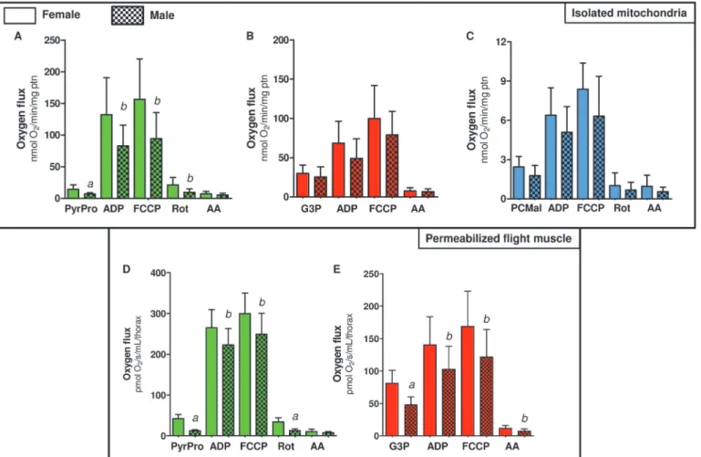
look on these data reveal very interesting patterns, such as the extremely high respiratory rates ofDrosophilamitochondria when use complex I substrates [12,29,31,60], as well as when Locustause G3P [32,33], pointing out the preferential substrates use to fuel flight activity. Cu-riously, our previous data onA.aegyptiindicated that this insect utilized almost equally com-plex I (using pyr+pro) and G3PDH (G3P-dependent) substrates to fuel oxygen consumption [34]. Considering complex I substrates,A.aegyptiflight muscle respiratory rates were markedly lower compared to most insect species, resembling the rates observed in bumblebee [62] and vertebrate mitochondria [64–66]. Thus, in order to improve our understanding of mitochon-drial respiratory capacities and substrates dependences inA.aegyptiflight muscle mitochon-dria, we utilized HRR-SUIT protocols for this purpose [52].
S1 Fig.shows representative oxygen flux traces of flight muscle mitochondria of bothA.
aegyptifemales and males during typical HRR-SUIT experiments. The routine described in the methods section was applied to assess the contribution of three different substrates combina-tions: 10 mM Pyr+Pro; 20 mM G3P, 10μM PC + 5 mM Mal on oxygen fluxes in isolated
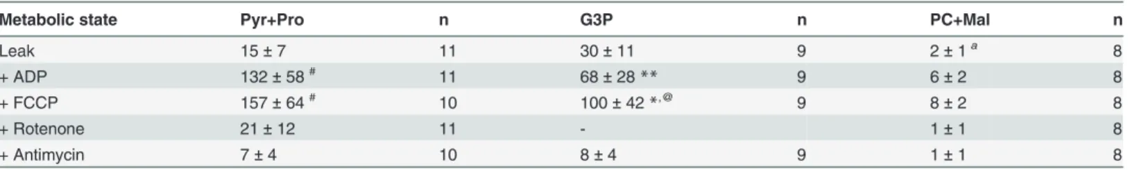
mito-chondria from females (Table 3) and males (S2 Table) ofA.aegypti. When mitochondria from females (Table 3) were incubated only with substrates ("Leak") the respiratory rates were in general low. This metabolic state is defined by a non-phosphorylating respiratory condition that is essentially limited by the magnitude of the protonmotive force (pmf), and the respirato-ry rates are compensated by the proton leak, relieving the inhibitorespirato-ry effect of high pmf on the oxygen flux. The highest oxygen fluxes on both phosphorylating (ADP) and uncoupled (FCCP) metabolic states in female mitochondria were obtained when using Pyr+Pro as sub-strates, followed by G3P, and PC+Mal. Interestingly, the high respiratory rates induced by Pyr +pro may explain the complete depletion of glycogen stores in fat body and flight muscle after flight to exhaustion [67]. On the other hand, the low rates of oxygen fluxes induced by PC+Mal strongly indicates that fatty acid oxidation is not a major pathway to provide the energy re-quired to sustainA.aegyptiflight activity, which is in contrast to other insect species [63]. The limited capacity ofA.aegyptiflight muscle mitochondria to use fatty acid oxidation to sustain
Table 3. Contribution of different substrates to respiration of isolated mitochondria from flight muscle ofA.aegyptifemales.
Metabolic state Pyr+Pro n G3P n PC+Mal n
Leak 15±7 11 30±11 9 2±1a 8
+ ADP 132±58# 11 68±28
** 9 6±2 8
+ FCCP 157±64# 10 100±42
*,@ 9 8±2 8
+ Rotenone 21±12 11 - 1±1 8
+ Antimycin 7±4 10 8±4 9 1±1 8
Values were expressed as mean±SD of nmol oxygen consumed/min/mg protein with the following substrates: 10 mM pyruvate + 10 mM proline (Pyr +Pro), 20 mMsnglycerol-3 phosphate (G3P) or 10μM palmitoylcarnitine + 5 mM malate (PC+Mal). Addition of OXPHOS modulators were indicated as "+" in thefirst column as following: 2 mM ADP (+ADP), 10μM cytochrome c (not shown), 2μM FCCP (+FCCP), 0.5μM rotenone (+Rotenone), andfinally 2.5μg/mL antimycin A (+ Antimycin). For all G3P measurements, experiments started after the addition of 0.5μM rotenone. Statistical analyses were carried out only between the groups of different substrates and mitochondrial metabolic state and were performed by using either Kruskal-Wallis test followed bya posterioriDunn´s test (indicated by superscript letters) or by ANOVA anda posterioriTukey´s test (indicated by superscript symbols). Significant differences in“Leak”were
ap<0.0001, relative to Pyr+Pro and G3P. In
“ADP”, significant differences were **p<0.01relative to Pyr+Pro and PC+Mal,
#p<0.001relative to PC+Mal. In
“FCCP”, significant differences were *p<0.05relative to Pyr+Pro,#p<0.001relative to PC+Mal, @p<0.001relative to PC+Mal.
respiration shown in Tables3andS2is in line with the undetectable capacity of PC to promote cytochromecreduction (Table 2). It is long known that Dipteran insects, the order which be-longA.aegyptiand other mosquitoes, utilize mostly carbohydrate (glucose) and aminoacid (proline) as substrates to sustain flight activity, exhibiting respiratory quotients close to unity [67,68]. Indeed, glycogen stores were depleted in the fat body and flight muscle ofCulex mos-quitoes after flight to exhaustion, despite the fat deposits remained stable [67]. Later, it was demonstrated that particulate fractions ofAedesflight muscle were unable to oxidize -hydroxy-butyrate [47]. More recently, a comprehensive study demonstrated thatAnopheles stephensi mosquitoes were unable to use ketone bodies, as well as octanoate and octanoylcarnitine to sus-tain respiration [35]. Interestingly, comparisons of fatty acid oxidation of these mitochondria with those from locust and mammalian muscle revealed that octanoylcarnitine oxidation is fairly high in these latter two, but completely absent inAnophelesmitochondria [35]. Also, de-spite carnitine play a key role in allowing fatty acid oxidation in flight muscle of some insects [69], its presence and metabolism cannot be directly assumed as a proxy of fatty acid oxidation capacity. A good example in this regard is the blowflyPhormia regina, which is unable to oxi-dize fatty acids to sustain respiration [70], but exhibit high levels of carnitine as well as an ac-tive acetyl carnitine transferase [70]. Unexpectedly, carnitine in this insect revealed to be important for pyruvate metabolism by allowing acetylcarnitine formation from pyruvate decar-boxylation, which prevent CoA and ATP depletion [70]. In this sense, we think that limited fatty acid oxidation inA.aegyptiflight muscle mitochondria is not related to CoA depletion, since most experiments conducted here (with the exception ofTable 2) were carried out in the presence of malate, which generates oxaloacetate and then allow CoA recycling by promoting CS reaction. Therefore, the low contribution of fatty acid to respiration inA.aegypti mitochon-dria is not related to a specific substrate, co-factors depletion or the availability/utilization of other fatty acids. Rather, our data strength the general trend observed in all Dipteran insects that fatty acid oxidation would play a minor (if any) role on respiration in flight muscle mito-chondria of these particular group of insects.
Proline oxidation plays a key energetic role to sustain flight activity in many insect species [37,71] includingA.aegypti[13,34]. In this sense, Figs.2andS5shows that the contribution of different dehydrogenases on the maximum respiratory rates is essentially maintained by com-plex I, using pyr+pro as substrates, and G3PDH activities (Fig. 2A: females>94%;S5A Fig.:
males>97% of total), whereas the direct contribution of ProDH, complex I (when using PC
+Mal as substrates) and ETF:QOR together had a minor role on respiratory rates (Fig. 2A: females>5.7%;S5A Fig.: males>2.8% of total). Indeed, inTable 3we demonstrate that at
than in females, suggesting that metabolism of this aminoacid is distinct among sexes. The sex-ual differences related to mitochondrial physiology, and the respiration driven by proline oxi-dation will be addressed later in this manuscript.
d) Mitochondrial physiology in A. aegypti flight muscle can be studied in
situ without organelle isolation
We next investigated the possibility to study mitochondrial physiologyin situusing whole flight muscle from individualA.aegyptiinsects, instead of isolated mitochondria. Similar ap-proaches have also been applied to study flight muscle mitochondria inDrosophila[73–76] and revealed the enormous potential of this methodology to investigate mitochondrial function in a more physiological way. This approach has a number of advantages, allowing:i)the study of mitochondrial function in the natural cellular environment;ii)the preservation of contacts between mitochondria and other organelles;iii)the assessment of mitochondrial oxygen con-sumption using reduced number of insects, andiv)the bypass of time-consuming and stressful methods employed to obtain enriched mitochondrial preparations. For this sake, we developed a procedure (seemethodssection) to measurein situsubstrate-induced oxygen consumption rates on wholeA.aegyptiflight muscle, based on the literature [74–76]. During our first experi-ments, we observed that physical permeabilization of flight muscle, provided by the magnetic stirring of the respirometer, was enough to allow free access of substrates and OXPHOS
Fig 2. Complex I and G3PDH represent the major electron donor sites to support respiration in femaleA.aegyptiflight muscle.Oxygen
consumption rates from femaleA.aegyptiisolated mitochondria (A) and permeabilized flight muscle (B) were calculated from values shown in Tables3and 4. Data are expressed as mean±SD of at least seven different experiments. Comparisons between groups were done by Kruskal-Wallis anda posteriori
Dunn's tests. Figure (A):ap<0.05 relative to Complex I Pyr+pro;bp<0.05 relative to G3P;cp<0.01, relative to Complex I Pyr+pro;dp<0.001, relative to
Complex I Pyr+pro and G3P. Figure (B):ap<0.001 relative to complex I;bp<0.001 relative to G3P.
inhibitors onA.aegyptiflight muscle, without affecting mitochondrial structure (data not shown). In order to avoid respiratory limitation in flight muscle bundles due to restricted oxy-gen diffusion, we maintained the oxyoxy-gen tension of about 450 nmol/mL during all measure-ments [52].S2 Fig.show two representative HRR-SUIT oxygen flux traces of a singleA.aegypti thorax from female (S2A Fig.) or male (S2B Fig.) using Pyr+Pro as substrates. Interestingly, the effects of all OXPHOS modulators on oxygen consumption observed on isolated mitochondria
(S1 Fig.) were replicated using permeabilized flight muscle, in both sexes using Pyr+Pro (S2
Fig.) or G3P as substrates (S3 Fig.). We then investigated the respiratory pattern of permeabi-lized flight muscle using only these two substrates, since they induced the highest cytochromec reduction and respiratory rates in isolated mitochondria (Tables2,3and Figs.2A,S2,S5A). We assessed the quality of our measurements in terms of structural intactness of mitochondria, by evaluating the effects of two compounds on oxygen fluxes:i)ADP, which would reflect the OXPHOS functionality, andii)cytochromec, which increase the respiratory rates when the structure of mitochondrial outer membrane is compromised [52]. InTable 4, we observed that when female flight muscles were incubated only with substrates ("Leak" condition) the respira-tory rates induced by G3P were significantly higher when compared to Pyr+Pro (p<0.001),
fol-lowing the same pattern with isolated mitochondria (Table 3, "Leak" at G3Pvs. Pyr+Pro). "Leak" respiratory rates were also significantly higher in male flight muscle when using G3P compared to Pyr+Pro (p<0.001) (S3 Table). Also, ADP significantly increased the respiratory
rates, regardless the substrate and sex, and cytochromeccaused no significant change on oxy-gen fluxes, indicating the preserved structure of mitochondria in permeabilized flight muscle (S2andS3Figs). The respiratory rates during phosphorylating (ADP) and uncoupled (FCCP) conditions were significantly higher (p<0.001) when using Pyr+pro as substrates than with
G3P (Table 4). Interestingly, maximum respiration (uncoupled by FCCP) induced by G3P cor-respond to only 43.7% of Pyr+Pro respiratory rates, strengthening the preference of these sub-strates to sustain the high energy demands posed by flight. We also observed that under ADP phosphorylation, the respiratory rates of permeabilized flight muscle using Pyr+Pro and G3P represented 88.3% and 88% of uncoupled oxygen consumption, respectively, indicating that ATP synthesis inA.aegyptiflight muscle demand a large proportion of the maximum respira-tory capacity regardless the substrate. Rotenone and antimycin-insensitive oxygen consump-tion rates represented 11.4% and 3.4%, respectively, of uncoupled respiraconsump-tion, indicating that at least 88% of the electrons in this substrate combination are channeled to the electron transport system by complex I. Indeed, Figs.2BandS5Bshows that the contribution of different
Table 4. Contribution of different substrates to sustain respiration in permeabilized flight muscle fromA.aegyptifemales.
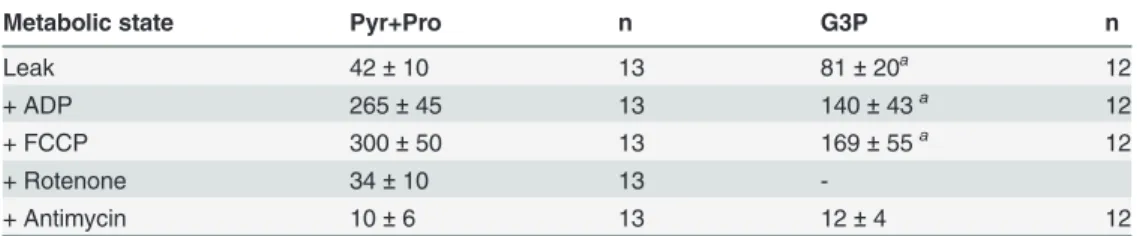
Metabolic state Pyr+Pro n G3P n
Leak 42±10 13 81±20a 12
+ ADP 265±45 13 140±43a 12
+ FCCP 300±50 13 169±55a 12
+ Rotenone 34±10 13
-+ Antimycin 10±6 13 12±4 12
Values were expressed as mean±SD of pmol O2/s/mL/thorax infive different mitochondrial metabolic states using: 10 mM pyruvate + 10 mM proline, 20 mMsnglycerol-3 phosphate, followed by the addition of 2 mM ADP (ADP), 10μM cytochrome c (not shown), 2.5μM FCCP, 0.5μM rotenone, 2.5μg/mL antimycin A. Statistical analyses were performed using Mann-Whitney test.
ap<0.001relative to Pyr+Pro.
dehydrogenases on the maximum respiratory rates in permeabilized muscle, which is essential-ly maintained by complex I, using Pyr+pro, and G3PDH activities in both sexes (Fig. 2B: females>94%;S5B Fig.: males>98% of total), whereas the direct contribution of ProDH to
respiration plays a minor role (Fig. 2B: females>5.3%;S5B Fig.: males>1.4% of total).
Simi-larly to what we found on isolated mitochondria, proline oxidation coupled to respiration was more prominent in females than in males, strengthening the concept that females were more adapted to utilize this aminoacid to sustain respiration [13,25]. Finally, a comparison of respi-ratory data obtained for whole flight muscle and isolated mitochondria for both sexes using Pyr+pro or G3P (S6 Fig.) revealed the striking proportionality of oxygen fluxes obtained in both approaches, clearly indicating that mitochondrial physiology inA.aegyptiflight muscle can be assessedin situwithout organelle isolation.
e) Flight muscle exhibit similar mitochondrial bioenergetic efficiencies
and distinct capacities among substrates
Mitochondrial ATP production is determined by their bioenergetic efficiency (defined as the ATP produced in mitochondria per molecule of nutrient) and their ATP synthesis capacity (defined as the rate of ATP produced in mitochondria per unit of time), which are both regu-lated by the cellular energy demand and supply. Assuming that insect flight plays a central eco-logical role to reproduction and dispersal, representing one of the most energy demanding processes in Animal Kingdom [77], flight muscle high respiratory capacity must be tightly cou-pled to ATP synthesis. In this regard, one can speculate that efficiency and capacity of mito-chondrial ATP production would vary among different substrates, being regulated byi)the substrate transport to mitochondria;ii)the oxidation potential provided by the mitochondrial dehydrogenases and the electron transport system complexes andiii)the degree of electron transport system coupling to OXPHOS.
literature to investigate the effect of hypoxia on mitochondrial physiology in permeabilized human skeletal muscle fibers during exercise [52,58].S7 Fig.shows the correlation analyses carried out for female (S7A–C Fig.) and male (S7D–F Fig.) isolated mitochondria using Pyr +Pro (S7A and S7D Fig.), G3P (S7B and S7E Fig.) and PC+Mal (S7C and S7F Fig.) as sub-strates. We observed that in all cases, there is a linear correlation between the OXPHOS and maximum uncoupled respiratory rates, which varied in a direct proportion, indicating pre-served coupling between these two distinct mitochondrial metabolic states regardless the sub-strate utilized and sex. We also conducted the same analyses on permeabilized flight muscle of both sexes using Pyr+Pro (S7G and S7I Fig.) and G3P (S7H and S7J Fig.) and a pattern very similar to that of isolated mitochondria was observed. A closer look on these data revealed in-teresting features of mitochondrial functionality inA.aegyptiflight muscle.Table 5shows the compiled bioenergetic efficiency (slope) and capacity (OXPHOS) values of female mitochon-dria for all comparisons among the substrates and preparations. First, the bioenergetic capaci-ties are remarkably distinct among the substrates (as previously shown in Tables3and4), from the lowest values with PC+Mal to the highest with Pyr+Pro, indicating that the extent of sub-strate oxidation play a central role to bioenergetic capacity. Second, the bioenergetic efficiency, determined as the slope of correlation analyses, is high, ranging from 0.46 (G3P) to 0.74 (Pyr +Pro) both in permeabilized flight muscle (Table 5). These data suggest that bioenergetic ca-pacity among the substrates is essentially driven by their potential to be oxidized by the mito-chondrial dehydrogenases and the electron transport system complex activities, and not by the degree of mitochondrial inner membrane permeability, since the bioenergetic efficiency (slope) is roughly preserved. In males, a very similar pattern of bioenergetic efficiency and capacity among substrates and preparations was achieved (S7D-F, S7I, S7J Fig.andS4 Table). The bio-logical significance of these findings is that, regardless the sexual differences ofA.aegypti, sub-strates oxidation provided by mitochondrial dehydrogenases and the electron transport system activity define the bioenergetic capacity, but not efficiency as the degree of mitochondrial inner
Table 5. Mitochondrial bioenergetic efficiency and capacity inA.aegyptifemales flight muscle using different substrates.
Isolated mitochondria
Bioenergetic efficiency (Slope) Correlation coefficient p Bioenergetic capacity (OXPHOS)
Pyr+Pro 0.67±0.17 0.78b 0.004 118±57
*
G3P 0.55±0.08 0.77a 0.0002 39±25
**
PC+Mal 0.72±0.23 0.79b 0.021 4±1
Flight muscle
Pyr+Pro 0.74±0.14 0.83b 0.0004 223±45#
G3P 0.46±0.06 0.84b <0.0001 59±27
Values of bioenergetic efficiency (slope) were expressed as mean±SD of OXPHOS versus maximum uncoupled respiratory rate linear regression and correlation analyses made inS7 Fig.Correlation coefficient values (Spearman or Pearson) were depicted as superscript letters“a”or“b”, respectively. P values represent the statistical significance of linear regression slopes in each group. Bioenergetic capacity (OXPHOS) values represent the respiratory rates data induced by ADP and calculated by subtracting the ADP rates by their equivalent "leak" values shown inTable 3(for isolated mitochondria) and Table 4(forflight muscle). Statistical analyses on bioenergetic capacity in isolated mitochondria were performed by using Mann Whitney test (indicated by superscript astersiks) as well as ANOVA anda posterioriTukey´s test (indicated by superscript symbols) forflight muscle. Significant differences in isolated mitochondria were
*p<0.005relative to G3P and PC+Mal, and
**p<0.0001relative to PC+Mal. Inflight muscle, significant difference was #p<0.001relative to G3P.
membrane permeability is preserved. Also, these data strongly suggest that proline and glucose metabolism represent the main energy-providing pathways to sustain flight in this insect [13,
28,34,47,67].
f) Mitochondrial metabolic preference towards proline oxidation in
females
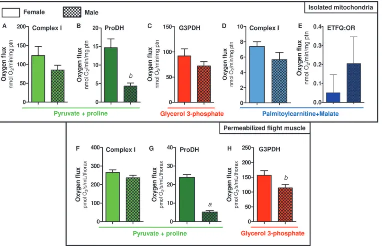
We next investigated whether mitochondrial oxygen consumption would be distinct among sexes inA.aegyptiflight muscle. Previous reports have already addressed the sex-associated mi-tochondrial structural and functional differences in a variety of organisms and tissues [79–84]. Although some reports support the concept of sex-associated differences in mitochondrial function [76,79–83], consensus about a general trend is lacking. For example, despite the levels of antioxidant defenses in rats are reduced, and redox imbalance markers are higher in males [84], mitochondrial functional parameters were quite similar in three mice tissues regardless the sex [83]. InDrosophilamales, the rate of mitochondrial H2O2formation is significantly lower than in females, which is consistent with higher antioxidant enzyme activities (catalase, Mn-SOD and Cu/Zn-SOD) in males [85]. In addition, OXPHOS efficiency and proton leak were higher in females compared to males, indicating that mitochondrial functional differences among sexes may reflect the increased energy demand posed by sex-specific activities, such as reproduction in females. InA.aegyptiflight muscle, we observed a general trend of lower respi-ratory rates in males compared to females, in all substrates tested (Fig. 3). However, significant-ly reduced respiratory rates in males were onsignificant-ly observed when using Pyr+pro (Fig. 3A and 3D) and G3P (Fig. 3E). Respiratory rates of male isolated mitochondria using Pyr+pro were about 38% lower than in females, while in G3P and PC+Mal these rates were about 22% lower com-pared to females. Interestingly, we observed that respiratory rates of female permeabilized flight muscle were significantly higher than in males, using both Pyr+pro (Fig. 3D) or G3P (Fig. 3E) as substrates.
A closer look on the data shown inFig. 3reveal that the contribution of proline oxidation to respiration was strikingly higher in females. The respiratory rates induced by Pyr+pro in both isolated mitochondria (Fig. 3A) and permeabilized flight muscle (Fig. 3D) were significantly higher in females than in males (2.06 and 3.5 times higher than males, respectively). In addi-tion, the rotenone-resistant respiration was higher in females (2.2 and 2.7 times higher than males, respectively) in both preparations (Fig. 3A and 3D). Indeed, assignment of mitochondri-al dehydrogenases that provide electrons to the electron transport system (Fig. 4) indicated that respiration associated to ProDH activity was 3.4 times higher in female isolated mitochon-dria compared to males (Fig. 4B). This effect was specific to the respiration associated to ProDH since there were no apparent differences among the sexes in all three other sites of elec-tron transport system (complex I, G3PDH and ETF:QOR)(Fig. 4A,4C,4D and 4E). A very simi-lar trend was observed when the contribution of different dehydrogenases was assessed on permeabilized flight muscle (Fig. 4F-H). Noteworthy is the specifically high contribution of proline oxidation to respiration in female mitochondria (Fig. 4G), which was about 4.6 times higher than in males. Since blood feeding habit inA.aegyptiis restricted to females, it seems that proline metabolism would play a key role in female physiology, as the increment of respi-ratory rates induced by this aminoacid is higher in obligatory blood feeding Diptera when com-pared to other facultative blood-suckers and even to non hematophagous insects [25].
flight-induced reductions in proline levels in the hemolymph and thorax of maleA.aegyptiwere much less pronounced than observed in females [13], indicating that oxidation of this aminoa-cid in flight muscle is more intense in females. Conceivably, proline transport across inner mi-tochondrial membrane in males would be limited, directly affecting respiration, since there were no changes in proline-induced cytochromecreduction among sexes (Table 2). In this sense, although in the present work mitochondrial function was assessed only in sugar fed insects, it seems possible that substrates preferences may change after blood ingestion, due to the higher protein content in this dietary source. The relatively high respiratory rates in-duced by G3P inA.aegyptiflight muscle (Fig. 4C and 4H) suggest increased dependence of this tissue to glucose utilization, as in insect´s flight muscle the cytosolic isoform of G3PDH is largely responsible for the re-oxidation of extramitochondrial glycolysis-derived NADH in-stead of classical lactate dehydrogenase [86,87]. However, despite the advantages of assessing mitochondrial physiology on permeabilized flight muscle, we are aware about the potential limitations with this approach in terms of the respiratory capacities among sexes, since a clear sexual size dimorphism exists inA.aegypti(S1 Table). In this regard, correlating the uncoupled
Fig 3. Comparative analyses of respiratory rates induced by different substrates amongA.aegyptisexes.Oxygen consumption rates from isolated mitochondria (A-C) and whole permeabilized flight muscle (D and E) from females (solid bars) and males (hatched bars) were plotted from values shown in Tables3,4,S2andS3. Data are expressed as mean±SD of at least six different experiments. Comparisons between groups were done by Student´s t- test. Figure (A):ap<0.005 andbp<0.05 relative to their equivalent metabolic state in female. Figure (D):ap<0.0001 andbp<0.05 relative to their equivalent
metabolic state in female. Figure (E):ap<0.001 andbp<0.05 relative to their equivalent metabolic state in female.
respiratory rates of whole flight muscle from individual insects of both sexes using Pyr+pro as substrates, by their respective whole body weight, gives a linear positive relationship (S8 Fig.). This indicates that permeabilized flight muscle respiration varies in a direct proportion with the insect mass, regardless the sex. Since body weight in females is higher than in males (S1
Table), this could explain the differences in respiratory rates observed in permeabilized flight
muscle among sexes. We think that this would be the case for the sexual comparisons of abso-lute values observed for most metabolic states in permeabilized flight muscle (Figs.3E,4F, and 4H), but not for the ProDH-dependent respiration (Figs.3Dand4G). Indeed, the sexual differ-ences in respiratory rates provided specifically by ProDH (Fig. 4G, females 4.6 times higher than males) overpass the predicted sexual difference in respiratory rates due only to the weight
(S1 Table, females 1.7 times higher than males). Therefore, we conclude that there is a clear
preference towards proline oxidation in female flight muscle mitochondria compared to males. The biological significance of these data is that specific reduced proline oxidation and respira-tory rates observed in male flight muscle may explain their limited flight capacity in nature when compared to females [88].
Fig 4. Preference towards proline oxidation inA.aegyptifemale mitochondria.Oxygen consumption rates from isolated mitochondria (A-E) and whole permeabilized flight muscle (F-H) from females (solid bars) and males (hatched bars) were calculated from values shown inFig. 3. Data are expressed as mean±SD of at least seven different experiments. Comparisons between groups were done by Student´s t tests. Figure (B):bp<0.001 relative to female;
Figure (G):ap<0.0001 relative to female; Figure (H):bp<0.038 relative to female.
g) An unique mitochondrial redox feature: H
2O
2production is essentially
driven by pyruvate+proline and G3P oxidation, with equivalent
contribution among these substrates
The contribution of mitochondria to cell physiology goes far beyond their classical role in ener-gy metabolism, directly participating in cell signaling, apoptosis, and on redox homeostasis, representing an important source of ROS. Indeed, superoxide (O2•) is the primary ROS pro-duced in at least ten different sites in mitochondria (see purple arrows inFig. 1and refs [29,31,
37,89–93]). Few studies were dedicated to understand the contribution of mitochondria to
cel-lular ROS production in insects, despite their implication in a myriad of biological processes in these organisms including aging [12,94–98], development [99], hypoxia tolerance [100], apo-ptosis [101], muscular and neuronal function [102], ecdysteroid synthesis [103], as well as on immune response [44–46]. Regarding the mechanisms involved on mitochondrial ROS genera-tion in insects, early work conducted inMusca domesticaflight muscle demonstrated that hy-drogen peroxide (H2O2) generation was mostly supported by G3P (16-times higher) than Pyr +pro oxidation [94]. Later, Miwa and co-workers have showed that inDrosophilaflight muscle mitochondria H2O2formation occurred mainly by G3PDH and the centeroof complex III (12-times higher), followed by complex I ROS generation to the mitochondrial matrix side (see
Table 2of ref [29]). Recent evidence also point out that mitochondrial proline oxidation
through ProDH contributes to indirect O2•production particularly at complexes I and II [37]. Given the relevance of mitochondrial redox metabolism on insect physiology, inTable 6we compiled the mitochondrial H2O2production data available on insects and compared with ver-tebrate muscle mitochondria [12,29,31,90,94–98,101–106]. Interestingly, general trends can be pointed out here, such as:i)the rates of H2O2formation supported by G3P were strikingly higher than by complex I substrates, regardless the species;ii)H2O2production rates by G3P in insects are much higher than in vertebrate skeletal muscle, but close to the rates reported for heart mitochondria. Considering that reported respiratory rates induced by G3P inDrosophila are 52,7% lower than those produced by Pyr+pro oxidation (Table 1), while H2O2production rates by G3P are 567% higher than by complex I (Table 6), it seems plausible that electron flow through G3PDH is more "leaky" than by complex I, resulting in increased O2•formation at both sides of mitochondrial inner membrane [12,29,31,90,94–98,101–106]. In fact, the con-tribution of different substrates to mitochondrial H2O2formation has been investigated for de-cades and the intrinsic complexity of the system makes the determination of absolute values
Table 6. Comparison of mitochondrial H2O2production by insect flight muscle and vertebrate muscles.
NADH-dependent G3P-dependent
Fruitfly(Drosophila)[12,29,31,96] 265 1503 Housefly(Musca)[95–98]
70 1195
Fleshfly(Sarcophaga)[97] - 1950
Blowfly(Calliphora)[97] - 2150
Vertebrate heart[90,104–106] 158 1687
Vertebrate skeletal muscle[104,106] 165 590
Mitochondrial hydrogen peroxide formation was expressed as pmol of H2O2/min/mg protein during non-phosphorylating conditions. In the case ofDrosophila,Muscaand vertebrate muscles, in which multiple articles were utilized for calculations, an average value was obtained. Superscript letters indicate the data source.
highly variable, depending on many parameters such as the organism model, the experimental conditions, substrates transport and oxidation, theΔѰ
mmagnitude, and mitochondrial dy-namics [104,107–110].
Our group have previously shown that blood feeding promoted not only mitochondrial fu-sion inA.aegyptiflight muscle, but also a drastic reduction in cytochromecoxidase activity, mitochondrial oxygen consumption and H2O2formation [34]. We speculated that functional and structural mitochondrial remodeling upon blood feeding would avoid the interaction of mitochondrial-derived ROS with blood derived products, such as heme and iron, which could lead to redox imbalance and eventually tissue damage [34,102,111]. Thus, in order to gain a deeper insight on the contribution of mitochondria toA.aegyptiredox metabolism, we as-sessed the H2O2production on isolated flight muscle mitochondria.S4 Fig.show representa-tive fluorimetric traces of H2O2formation by male and female mitochondria fromA.aegypti flight muscle using Pyr+pro as substrates. H2O2generation rates during phosphorylating con-ditions proceeded stably, which were boosted after F1FoATP synthase inhibition by oligomy-cin, and subsequently reduced by OXPHOS uncoupling promoted by the proton ionophore FCCP. Then, for Pyr+pro and PC+Mal, complex I inhibitor rotenone was added, causing small increases on H2O2generation rates, which were further increased upon complex III inhibition by antimycin A, and resulting in the maximal rates of H2O2production [108,112]. When using G3P as substrate, experiments started by the addition of 0.5μM rotenone.Table 7shows
that mitochondrial H2O2formation rates in both sexes were in general 20–80% higher during non-phosphorylating conditions (oligomycin) than after uncoupling by FCCP. However, these differences were only significant when using G3P (on both sexes) or PC+Mal (only females), indicating their higher dependence onΔѰmto generate O2•. This contrasts to data obtained with mice skeletal muscle mitochondria oxidizing long chain fatty acids, which revealed to be only slightly affected by reductions in theΔѰm[113]. Mitochondrial H2O2production rates in A.aegyptiflight muscle induced by Pyr+pro or G3P during non-phosphorylating (oligomycin) conditions were indistinguishable among each other, and significantly higher than with PC +Mal (Table 7). Indeed, a comprehensive investigation in different rat tissues revealed that fatty acid oxidation plays a major role in mitochondrial H2O2production only in kidney and liver [104]. These data indicate that fatty acid oxidation plays a minor role on cytochromec re-duction (Table 1), respiration (Tables3,S2,S4Figs., Figs.S5A,S7C, S7F,2A,3C,4D, 4E) and H2O2formation (Table 7) inA.aegyptiflight muscle mitochondria. Comparative analyses be-tween Tables6and7show that NADH-dependent H2O2generation inA.aegypti mitochon-dria is higher than any other organism (about 49% and 464% higher thanDrosophilaand Musca, respectively). Considering that the respiratory rates mediated by complex I substrates inA.aegyptiflight muscle were lower than in other insects species (Tables1,3andS2), we sug-gest that redox reactions that provide NADH to complex I such as pyruvate dehydrogenase andα-ketoglutarate dehydrogenase, might be involved in controlling both respiration and H2O2generation. On the other hand, mitochondrial H2O2generation rates inA.aegyptiusing G3P were strikingly lower when compared to other insects (80% and 72% lower than Calli-phoraandDrosophila, respectively) and even to vertebrate mitochondria (28% and 75% lower than skeletal muscle and heart, respectively). InDrosophilamitochondria, G3P-induced H2O2 production was 7.6 times higher than with Pyr+Pro (seeTable 2on ref [29]), and was also less ΔѰ
mdependent than complex I [29]. Interestingly, as previously pointed out (Tables1,3and
+pro and G3P are the main substrates that drive mitochondrial H2O2production inA.aegypti flight muscle mitochondria, which were both equivalent in terms of their capacity to generate ROS (Table 7). This feature, instead of the usual preference towards G3P oxidation to produce H2O2(Tables6and7), represents a unique mitochondrial redox feature present inA.aegypti flight muscle.
h) The major sites of superoxide production mediated by proline and
G3P oxidation takes place at distinct points other than site I
FConsidering that mitochondrial electron leak take place in at least ten different sites [29,31,37,
89–91,93,104,106,112–114]), we next investigated the topology of H2O2production inA.
aegyptiflight muscle mitochondria.Table 8shows the effects of rotenone, and subsequently an-timycin A, on H2O2generation in mitochondria using Pyr+pro, G3P and PC+Mal under un-coupled (FCCP) conditions. This approach has some advantages in terms of assessing mitochondrial redox metabolism, since uncoupling keep the electron transport system en-zymes in a state that would not be inhibited by their products, as well as to consume NAD(P) H, which lowers the transhydrogenase activity and then the pool of reduced glutathione. A gen-eral trend observed is that inhibition of complex I by rotenone caused only slight increases (about 25%) on Pyr+pro-induced H2O2formation, regardless the sex. It is known that complex I produce O2•at IFand IQsites [114], which are stimulated by increased NADH/NAD+ratio and high protonmotive force (pmf), respectively. Since O2•generation at site IQrequires high pmf to allow reverse electron transfer to complex I, and inTable 8all measurements were con-ducted in the presence of FCCP, we conclude that induction of H2O2generation caused by ro-tenone can only be accounted by the IFsite of complex I. Significant increases in rotenone-induced H2O2generation were only achieved in female mitochondria when using PC+Mal as Table 7. Contribution of different substrates to mitochondrial H2O2production inA.aegyptiflight muscle.
Female
Modulator Pyr+Pro n G3P n PC+Mal n
Oligomycin 371±112 5 369±82** 9 112±28a, # 5
+ FCCP 207±104 5 235±42 9 66±23c 5
Male
Modulator Pyr+Pro n G3P n PC+Mal n
Oligomycin 419±94 5 473±62* 9 128±19b 5
+ FCCP 271±111 5 396±61 9 98±19d 5
Values were expressed as mean±SD of pmol hydrogen peroxide produced/min/mg protein in two different mitochondrial metabolic states using: 10 mM pyruvate + 10 mM proline (Pyr+pro), 20 mMsnglycerol-3 phosphate (G3P), or 10μM palmitoylcarnitine + 5 mM malate (PC+Mal) followed by 2 mM ADP, 4μg/mL oligomycin (Oligo), and 2μM FCCP. For all G3P experiments, measurements were carried out after addition of 0.5μM rotenone. Statistical analyses were carried out between the groups of different substrates and mitochondrial metabolic state within the same sex, and were performed by using Kruskal-Wallis test followed bya posterioriDunn´s test (indicated by superscript letters). Significant difference in“Oligo”was
ap = 0.0052, relative to Pyr+pro and G3P, bp = 0.0045, relative to G3P. Signi
ficant differences in“FCCP”was;
cp = 0.0065, relative to G3P;;
dp = 0.0016, relative to G3P. Analyses were also conducted between the two mitochondrial metabolic states (oligo vs. FCCP) within the same substrate
and sex and were performed by using Mann Whitney test (indicated by superscript symbols). Significant differences in G3P were**p<0.0001 relative to
female FCCP;
*p= 0.016 relative to male FCCP. Significant difference in PC+Mal were #p= 0.03 relative to female FCCP;