D
espite being more remarkable at adult age, the aging process begins since birth, and continues throughout the whole life, due to genetic and ex-ternal factors, such as UV exposure and smoking. The face, in most cultures, is one of the most exposed areas of our bodies, so that the aging process is not only earlier, but more easily recognizable. The aging process comprises all layers of the face: skin, subcutaneous fat, muscles, and skeleton, and the signs of aging depend mainly on which layer is mostly affected. Aging may affect the skin in terms of quantity (skin sagging) and/or quality (changes in skin hydration, appearance of spots and wrinkles). There is a reduction of the collagen content of the skin, not only due to decrease in synthesis, as a process of intrinsic aging, but also due to extrinsic factors, such as UV radiation, which may generate reactive oxygen species (ROS), leading toan increment in collagen breakdown by upregulating en-zymes called matrix metalloproteinases.1 The decrease of collagen content may also affect deeper layers of the face. The loss of the subcutaneous facial fat compartments with age leads to the appearance of increased skin laxity or prominent folds around the nasolabial region, periorbital region, and jowl.2 The muscles may present hypo- or hy-pertonicity (i.e., glabella, masseter) depending upon the use and area of the face. The bone is a dynamic, sensitive, ever-changing tissue. Bone growth takes place from birth until the hormonal stimulus ceases, with long bone epiphy-sis consolidation usually around 15–18 years of age. On the other hand, bone remodeling continues throughout life, determined not by intrinsic factors, but mainly by regional changes in the soft tissues related to each bone, such as muscles, tongue, lips, skin, brain, among others.3,4 Bone remodeling serves to adjust bone architecture to meet changing mechanical needs, repair microdamages, and guarantee calcium homeostasis.5 Therefore, it is transitory and does not involve the totality of the bone. The process of bone remodeling involves the removal of mineralized bone by osteoclasts and the formation of bone matrix through the osteoblasts. Both processes of bone remodel-ing occur throughout life, although the balance between them changes according to the period of life. Bone for-mation is more prominent in childhood; the processes From the *Department of Anthropology, Civil Police Department;
†Department of Anatomy, School of Medicine of Barbacena; ‡Department of Anatomy, Federal University of Minas Gerais (UFMG); §Department of Anatomy, School of Health and Human Ecology (FASEH); ¶Department of Anatomy, Federal University of Ouro Preto (UFOP), Minas Gerais, Brazil; and ‖University of Itaúna, Minas Gerais, Brazil.
Received for publication December 5, 2016; accepted February 24, 2017.
Copyright © 2017 The Authors. Published by Wolters Kluwer Health, Inc. on behalf of The American Society of Plastic Surgeons. This is an open-access article distributed under the terms of the Creative Commons Attribution-Non Commercial-No Derivatives License 4.0 (CCBY-NC-ND), where it is permissible to download and share the work provided it is properly cited. The work cannot be changed in any way or used commercially without permission from the journal. DOI: 10.1097/GOX.0000000000001297
Background: The aging process of the face comprises all layers: skin, subcutaneous fat, muscles, and skeleton, and the signs of aging depend mainly on which layer is mostly affected.
Objective: To evaluate the aging facial skeleton, as well as establish the sexual dif-ferences, areas with a strong predisposition to resorption, and aesthetic repercus-sion for better treatment approach.
Methods: Skulls from the Forensic Anthropology Department of the Institute of Forensic Medicine of Belo Horizonte, Brazil, were classified according to gender and age group (i.e., <20 years, 20–50 years, >50 years). Structural changes were classified according to gender and age group.
Results: Of the 241 skulls included, 192 were male skulls and 49 female. Sexual dimorphism and age-related peculiarities are described herein.
Conclusions: The knowledge of the anatomy of the aging face, taking into con-sideration all the layers (skin, fat pads, muscles, and bones), as a whole, for the treatment of folds and shadows is vital for a better and more natural final aes-thetic outcome. (Plast Reconstr Surg Glob Open 2017;5:e1297; doi: 10.1097/ GOX.0000000000001297; Published online 27 April 2017.)
Luiz Eduardo Toledo Avelar, MD* Márcio Alberto Cardoso, MD†‡§ Leonardo Santos Bordoni, PhD‡§¶ Lorena de Miranda Avelar§‖
João Victor de Miranda Avelar§‖
Aging and Sexual Differences of the Human Skull
Disclosure: The authors have no financial interest to declare in relation to the content of this article. This study was supported by departmental resources. The Article Process-ing Charge was paid for by an institutional grant from Gal-derma Brazil. GalGal-derma had no role in the design, collection, management, analysis, and interpretation of data.
PRS Global Open
•
2017
are balanced in adults, although bone resorption is more prominent in the elderly. Among the theories of bone re-modeling, the Functional Theory correlates bone remod-eling to response to demand. When the bone is submitted to traction by a hypertrophic muscle or subcutaneous fat
distension, a local demand is generated and bone is pro-duced in that area. On the other hand, continuous pres-sure on the bone (e.g., silicon chin prosthesis) or intense muscle, ligament or skin laxity, may lead to bone resorp-tion.3,4 The objective of this study is to evaluate the aging facial skeleton, and establish the sexual differences, areas with a strong predisposition to resorption and aesthetic re-percussion for a better treatment approach.
METHODS
The study was approved by the national research ethic committee (number CAAE: 55561816.5.0000.5119).
Between 2010 and 2015, skulls from the Forensic An-thropology Department of the Institute of Forensic Medi-cine of Belo Horizonte, Brazil, were evaluated and only intact skulls, with available clinical data, were included in the study. The skulls were further classified according to gender and age group (i.e., <20 years, 20–50 years, >50 years). Structural changes were then evaluated according to gender and age group.
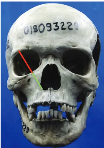
Measurement of the distance from the superior orbital rim to the inferior orbital rim (OM) (Fig. 1)6 and from the inferior orbital rim to the lateral inferior portion of the pyriform aperture was performed (MM). The MM/ OM ratio was calculated.
RESULTS
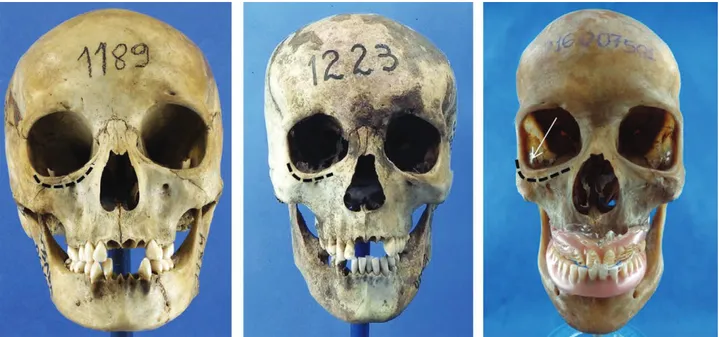
Overall, 241 skulls were included, being 192 male and 49 female (Fig. 2). The distribution among age groups and gender is described in Table 1.
Sexual Dimorphism
Among women, the forehead is straighter (Fig. 2, Table 2), the glabella is more curved and less pronounced, and the supraorbital rim is less noticeable than in men.
The midface in women presents more subtle angles, whereas the midface in men presents more pronounced zygomatic bones (Fig. 3). In men the mandible is larger, stronger, and more faceted and has more clear-cut angles Fig. 1. anthropometric lines: OM, orbital medial (red line); MM,
maxil-lary medial (green line). the MM/OM ratio depicts the increase in orbit and pyriform aperture size, due to maxillary resorption during aging.
than in women. Also, the chin is larger, giving men a square-shaped face, whereas women tend to have a trian-gular or heart-shaped face.
Aging Skull
The pyriform aperture becomes larger with aging, ex-periencing remodeling of lateral and inferior walls, where-as the upper and olfactory cavities remain intact, so that the nasal cavity presents a lateral and anterior expansion and the palatus is reallocated inferiorly. The maximum nasal aperture (the length of the base of the pyriform ap-erture) increases with age (Fig. 4).
With the process of aging, the floor of the orbit ex-pands inferiorly and laterally, losing the round shape
observed in younger skulls (Fig. 4). The lacrimal bone works as a pivot: its inferior portion rotates laterally, leading to an inferior slide of the maxilla. The maxilla is more anterior and more prominent in younger skulls, whereas in older skulls an anterior and inferior bone resorption takes place, giving the appearance of a retru-sion of the face (Fig. 5). The zygoma presents an an-terior resorption (Fig. 6), whereas the zygomatic arch suffers a posterior and anterior remodeling, increasing the temporal fossa. The MM/OM ratio increases from <20 years to 20–50 years and decreases in >50 years group (Table 3).
The mandible undergoes a posterior and superior bone formation, although it suffers an anterior and infe-rior resorption (Fig. 5). Without the presence of teeth, the mandible experiences further atrophy. The angle of the mandible increases with age (97 degrees in younger skulls to 135 degrees in older skulls), mainly due to resorption of the inferior border of the angle, next to body–ramus junction (Fig. 7). Also, the chin becomes more anterior, oblique, and shorter with age (Fig. 7).
Not all the bones in the skull suffer resorption. Al-though the midface recedes, the forehead suffers
contin-Table 1. Distribution of Skulls According to Gender and Age Group
Age Group Male Female
<20 y 23 08
20–50 y 120 28
>50 y 49 13
Total 192 49
Table 2. Sexual Dimorphism Among Skulls
Area Female Male
Forehead More straight More oblique
Glabella Curved and subtle Frontal-nasal suture is more prominent Supraorbital rim Less conspicuous More striking
Midface Subtle angles Irregular surface and little anterior projection Zygoma More prominent and curvilinear Less prominent
Mandible Lighter, with subtle angles Larger, stronger, with clear-cut angles Chin Smaller and rounded shape Larger and square-shaped
PRS Global Open
•
2017
uous expansion, due to bone deposition in the external wall of the frontal bone, especially in the supraorbital rim (Fig. 8).
DISCUSSION
The sexual dimorphism among human skulls is a well-established subject in anthropology. These differences have aided in the distinction of male and female skulls (Fig. 2). Nevertheless, it is the array (Table 2), and not single characteristic that allows gender determination of skulls.7,8
The correct knowledge of gender differences of the hu-man skull is vital in the aesthetic approach to aging, because exaggerated filling of particular areas may render a mascu-line appearance to women, for example. A prominent supra-orbital region is characteristic of men (Fig. 2), so that filling these areas in women must be careful and subtle, only to re-store the retro-orbicular fat loss. On the other hand, when treating the aging midface, it is imperative to remember that a more curvilinear prominent cheekbone is peculiar to wom-en, whereas men present a more angled face, due to strong muscle insertions.
Fig. 4. Orbit and piriform aperture. the rounded orbit shape initially increases its size, while maintaining the shape. later the latero-inferior border presents a more pronounced resorption. the maximum measurement of piriform aperture increases with aging, being prominent in skulls >50 years.
Furthermore, deep knowledge of the anatomy of the aging face allows a better approach to the underlying cause of the fold, rendering better and more natural sults than just simply filling up the fold. The maxillary re-trusion, associated with the decrease in collagen content of the skin and loss of fat compartments, works as a true slide for soft-tissue descent (Figs. 5, 6).9 Because the zy-goma works as a support of the soft tissue of the midface, restoring the volume of the zygomatic prominence would
not only improve the local area, giving a youthful and more natural appearance, but would also improve10,11 and minimize the need to treat the nasolabial fold itself. The changes in MM/OM ratio observed in this study reflect the increase in orbit and pyriform aperture size, at the ex-pense of the maxillary bone in the process of aging, cor-roborating the findings of previous authors.6,12,13 High G prime hyaluronic acid or poly-L-lactic acid injections deep in the supraperiosteal area and in the pyriform fossa help soften the lengthening of the nose, drooping of the nose tip, and enlargement of the nostrils.
The orbit does not age homogeneously. The superi-or-medial and inferior-lateral aspects of the orbit have the greatest tendency to resorb, which is associated with soft-tissue laxity that occurs with aging, leading to an increased prominence of the medial fat pad and length-Fig. 6. Zygoma. Considering the skull in anatomic position, the zygoma becomes more retropositioned with aging, with an increasing angle between an imaginary vertical line and the anterior border of the zygoma.
Table 3. MM/OM Ratio
Age Group Male (n = 192) Female (n = 49)
<20 y 0.75 0.78
20–50 y 0.77 0.77
>50 y 0.68 0.71
N = 241.
PRS Global Open
•
2017
ening of the lid–cheek junction, giving the patient a sad appearance.13–16
The facial remodeling depends entirely on growth, de-velopment, and overlying soft-tissue function. All bone vol-ume or size changes throughout life occur in response to functional demand of the face.17,18 Indeed, loss of teeth, due to infection, trauma, or aging, increases local bone resorp-tion and is listed among the major bone factors that impact aesthetic outcome.19,20 The decrease in mandible volume and shortening of the chin, due to decrease in bone support, lead to further soft-tissue excess and sagging (Fig. 7).21,22
CONCLUSIONS
The clinical approach to the aging face has changed in the past years.2 The knowledge of the anatomy of the aging face, taking into consideration all the layers (skin, fat pads, muscles and bones), as a whole, for the treatment of folds and shadows, is vital for a better and more natural final aesthetic outcome.
Luiz Eduardo Toledo Avelar, MD Department of Anthropology and Plastic Surgery Police Department of Minas Gerais Avenida do Contorno, 4852 30110-100 Belo Horizonte-MG, Brazil E-mail: [email protected]
REFERENCES
1. Helfrich YR, Sachs DL, Voorhees JJ. Overview of skin aging and photoaging. Dermatol Nurs. 2008;20:177–183; quiz 184.
2. Farkas JP, Pessa JE, Hubbard B, et al. The science and theory be-hind facial aging. Plast Reconstr Surg Glob Open 2013;1:e8–e15. 3. Enlow DH. The Human Face. New York: Harper and Row; 1968. 4. Enlow DH, Hans MG. Noções Básicas de Crescimento Facial. 1st ed.
São Paulo: Santos;1998.
5. Rucci N. Molecular biology of bone remodelling. Clin Cases Miner Bone Metab. 2008;5:49–56.
6. Pessa JE, Zadoo VP, Yuan C, et al. Concertina effect and facial aging: nonlinear aspects of youthfulness and skeletal remodel-ing, and why, perhaps, infants have jowls. Plast Reconstr Surg. 1999;103:635–644.
7. Rosas A, Bastir M, Martínez-Maza C, et al. Sexual dimorphism in the Atapuerca-SH hominids: the evidence from the mandi-bles. J Hum Evol. 2002;42:451–474.
8. Bartlett SP, Grossman R, Whitaker LA. Age-related changes of the craniofacial skeleton: an anthropometric and histologic anal-ysis. Plast Reconstr Surg. 1992;90:592–600.
9. Pessa JE, Zadoo VP, Mutimer KL, et al. Relative maxillary retru-sion as a natural consequence of aging: combining skeletal and soft-tissue changes into an integrated model of midfacial aging.
Plast Reconstr Surg. 1998;102:205–212.
10. Shaw RB Jr, Kahn DM. Aging of the midface bony elements: a three-dimensional computed tomographic study. Plast Reconstr Surg. 2007;119:675–681; discussion 682.
11. Gosain AK, Amarante MT, Hyde JS, et al. A dynamic analysis of changes in the nasolabial fold using magnetic resonance imag-ing: implications for facial rejuvenation and facial animation sur-gery. Plast Reconstr Surg. 1996;98:622–636.
12. Fitzgerald R, Vleggaar D. Facial volume restoration of the aging face with poly-l-lactic acid. Dermatol Ther. 2011;24:2–27.
13. Mendelson B, Wong CH. Changes in the facial skeleton with ag-ing: implications and clinical applications in facial rejuvenation.
Aesthetic Plast Surg. 2012;36:753–760.
14. Mendelson BC, Hartley W, Scott M, et al. Age-related changes of the orbit and midcheek and the implications for facial rejuvena-tion. Aesthetic Plast Surg. 2007;31:419–423.
15. Lambros, V. Observations on periorbital and midface aging.
Plastic Reconstr Surg. 2007;120:1367–1376; discussion 1377. 16. McCord CD, Boswell CB, Hester TR. Lateral canthal anchoring.
Plast Reconstr Surg. 2003;112:222–237; discussion 238.
17. Pessa JE. An algorithm of facial aging: verification of Lambros’s theory by three-dimensional stereolithography, with refer-ence to the pathogenesis of midfacial aging, scleral show, and the lateral suborbital trough deformity. Plast Reconstr Surg. 2000;106:479–488; discussion 489.
18. Pessa JE, Desvigne LD, Lambros VS, et al. Changes in ocular globe-to-orbital rim position with age: implications for aesthetic blepha-roplasty of the lower eyelids. Aesthetic Plast Surg. 1999;23:337–342. 19. Sutton DN, Lewis BR, Patel M, et al. Changes in facial form
relative to progressive atrophy of the edentulous jaws. Int J Oral Maxillofac Surg. 2004;33:676–682.
20. Wong CH, Mendelson B. Newer understanding of specific ana-tomic targets in the aging face as applied to injectables: aging changes in the craniofacial skeleton and facial ligaments. Plast Reconstr Surg. 2015;136(5 Suppl):44S–48S.
21. Coleman SR, Grover R. The anatomy of the aging face: volume loss and changes in 3-dimensional topography. Aesthet Surg J. 2006;26(1S):S4–S9.
22. Pessa JE, Slice DE, Hanz KR, et al. Aging and the shape of the mandible. Plast Reconstr Surg. 2008;121:196–200.