J. Serb. Chem. Soc. 76 (2) 211–220 (2011) UDC *Haberlea rhodopensis:577.121:57–188
JSCS–4113 Original scientific paper
GC–MS profiling of bioactive extracts from
Haberlea
rhodopensis
: an endemic resurrection plant
STRAHIL H. BERKOV1, MILENA T. NIKOLOVA2, NEVENA I. HRISTOZOVA3,
GEORGI Z. MOMEKOV4, ILIANA I. IONKOVA4 and DIMITAR L. DJILIANOV1*
1AgroBio Institute, 8 Dragan Tzankov Blvd., 1164-Sofia, 2Institute of Botany, Bulgarian
Academy of Sciences, 23 Acad. G. Bonchev Str., 1113-Sofia, 3Faculty of Biology, Sofia
University, 8 Dragan Tzankov Blvd., 1164-Sofia and 4Faculty of Pharmacy, Medical
University of Sofia, 2 Dunav Str., 1000- Sofia, Bulgaria
(Received 24 March, revised 6 July 2010)
Abstract: GC–MS metabolic profiling of the apolar and polar fractions from methanolic extracts of Haberlea rhodopensis revealed more than one hundred compounds (amino acids, fatty acids, phenolic acids, sterols, glycerides, sac-charides, etc.). Bioactivity assays showed that the polar fractions possessed strong free radical scavenging activity (IC50 = 19.95±14.11 μg ml-1 for fresh
leaves and 50.04±23.16 μg ml-1 for desiccated leaves), while both the polar and
apolar fractions failed to provoke any significant cytotoxic effects against the tested cell lines. Five compounds possessing antiradical activity were identified – syringic, vanillic, caffeic, dihydrocaffeic and p-coumaric acids.
Keywords: Haberlea rhodopensis; metabolites; free radical scavenging activity.
INTRODUCTION
Haberlea rhodopensis
Friv. is a very rare Balkan endemite belonging to the
group of extremely desiccation-tolerant (ressurection) plants which are capable
of withstanding long periods of almost full desiccation and to recover quickly on
water availability.
1,2Carbohydrates and phenols were found to play an important
role in the survival of plants under extreme conditions.
3Phenolic compounds,
accumulated in high amounts in ressurection plants, are assumed to protect the
membranes against desiccation and free radical-induced oxidation.
4,5Ethnobotanical data that
Haberlea
leaves were used for the treatment of
wounds and diseases of stock in the Rhodope region of Bulgaria stimulated our
interest in this plant species. Similarly,
Myrothamnus flabelifolia
, a desiccation-
-tolerant plant accumulating gallotannins, is used in traditional folklore and
me-dicine in southern Africa due to its wound-healing properties.
5Alcoholic extracts
of
H. rhodopensis
were found to possess strong antioxidant and antimicrobial
activities.
6,7Preliminary phytochemical studies indicated that this plant contains
flavonoids, tannins and polysaccharides,
7in addition to previously reported
li-pids
8and saccharides.
3The aim of the present study was to perform metabolic profiling of this
resurrection plant in parallel with antioxidant and cytotoxicity activity assays in
an attempt to make a preliminary evaluation of its potential for application in
phytotherapy.
EXPERIMENTAL
Plant material
Micropropagated plants, obtained by an in vitro propagation system developed in our laboratory,9 were used in this study to avoid possible damage of natural habitats and problems
resulting from handling material of unknown age, size and stage of plant growth. The plants were maintained routinely in culture rooms under a 16/8 h light/dark photoregime, at 22 °C with a light intensity of 75 µmol m-2 s-1. The micropropagated Haberlea plants possess the
same resurrection behaviour as plants taken from natural habitats.6 Leaves from
well-deve-loped plantlets (about 3 months in culture) were taken out from the culture vessels and left to dry to the full air-dried stage in a culture room under controlled conditions (22–25 °C and 60 % relative humidity in the dark) or lyophilized to obtain the desiccated and fresh leaf samples, respectively.
Sample preparation
For metabolite analysis,50 mg (DW) of leaf samples were macerated in 500 µl of metha-nol in Eppendorf tubes. 20 µl of nonadecanoic acid (C19:0, 2 mg ml-1) and ribitol (2 mg ml-1)
were added as internal standards and the material was extracted for 30 min at 70 °C. Sub-sequently, 500 µl of chloroform was added and the material was extracted for a further 5 min at room temperature with vortex. Then, 300 µl of distilled water was added and the extract was centrifuged at 13,000 rpm for 10 min to separate the apolar and polar fraction. 300 µl ali-quots of the polar and apolar fractions were dried by lyophilisation. Dried polar and apolar fractions were dissolved in 50 µl of pyridine and derivatized with N,O -bis(trimethylsilyl)-trifluoroacetamide (BSTFA, 50 µl) for 90 min at 40 °C. The derivatized extracts were dissol-ved in 100 µl chloroform and injected into the GC–MS system. BSTFA and pyridine were purchased from Sigma-Aldrich (St. Louis, MO, USA).
For the bioactivity assays, about 420 mg of dry plant material was extracted in a similar manner to the above-described method, using 4 ml of both methanol and chloroform and 2.5 ml of water to obtain the polar and apolar fractions. No internal standards were added.
GC–MS analyses
The GC–MS analyses were performed on a Hewlett Packard 7890 instrument coupled with MSD 5975 equipment (Hewlett Packard, Palo Alto, CA, USA) operating in EI mode at 70 eV. An HP-5 MS column (30 m×0.25 mm×0.25 µm) was used. The temperature pro-gramme was: 100–180 °C at 15 °C min-1 and 180–300 °C at 5 °C min-1 with a 10 min hold at
Metabolites identification
The compounds contained in the polar and apolar fractions were identified as TMS deri-vatives with the help of the NIST 05 database (NIST Mass Spectral Database, PC-Version 5.0 – 2005, National Institute of Standardization and Technology, Gaithersburg, MD, USA), and other plant-specific databases: the Golm Metabolome Database (http://csbdb.mpimp-golm.mpg.de/ /csbdb/gmd/home/gmd_sm.html) and the lipid library (http://www.lipidlibrary.co.uk/ms/ ms01/ /index.htm), as well as literature data10 based on the matching of the mass spectra and the
Kovats retention indexes (RI). A syringic acid standard, purchased from Sigma-Aldrich (St. Louis, MO, USA), was co-chromatographed for confirmation of the major phenolic acid in the polar fraction. The measured mass spectra were deconvoluted using AMDIS 2.64 software before comparison with the databases. The groups of unidentified compounds were deter-mined based on the specific mass spectral fragmentation and in comparison with the mass spectra of the known metabolites. All unknown compounds, comprising more than 0.1 % of the TIC (total ion current), were used to calculate the relative contribution of each metabolite group. The response ratios were calculated for each analyte relative to the internal standard (ribitol for the polar and nonadecanoic acid for the apolar metabolites) using the calculated areas for both components. The RI values of the compounds were measured with a standard n -hydrocarbon calibration mixture (C9–C36) (Restek, Cat No. 31614, supplied by Teknokroma, Spain) using AMDIS 2.64 software.
Determination of the free radical scavenging activity
The stable radical 1,1-diphenyl-2-picrylhydrazyl (DPPH) was used for the determination of the free radical scavenging activity of the extracts.11 Different concentrations of the extracts
(5, 10, 20, 50, 100 and 200 μg mL-1 in methanol) were added to an equal volume (2.5 mL) of
a methanolic solution of DPPH• (0.3 mM, 1 mL). After 30 min at room temperature, the Ab
values were measured at 517 nm using a Jenway 6320D spectrophotometer and converted into the percentage antioxidant activity using the following equation: DPPH• anti-radical
scaveng-ing capacity (%) = 100((Ab(sample) – Ab(blank)×100/Ab(control)). Methanol (1.0 mL) plus plant extract solution (2.5 mL) was used as the blank, while DPPH• solution plus methanol
was used as the control. The extracts were measured in triplicate on two different days. The results are presented as the mean±standard error of IC50.
Cytotoxic activity
The cell lines used in this study, namely HL-60 (acute myelocyte leukaemia), its multi- -drug resistant sub-line HL-60/Dox, SKW-3 (KE-37 derivative) (T-cell leukaemia), and MDA-MB-231 (breast cancer), were purchased from the German Collection of Microorga-nisms and Cell Cultures (DSMZ GmbH, Braunschweig, Germany). They were cultured under standard conditions – RPMI-1640 liquid medium supplemented with 10 % foetal bovine se-rum (FBS) and 2 mM L-glutamine, in cell culture flasks, housed at 37 °C in an incubator “BB 16-Function Line” Heraeus (Kendro, Hanau, Germany) with a humidified atmosphere and 5 % CO2. The cell cultures were maintained in the logarithmic growth phase by supplementation with fresh medium two or three times weekly. The mdr-phenotype of HL-60/Dox was main-tained by culturing cells in the presence of 0.2 μM doxorubicin. In order to avoid synergistic interactions, HL-60/Dox were maintained in an anthracycline-free medium (90 % RPMI 1640, 10 % FCS) for at least 72 h prior to the cell viability experiments.
Cellular viability after exposure to the tested compounds was assessed using the standard MTT-dye reduction assay as described by Mosmann12 with some modifications.13 The
pro-duct via mitochondrial succinate dehydrogenase in viable cells. The exponentially growing cells were seeded in 96-well flat-bottomed microplates (100 μl well-1) at a density of 1×10⁵
cells per ml and after 24 h incubation at 37 °C, they were exposed to various concentrations of the tested compounds for 72 h. At least 8 wells were used for each concentration. After the incubation with the test compounds, 10 μl MTT solution (10 mg ml-1 in PBS) aliquots were
added to each well. The microplates were further incubated for 4 h at 37 °C, after which the formed MTT-formazan crystals were dissolved by adding 100 μl well–1 of 5 %
HCOOH-aci-dified 2-propanol. The MTT-formazan absorption was determined at 580 nm using a micro-processor-controlled microplate reader (Labexim LMR-1). Cell survival fractions were cal-culated as percentage of the untreated control.
Data processing and statistics
The cell survival data were normalized as percentage of the untreated control (set as 100 % viability). The IC50 values (concentrations causing 50 % scavenge of the DPPH•) were
cal-culated using non-linear regression analysis (GraphPad Prism Software). The statistical pro-cessing of the biological data included the Student’s t-test, whereby values of p≤ 0.05 were considered as statistically significant.
RESULTS AND DISCUSSION
The free radical scavenging activity assays (Table I) showed that the polar
fractions of both desiccated and fresh samples possessed strong antioxidant
activity; the fraction from the desiccated leaves being more active (
p
< 0.05).
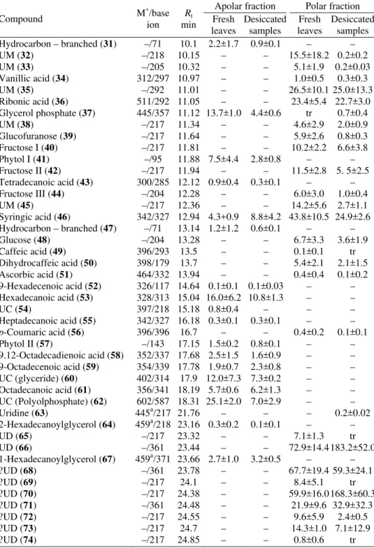
Mainly saccharides, as well as organic acids, phenolic acids,
phosphate-contain-ing compounds and amino acids, showphosphate-contain-ing high levels of biological variability
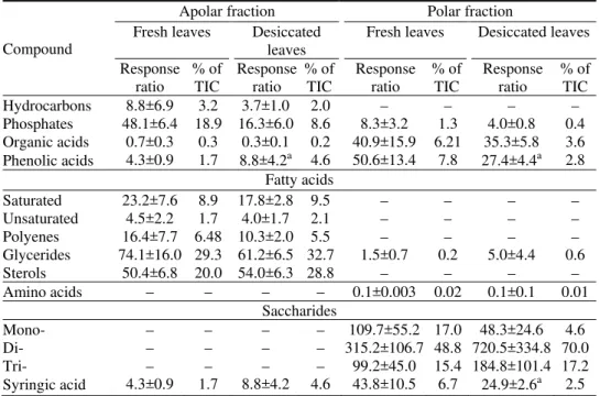
14were found by GC–MS (Table II). The total amounts of disaccharides and
tri-saccharides were about two-times more in the samples obtained from the
desic-cated as compared to those from the fresh samples (Table III). The amounts of
phosphate-containing compounds tended to decrease in the desiccated leaves,
while glycerides, mainly glycerol, increased. There were no detectable changes
in the total amount of amino acids.
TABLE I. Free radical scavenging activity of H. rhodopensis fractions
Fractions of H. rhodopensis IC50±SE / μg ml -1
Polar fraction of desiccated leaves 19.95±3.42b
Polar fraction of fresh leaves 50.04±3.44b
Apolar fraction of desiccated leaves >200
Apolar fraction of fresh leaves >200
Quercetina 3.23±0.39
Syringic acida 4.40±0.37
a
Reference compound; bsignificantly different at p < 0.05
The antiradical activity of plant extracts may be explained by the presence of
phenolic compounds.
15,16Five free phenolic acids were detected in these
effectively inhibiting linoleic acid (fatty acid) peroxidation.
15The concentration
of syringic acid and of the other free phenolic compounds were found to be about
3-times less (
p
< 0.05) in the desiccated (2.8 % of the total ion current, TIC) than
in the fresh leaves (7.8 % of the TIC) of
H. rhodopensis.
Similar results
concern-ing the total phenolic acids were reported for
Ramonda serbica
, another
resur-rection plant. The predominant phenolic compounds found in
R. serbica
,
how-ever, were protocatechuic and chlorogenic acids.
4TABLE II. Metabolites found in H. rhodopensis fractions; identification: Golm database, NIST05, the lipid library, Madeiros and Simoneit10 and standard; results represent the means±SE of the response ratios of measurements on 3 different fractionations from different samples (50 mg). The response ratios represent peak area ratios using ribitol (40 μg) for polar and nonadecanoic acid (40 μg) for apolar metabolites as quantitative internal standards
Compound M
+
/base ion
Rt
min
Apolar fraction Polar fraction Fresh
leaves
Desiccated samples
Fresh leaves
Desiccated samples Glycolic acid (1) 205a/147 3.69 – – 0.1±0.1 tr
L-Valine (2) 174/72 3.77 – – tr 0.1±0.03
Lactic acid (3) 234a/147 4.35 – – tr tr
Phosphoric acid methyl ester (4) 256/241 4.76 – – tr tr
Malonic acid (5) 248/147 4.98 – – tr
4-Hydroxybutanoic acid (6) 233a/147 5.29 – – tr tr
L-Serine (7) –/132 5.54 – – tr tr
Hydrocarbon – branched (8) –/57 4.96 0.2±0.2 0.1±0.02 – – Glycerol (9) 293a/147 5.71 1.5±0.6 1.0±0.3 0.6±0.1 4.1±4.2 Phosphoric acid (10) 314/299 5.75 9.4±3.5 4.9±2.5 8.2±3.2 3.4±0.5
Succinic acid (11) 262/147 6.07 0.1±0.1 tr 2.0±1.0 0.5±0.1 Hydrocarbon – branched (12) –/71 6.16 0.6±0.4 0.2±0.1
2,3-Dihydroxypropanoic acid (13)
307/147 6.28 – – 0.5±0.4 tr
UC (14) –/145 6.32 2.9±0.4 – – –
Fumaric acid (15) –/245 6.39 – – 0.1±0.1 tr
3,4-Dihydroxyl-2-furanone (16) 262/147 6.71 – – 0.3±0.1 tr Hydrocarbon – branched (17) –/57 7.03 0.4±0.01 0.3±0.1 – –
Hexadecane (18) –/57 7.31 0.3±0.2 0.2±0.1 – –
Hydrocarbon – branched (19) –/57 7.39 0.5±0.3 0.2±0.2 – – Hydrocarbon – branched (20) –/71 7.51 0.2±0.1 tr – – Hydrocarbon – branched (21) –/71 7.72 3.4±2.8 1.2±0.3 – – Malic acid (22) 335a/147 7.73 0.5±0.3 0.3±0.03 10.4±5.5 4.0±0.7
Erythritol (23) –/217 7.97 – – 0.7±0.5 0.2±0.1
UCb (24) –/355 8.62 1.9±0.5 – – –
Glutamine (25) 363/246 9.13 0.1±0.1 – – –
UC (26) –/220 9.2 – – 18.1±9.2 6.8±1.6
Xylonic acid (27) 364/217 9.34 – – 4.0±3.0 7.8±1.8
UC (28) –/355 9.34 1.9±0.2 – – –
TABLE II. Continued
Compound M
+
/base ion
Rt
min
Apolar fraction Polar fraction Fresh
leaves
Desiccated samples
Fresh leaves
Desiccated samples Hydrocarbon – branched (31) –/71 10.1 2.2±1.7 0.9±0.1 – –
UM (32) –/218 10.15 – – 15.5±18.2 0.2±0.2
UM (33) –/205 10.32 – – 5.1±1.9 0.2±0.03
Vanillic acid (34) 312/297 10.97 – – 1.0±0.5 0.3±0.3
UM (35) –/292 11.01 – – 26.5±10.1 25.0±13.3
Ribonic acid (36) 511/292 11.05 – – 23.4±5.4 22.7±3.0
Glycerol phosphate (37) 445/357 11.12 13.7±1.0 4.4±0.6 tr 0.7±0.4
UM (38) –/217 11.34 – – 4.6±2.9 2.0±0.9
Glucofuranose (39) –/217 11.64 – – 5.9±2.6 0.8±0.3
Fructose I (40) –/217 11.81 – – 10.2±2.2 6.6±3.8
Phytol I (41) –/95 11.88 7.5±4.4 2.8±0.8 – –
Fructose II (42) –/217 11.94 – – 11.5±2.8 5. 5±2.5
Tetradecanoic acid (43) 300/285 12.12 0.9±0.4 0.3±0.1 – –
Fructose III (44) –/204 12.28 – – 6.0±3.0 1.0±0.4
UM (45) –/217 12.36 – – 14.2±5.6 2.7±1.1
Syringic acid (46) 342/327 12.94 4.3+0.9 8.8±4.2 43.8±10.5 24.9±2.6 Hydrocarbon – branched (47) –/71 13.14 1.2±1.2 0.6±0.1 – –
Glucose (48) –/204 13.28 – – 6.7±3.3 3.6±1.9
Caffeic acid (49) 396/293 13.5 – – 0.1±0.1 tr
Dihydrocaffeic acid (50) 398/179 13.7 – – 5.4±2.1 2.1±1.5
Ascorbic acid (51) 464/332 13.94 – – 0.4±0.4 0.1±0.2
9-Hexadecenoic acid (52) 326/117 14.64 0.1±0.1 0.1±0.03 – – Hexadecanoic acid (53) 328/313 15.04 16.0±6.2 10.8±1.3 – –
UC (54) 397/218 15.18 0.8±0.4 – – –
Heptadecanoic acid (55) 342/327 16.18 0.3±0.1 0.3±0.1 – –
p-Coumaric acid (56) 396/396 16.7 – – 0.4±0.2 0.1±0.1
Phytol II (57) –/143 17.15 1.5±0.2 0.8±0.1 – –
9.12-Octadecadienoic acid (58) 352/337 17.68 2.5±1.5 1.6±0.9 – – 9-Octadecenoic acid (59) 354/339 17.78 1.9±0.7 2.3±0.8 – – UC (glyceride) (60) 402/314 17.9 12.0±7.3 7.3±0.2 – – Octadecanoic acid (61) 356/341 18.19 5.7±0.6 6.2±1.3 – – UC (Polyolphosphate) (62) 602/587 18.31 25.1±2.0 7.0±2.9 – –
Uridine (63) 445a/217 21.76 – – – 0.2±0.02
2-Hexadecanoylglycerol (64) 459a/218 23.16 0.3±0.2 0.1±0.1 – –
UD (65) –/217 23.32 – – 7.1±1.3 tr
UD (66) –/361 23.44 – – 72.9±14.4 183.2±52.0
1-Hexadecanoylglycerol (67) 459a/371 23.66 2.7±1.0 3.2±0.5 – –
?UD (68) –/361 23.78 – – 67.7±19.4 59.3±24.1
?UD (69) –/217 24.1 – – 8.4±5.1 tr
?UD (70) –/217 24.38 – – 59.9±16.0 168.3±60.3
?UD (71) –/361 24.48 – – 21.9±9.6 32.9±32.3
?UD (72) –/217 24.55 – – 9.6±5.9 2.4±0.5
?UD (73) –/217 24.7 – – 14.3±1.0 7.1±12.9
TABLE II. Continued
Compound M
+
/base ion
Rt
min
Apolar fraction Polar fraction Fresh
leaves
Desiccated samples
Fresh leaves
Desiccated samples
UD (75) –/361 25.07 – – 11.0±7.1 55.5±30.0
UD (76) –/361 25.18 – – 47.3±59.5
Sucrose (77) –/361 25.26 – – 40.1±15.7 160.3±61.4
UD (78) –/217 25.74 – – 0.8±0.5 tr
UD (79) –/191 25.85 – – 1.0±0.7 2.0±1.8
1-Octadecanoylglycerol (80) 487a/399 26.39 3.6±1.2 4.5±0.7 0.9±0.6 1.0±0.2
Squalene (81) 409/69 26.83 0.4±0.1 0.4±0.1 – –
Tetracosanoic acid (82) 440/425 26.99 0.1±0.1 0.1±0.1 – –
UC(glycerol) (83) 530/193 29.14 27.9±0.2 21.9±4.4 – –
UT (84) –/217 30.27 1.1±0.4 0.7±0.2
Tocopherol (85) 502/502 31.05 7.0±2.9 6.2±1.0 – –
Cholesterol (86) 458/329 31.15 1.2±0.4 1.9±0.3 – –
UC(glycerol) (87) 558/133 31.57 27.6±6.1 24.3±0.6 – –
Campestrol (88) 472/382 32.45 9.2±1.1 10.1±1.4 – –
Stigmasterol (89) 484/83 32.82 0.6±0.1 0.6±0.3 – –
UT (90) –/217 33.46 – – 6.7±2.4 13.8±5.3
UT (91) –/217 33.57 – – 10.8±2.5 11.9±2.1
β-Sitosterol (92) 486/396 33.6 39.4±5.2 41.4±4.3 – –
UC (93) 396/381 33.79 – – 4.4±3.0 0.7±0.7
UT (94) –/361 33.83 – – – 26.2±22.1
UT (95) –/217 34.33 – – 14.9±10.9 29.8±6.1
UT (96) –/217 34.75 – – 3.0±1.4 4.2±3.8
UT (97) –/217 35.18 – – 3.7±2.4 6.9±1.9
UT (98) –/217 35.42 – – 7.8±5.7 21.4±12.9
UT (99) –/217 35.81 – – 9.8±2.4 33.4±33.4
UT (100) –/361 35.97 – – 10.8±7.3 24.9±7.1
Raffinose (101) –/361 36.21 – – 22.7±6.1 15.0±3.2
UT (102) –/217 36.61 – – 5.0±2.9 5.3±3.3
UT (103) –/217 37.04 – – 3.0±0.8 –
UC-diglyceride (104) –/129 38.36 16.0±1.6 9.6±7.7 – –
Total – – 255.6±
±39.7
187.0± ±12.3
648.1± ±114.4
1034.8± ±305.15
a
[M-15]+; bcompounds with “U” are unknown
As compared to desiccated leaves, the higher concentration of free phenolic
acids in the fresh leaves is not in correlation with their lower antiradical activity,
which indicates that other unidentified compounds contribute to the antiradical
activity of the polar fractions. The presence of flavonoids and tannins was
re-ported for
H. rhodopensis
6but, due to the limitations of GC–MS, such
com-pounds were not detected in the present study.
respectively), phosphate-containing compounds (19 and 9 %, respectively), free
fatty acids (11 and 12 %, respectively), polyenes (6%), phenolic acids and
hyd-rocarbons. The relatively weaker antioxidant activity could be explained by the
presence of small amounts of
α
-tocopherol and free phenolic acids.
TABLE III. Main groups of compounds in the extracts of H. rhodopensis; the results represent the means±SE of the response ratios and % of TIC (total ion current) of the measurements on 3 different fractionations from different samples (50 mg). The response ratios represents peak area ratios using ribitol (40 μg) for polar and nonadecanoic acid (40 μg) for apolar metabolites as quantitative internal standards
Compound
Apolar fraction Polar fraction Fresh leaves Desiccated
leaves
Fresh leaves Desiccated leaves
Response ratio
% of TIC
Response ratio
% of TIC
Response ratio
% of TIC
Response ratio
% of TIC
Hydrocarbons 8.8±6.9 3.2 3.7±1.0 2.0 – – – –
Phosphates 48.1±6.4 18.9 16.3±6.0 8.6 8.3±3.2 1.3 4.0±0.8 0.4 Organic acids 0.7±0.3 0.3 0.3±0.1 0.2 40.9±15.9 6.21 35.3±5.8 3.6 Phenolic acids 4.3±0.9 1.7 8.8±4.2a 4.6 50.6±13.4 7.8 27.4±4.4a 2.8
Fatty acids
Saturated 23.2±7.6 8.9 17.8±2.8 9.5 – – – –
Unsaturated 4.5±2.2 1.7 4.0±1.7 2.1 – – – –
Polyenes 16.4±7.7 6.48 10.3±2.0 5.5 – – – –
Glycerides 74.1±16.0 29.3 61.2±6.5 32.7 1.5±0.7 0.2 5.0±4.4 0.6
Sterols 50.4±6.8 20.0 54.0±6.3 28.8 – – – –
Amino acids – – – – 0.1±0.003 0.02 0.1±0.1 0.01
Saccharides
Mono- – – – – 109.7±55.2 17.0 48.3±24.6 4.6
Di- – – – – 315.2±106.7 48.8 720.5±334.8 70.0
Tri- – – – – 99.2±45.0 15.4 184.8±101.4 17.2
Syringic acid 4.3±0.9 1.7 8.8±4.2 4.6 43.8±10.5 6.7 24.9±2.6a 2.5
a
Significantly different at p <0.05
The polar and apolar fractions of
H. rhodopensis
were screened for their
cytotoxic activity against a panel of four human tumour cell lines, representative
for some important types of neoplastic diseases, including a multi-drug resistant
cell line (Table IV). Both tested fractions failed to evoke any significant
cytotoxic effects against any of the cell lines.
TABLE IV. Cytotoxic effects of the polar and apolar fractions of H. rhodopensis after 72 h continuous exposure (MTT-dye reduction assay); each value represents the arithmetic mean± ±SE from 8 independent experiments
Concentration mg/ml
HL-60 cells HL-60/Dox cells SKW-3 cells MDA-MB-231 cells
TABLE IV. Continued
Concentration mg/ml
HL-60 cells HL-60/Dox cells SKW-3 cells MDA-MB-231 cells
Hc 30 Hc 70 Hc 30 Hc 70 Hc 30 Hc 70 Hc 30 Hc 70
0.20 101±1 100±3 99±3 95±7 100±2 97±4 97±3 99±4
0.25 102±2 104±2 114±6 104±3 106±6 99±3 111±7 102±3
0.40 95±4 99±3 94±6 94±4 93±7 96±4 96±6 94±7
0.50 102±2 107±4 109±6 102±5 107±4 97±4 103±2 102±5
a
Polar fractions; bapolar fractions
CONCLUSIONS
In conclusion, the polar fractions of
H. rhodopensis
showed potent free
radi-cal scavenging activity. GC–MS metabolic profiling of the polar and apolar
frac-tions resulted in the detection of more than one hundred compounds, including
several phenolic acids. In depth quantitative analysis of the phenolic complex
(free and conjugated phenolic acids, flavonoids and polyphenols), however, is
needed to reveal the relationship between antiradical activity and metabolites in
the extracts of
H. rhodopensis
. The lack of any cytotoxic activity of the extracts
indicates that the plant may be used in phytotherapy for its antiradical properties.
In this respect, the desiccated leaves of
H. rhodopensis
are more suitable due to
their higher antiradical activity.
Acknowledgment. Financial support from Ministry of Education and Science, Sofia, Bul-garia (Grant D002-128/08 I. Ionkova) is acknowledged.
Ш Х
Љ Haberlea rhodopensis
STRAHIL H. BERKOV1, MILENA T. NIKOLOVA2, NEVENA I. HRISTOZOVA3, GEORGI Z. MOMEKOV4, ILIANA I. IONKOVA4 DIMITAR L. DJILIANOV1
1
AgroBio Institute, 8 Dragan Tzankov Blvd., 1164-Sofia, 2Institute of Botany, Bulgarian Academy of Sciences, 23 Acad. G. Bonchev Str., 1113-Sofia, 3Faculty of Biology, Sofia University, 8 Dragan Tzankov Blvd.,
1164-Sofia и4Faculty of Pharmacy, Medical University of Sofia, 2 Dunav Str., 1000-Sofia, Bulgaria
Haberlea rhodopensis
( - ,
, , , , , .).
-ђ
(IC50 = 19,95±14,11 μg ml-1 50,04±23,16 μg ml-1
),
-.
-: , , o , o
p- .
REFERENCES
1. J. Markovska, T. Tsonev, G. Kimenov, A. Tutekova, J. Plant Physiol.144 (1994) 100 2. K. Georgieva, S. Lenk, C. Buschmann, Photosynthetica46 (2008) 208
3. J. Muller, N. Sprenger, K. Bortlik, T. Boiler, A. Wiemken, Physiol. Plant. 100 (1997) 153 4. C. Sgherri, B. Stevanovic, F. Navari-Izzo, Physiol. Plant.122 (2004) 478
5. J. Moore, K. Westall, N. Ravenscroft, J. Farrant, G. Lindsey, W. Brandt, Biochem. J.385 (2005) 301
6. I. Ionkova, S. Ninov, I. Antonova, D. Moyankova, T. Georgieva, D. Djilianov,
Pharmazija1–4 (2009) 22
7. R. Radev, G. Lazarova, P. Nedialkov, K. Sokolova, D. Rukanova, Z. Tsokeva, Trakia J. Sci.7 (2009) 34
8. K. Stefanov, Y. Markovska, G. Kimenov, S. Popov, New Chem. 31 (1992) 2309
9. D. Djilianov, G. Genova, D. Parvanova, N. Zapryanova, T. Konstantinova, A. Atanassov,
Plant Cell Tissue Organ Cult.80 (2005) 115
10. P. Medeiros, B. Simoneit, J. Chromatogr. A1141 (2007) 271 11. C. Cimpoiu, J. Liq. Chromatogr. Relat. Technol. 29 (2006) 1125 12. T. Mosmann, J. Immunol. Met. 65 (1983) 55
13. S. Konstantinov, H. Eibl, M. Berger, Br. J. Haematol.107 (1999) 365
14. U. Roessner, C. Wagner, J. Kopka, R. Trethewey, L. Willmitzer, Plant J.23 (2000) 131 15. F. Liu, T. Ng, Life Sci. 66 (2000) 725


