PSMA-SPECIFIC LIGANDS IN PROSTATE CANCER
DIAGNOSIS AND THERAPY
Wei Jin, Ashutosh Barve, *Kun Cheng
Division of Pharmaceutical Sciences, School of Pharmacy, University of Missouri–Kansas City, Kansas City, Missouri, USA
*Correspondence to [email protected]
Disclosure: The authors have declared no conflicts of interest.
Received: 17.11.2015 Accepted: 06.01.2016
Citation: EMJ Urol. 2016;4[1]:62-69.
ABSTRACT
Prostate-specific membrane antigen (PSMA) is the most extensively studied biomarker and antigen of prostate cancer. It is overexpressed in almost all prostate cancers, and the expression level increases with prostate cancer progression. PSMA is also highly expressed in the neovasculature of solid tumours including prostate cancer. As a result, numerous PSMA-specific ligands have been discovered for prostate cancer diagnosis and therapy, and one of them has been approved for clinical use. Moreover, a number of other PSMA-specific ligands are currently evaluated in clinical studies. In this review we discuss four major types of PSMA-specific ligands, including antibody, aptamer, peptide, and small molecule inhibitor. Their emerging applications in prostate cancer diagnosis, targeted drug delivery, and therapy are also discussed.
Keywords: Prostate-specific membrane antigen (PSMA), diagnosis, targeted drug delivery, antibody, aptamer, peptide.
INTRODUCTION
Prostate cancer is the most common male malignancy and the second-leading cause of cancer death in American men. Current therapies for prostate cancer include surgery, chemotherapy, cryosurgery, radiation, and hormonal therapy. These approaches are used either as monotherapy or in combination according to stages in the patients. A variety of prostate cancer speciic antigens have been explored for targeted drug delivery and diagnosis. Among them, prostate-speciic membrane antigen (PSMA) is the most extensively studied biomarker and antigen of prostate cancer.1
PSMA, also known as folate hydrolase, N-acetyl-
α
-linked acidic dipeptidase I, or glutamate carboxypeptidase II, is a Type 2 transmembrane glycosylated protein. Belonging to the group of zinc-dependent exopeptidases with a binuclear zinc site, PSMA contains a glycosylated extracellular domain of 707 amino acids, a transmembrane domain of 24 amino acids, and an intracellular domain of 19 amino acids.1 The crystal structure of PSMA reveals a symmetric dimer with each polypeptide chain containing three functional and structural domains: a C-terminal/helical domain, an apical domain, and a protease domain.2,3 Although the exact physiologic function of PSMAEDITOR’S PICK
In this review, Wei Jin and colleagues from the University of Missouri-Kansas City, Missouri, USA, have summarised the ligands associated with prostate-specific membrane antigen (PSMA), which is a widely used biomarker and antigen in prostate cancer diagnosis. This topic is one of the most attractive and promising subjects in the diagnosis and therapy of prostate cancer, and
will be of great interest to our readers.
in the prostate is not fully understood, it is known that PSMA can cleave g-linked glutamates from polyglutamated folates (known as folate hydrolase activity) and
α
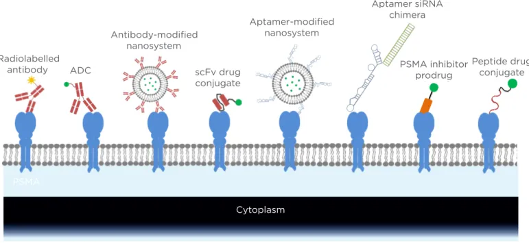
-linked glutamate from N-acetyl aspartyl glutamate (known as NAALADase activity).4PSMA is expressed in normal prostate epithelial cells, but its expression level in prostate cancer cells is much higher. Notably, PSMA is overexpressed in almost all prostate cancers, and the expression level increases with disease progression.1,5 Moreover, PSMA is overexpressed in the neovasculature of solid tumours, but not in normal tissue vasculature. PSMA expression is also detectable in several normal tissues, such as the small intestine, proximal renal tubules, and salivary glands,6 but the expression level of PSMA in prostate cancers is approximately 100 to 1000-fold higher.7,8 In addition, the site of PSMA expression in normal tissues (brush border/luminal location) is not exposed to direct blood circulation, leading to negligible interaction between PSMA-speciic ligands and PSMA in normal tissues.1,8 As a result, PSMA has been recognised as the most valuable prostate cancer-speciic antigen for prostate cancer diagnosis and therapy. The aim of this review is therefore to discuss the currently available PSMA-speciic ligands (antibody, single chain fragment variable [scFv], aptamer, peptide, and small molecule) that have been explored for prostate cancer diagnosis and therapy (Figure 1).
PSMA-Speciic Antibody and scFv
Antibody is by far the most common type of PSMA-speciic ligand in preclinical and clinical studies. PSMA was initially identiied by the murine monoclonal antibody (mAb) 7E11, which was discovered after immunisation of mice with preparations of PSMA-positive LNCaP cells.9 The mAb 7E11 has thereafter been used as the irst anti-PSMA antibody for various prostate cancer treatments and diagnoses. For example, ProstaScint® (111Indium-labelled mAb7E11) was approved by the FDA for the imaging of prostate cancer.10,11,12 However, mAb 7E11 binds to the intracellular domain of PSMA and therefore cannot bind viable prostate cancer cells, thus limiting its potential clinical applications.13 Consequently, numerous eforts have been made to discover other mAbs targeting the extracellular domain of PSMA.
Using hybridoma technology, Bander and co-
J415, and E99) that speciically bind to the extracellular domain of PSMA with Kd values in the low nM range (1.76–18 nM).14,15 Later, these PSMA-speciic mAbs were labelled with 131I and 111In for imaging studies in nude mice bearing LNCaP xenograft. These mAbs exhibit higher tumour uptake compared with radiolabelled 7E11. Moreover, 7E11 tends to accumulate in the areas of necrosis, whereas J591 and J415 mainly accumulate in viable tumours, suggesting the great therapeutic potential of mAbs in targeting the PSMA extracellular domain.16 Since then, several other groups have discovered numerous PSMA mAbs that target the extracellular domain of PSMA. All these mAbs show high ainity to puriied PSMA as well as viable prostate cancer cells.17,18
Among the mAbs shown to target the PSMA extracellular domain, mAb J591 has attracted the most attention as an anti-PSMA antibody for prostate cancer diagnosis and therapy because of its high ainity and speciicity to PSMA. Several J591-based imaging and therapeutic agents are currently being evaluated in clinical trials. For example, 89Zr-labelled J591 was developed as a positron emission tomography (PET) imaging agent to detect metastatic castration-resistant prostate cancer cells. The Phase I/II study reveals superior targeting of bone lesions compared with conventional imaging modalities or a combination of conventional imaging modalities.19 In another Phase II clinical trial, 177Lutetium-labelled J591 exhibits high tolerance, accurate tumour targeting, and dose-dependent prostate-speciic antigen responses in metastatic castration-resistant prostate cancer patients, suggesting its promising potential of targeting cytotoxic agents to PSMA.20 177Lutetium-J591 in combination with docetaxel is
in an ongoing Phase Iclinical study to evaluate its efectiveness against metastatic, castrate-resistant prostate cancer (Clinical Trial NCT00916123).
in a Phase II clinical trial. In addition to radiolabelled mAb and ADC, mAb J591 was used as a targeting ligand in the PEGylated liposome loaded with alpha-particle generator nuclide 225Ac as the anti-cancer agent. The J591-labelled liposome speciically binds to several PSMA-positive cell lines and accordingly induces cell apoptosis.22
Wetterauer et al.17 discovered three other PSMA-speciic mAbs (3/E7, 3/F11, 3/A12) from mice immunised with unpuriied LNCaP lysate. These mAbs demonstrate high ainity to puriied PSMA, viable LNCaP cells, and PSMA-transfected cells. Further studies showed that these three mAbs bind to diferent extracellular PSMA epitopes. Moreover, the mAbs 3/E7, 3/F11, and 3/A12 exhibit a similar binding ainity to J591 in C4-2 cells.23 The mAbs 3/E7, 3/F11, and 3/A12 were labelled with 64Cu and evaluated in mice bearing prostate cancer
xenografts using microPET. The imaging results reveal high uptake of the mAbs in PSMA-positive C4-2 xenograft at 24 and 48 hours post-administration but not in PSMA-negative DU-145 xenografts.24 The mAb 3/F11 was also evaluated as a radioimmunotherapeutic agent after labelling with 177Lu. The 177Lu labelled 3/F11 exhibits high uptake in C4-2 xenografts with a serum half-life of 7 days. A biodistribution study demonstrated maximum uptake in the tumour at 72 hours post-administration. Furthermore, a single dose of
177Lu-DOTA-3/F11 inhibits tumour growth and
improves the survival rate of the mice, indicating the great potential of using 3/F11 as a targeting ligand for prostate cancer therapeutics.25
Instead of immunising animals and using hybridoma technology to discover mAbs, scFv phage display has also been used to identify anti-PSMA antibody fragments. In one study, a scFv phage library was constructed from the hybridoma of the mAb 3/A12, which targets the PSMA extracellular domain. The scFv A5 was identiied after several rounds of biopanning on LNCaP cells and puriied PSMA, and exhibits high ainity to LNCaP cells with a
Kd of 38 nM.26 A recombinant immunotoxin
(A5-PE40) containing the toxic domain of Pseudomonas Exotoxin A (PE40) and scFv A5 was then constructed, and showed a very high cytotoxicity in LNCaP cells (IC50=20 pM).26 The same approach was also used to discover another anti-PSMA scFv D7 from the phage library that was constructed from the hybridoma 3/F11. Similarly, the recombinant immunotoxin (D7-PE40) shows high ainity to C4-2 cells and induces cell apoptosis with an IC50 of 140 pM.27 The immunotoxin D7-PE40 signiicantly reduces the size of established C4-2 xenografts in mice. Meanwhile, the immunotoxin D7-PE40 is well tolerated in mice up to a dose of 20
μ
g.27Radiolabelled antibody ADC
Antibody modified nanosystem
Aptamer modified nanosystem
PSMA inhibitor prodrug
Peptide dr conjugate ScFv drug
conjugate
PSMA
Figure 1: Targeting strategies of PSMA-speciic ligands in prostate cancer diagnosis and therapy.
PSMA: prostate-speciic membrane antigen; ADC: antibody drug conjugate; scFv: single chain fragment variable; siRNA: small interfering RNA.
Radiolabelled antibody ADC
Antibody-modiied nanosystem
scFv drug conjugate
Aptamer-modiied nanosystem
Aptamer siRNA chimera
Peptide drug conjugate PSMA inhibitor
prodrug
PSMA
Prostate Cancer-Speciic Aptamer
Aptamers are single-stranded oligonucleotides that have high ainity to various molecular moieties. Aptamers can be identiied through the systematic evolution of ligands by exponential enrichment (SELEX) from a large pool of random oligonucleotide sequences.28 Using SELEX against the PSMA extracellular domain, Lupold
and co-workers29 identiied two 40-mer RNA
aptamers, termed xPSM-A9 and xPSM-A10, with low nanomolar ainity. The aptamers xPSM-A9 and xPSM-A10 also inhibit the NAALADase activity of PSMA with a Ki of 2.1 nM and 11.2 nM, respectively.29
Due to its high ainity and speciicity to PSMA, the aptamer xPSM-A10 was radiolabelled with 64Cu and used as a PET imaging agent for targeted molecular imaging of prostate cancer.30 Moreover, the PSMA aptamers have been widely used for prostate cancer targeted drug delivery systems. For example, the aptamer xPSM-A9 was adopted as a PSMA-speciic ligand and conjugated to the surface of a liposome encapsulating doxorubicin.31 The aptamer-conjugated liposomes speciically internalise into PSMA-positive LNCaP cells and subsequently induce cell apoptosis, while neither uptake nor cytotoxicity is observed in PSMA-negative PC-3 cells. A biodistribution study demonstrated high accumulation of doxorubicin in tumour sites at 12 and 24 hours after intravenous administration. Accordingly, an enhanced anti- cancer efect was observed in the animals treated with the A9-conjugated liposome.31
Similarly, PSMA aptamers have been used in small interfering RNA (siRNA) delivery for prostate cancer therapy. Since the discovery of RNA interference, siRNAs have attracted much attention as novel therapeutic agents to treat various diseases. However, lack of eicient delivery systems is still the major hurdle that limits the clinical translation of siRNA. In an exploratory study of siRNA delivery, the A10 aptamer was directly linked to an siRNA to form the aptamer–siRNA chimera to achieve targeted delivery to PSMA-positive prostate cancers.32 The A10 aptamer-siRNA chimera not only shows selective uptake in PSMA-positive cancer cells in vitro but also demonstrates signiicant inhibition of LNCaP xenograft tumour growth in vivo. This is one of the few pioneering studies showing the in vivo activity of siRNA in animal studies.32
The PSMA aptamer was also explored as a therapeutic agent against advanced prostate cancer because several studies have suggested that PSMA’s enzymatic activity may play important roles in prostate cancer angiogenesis, progression, and metastasis.4,33 In one such study, the PSMA-speciic aptamer A9g (truncated form of the aptamer A9) speciically inhibits the migration and invasion of PSMA-positive prostate cancer cells in vitro. A further animal study of the aptamer A9g reveals potent inhibition of metastasis in a xenograft mouse model of metastatic prostate cancer. Data from this study suggest that PSMA inhibitors could be used alone as emerging therapeutic agents for advanced prostate cancer.34
Given the poor stability of RNA molecules under physiological condition, DNA aptamer targeting PSMA was discovered to overcome the poor stability of RNA aptamer. A 48-mer anti-PSMA DNA aptamer (SZTI01) containing a binding site for doxorubicin was identiied using SELEX against PSMA. A dimeric form of the aptamer SZTI01 was constructed to form a complex with doxorubicin. The aptamer/ doxorubicin complex is stable at physiological conditions and speciically binds to PSMA-positive C4-2 cells. The complex also induces high cytotoxicity in C4-2 cells but negligible activity in PSMA-null cells, indicating its high selectivity for PSMA-positive cells.35
Prostate Cancer-Speciic Peptide
target moiety is unknown.39 We recently designed a combinatorial biopanning strategy against a recombinant PSMA extracellular domain, LNCaP cells, and LNCaP xenografts in nude mice and identiied a PSMA-speciic peptide (GTI), which shows high ainity to PSMA-positive cells in vitro and in vivo. The Kd values of the GTI peptide to LNCaP and C4-2 cells are 8.22 and 8.91
μ
M, respectively (unpublished data). One potentialproblem that may limit the application of peptide ligands is their relative low binding ainity compared with antibodies and aptamers. Most peptides identiied from phage display show Kd values in the low micromolar range.40 Therefore, further modiication of peptide ligands, such
as D-form amino acid modiication,41 ligand
dimerisation, and tetramerisation38 may be used to further improve their ainity.
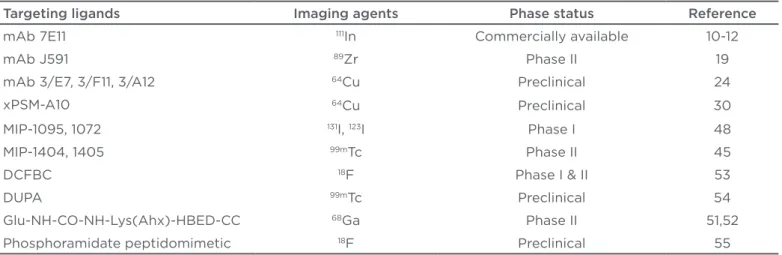
Table 1: Applications of prostate-speciic membrane antigen-speciic ligands in prostate cancer imaging.
Targeting ligands Imaging agents Phase status Reference
mAb 7E11 111In Commercially available 10-12
mAb J591 89Zr Phase II 19
mAb 3/E7, 3/F11, 3/A12 64Cu Preclinical 24
xPSM-A10 64Cu Preclinical 30
MIP-1095, 1072 131I, 123I Phase I 48
MIP-1404, 1405 99mTc Phase II 45
DCFBC 18F Phase I & II 53
DUPA 99mTc Preclinical 54
Glu-NH-CO-NH-Lys(Ahx)-HBED-CC 68Ga Phase II 51,52
Phosphoramidate peptidomimetic 18F Preclinical 55
mAb: monoclonal antibody.
Table 2: Applications of PSMA-speciic ligands in prostate cancer therapy.
Targeting ligands Therapeutic agents Delivery system Phase status Reference
mAb J591 177Lu-labelled Radiolabelling antibody Phase II 20
mAb J591 Monomethyl auristatin E Antibody drug conjugate Phase II 21
mAb J591 177Lu-labelled Combination with docetaxel Phase I NCT00916123*
mAb J591 177Lu-labelled Combination with
ketoconazole and hydrocortisone
Phase II NCT00859781*
mAb J591 25Ac Liposome Preclinical 22
mAb 3/F11 177Lu-labelled Radiolabelling antibody Preclinical 25
scFv fragment A5 scFv fragment D7
Pseudomonas exotoxin A (PE40)
Recombinant immunotoxin Preclinical 26-27
xPSM-A9 Doxorubicin Liposome Preclinical 31
xPSM-A10 PLK1 siRNA and
BCL2 siRNA
Aptamer siRNA chimeras
Preclinical 32
xPSM-A9 derivative xPSM-A9 derivative Aptamer alone Preclinical 34
DNA aptamer Doxorubicin Dimeric aptamer
doxorubicin conjugate
Preclinical 35
PSMA inhibitor DUPA TubH Prodrug Preclinical 54
*ClinicalTrials.gov Identiier (www.ClinicalTrials.gov)
Prostate Cancer-Speciic Small Molecule
N-acetyl-L-aspartyl-L-glutamate (NAAG) is an endogenous peptide found in the mammalian nervous system. It can be hydrolysed by the N-acetylated
α
-linked acidic dipeptidase (NAALADase, or NAAG peptidase) with a Km of 540 nM.42 Given the fact that PSMA is highly homologous to NAAG peptidase, several studies have been carried out to design NAAG analogues that can speciically bind to PSMA.43-46Based on their previous experience in designing NAAG analogues, Pomper et al.43 synthesised two radiolabelled small molecules, [11C]DCMC and [125I]DCIT. Both of these analogues exhibit high accumulation in LNCaP xenograft at 30 minutes post-administration, whereas the uptake in PSMA-negative MCF-7 and PC-3 derived tumours is negligible. Another group synthesised a series of glutamate-urea-X (X is a derivatised lysine) heterodimeric inhibitors with high ainity to PSMA in the low nM range.44 Two of the inhibitors, MIP-1072 and MIP-1095, eiciently inhibit PSMA enzymatic activity with Ki values of 4.6 nM and 0.24 nM, respectively. Moreover, MIP-1072 and MIP-1095 exhibit very high ainity to PSMA (Kd <4 nM).47 Speciic binding of 125I labelled MIP-1072 and MIP-1095 was demonstrated in mice bearing LNCaP xenografts.47 These promising data have led to the Phase I clinical trial of these small molecule inhibitors for imaging prostate cancer.48 Clinical studies demonstrate that 123I-MIP-1072 and 123I-MIP-1095 can detect lesions in the prostate
gland, soft tissues, and bones at 1–4 hours post-injection. Target-to background ratio is >10:1 at 4 and 24 hours for single photon emission computed tomograph/computed tomography (SPECT/CT).48 Two other PSMA small molecule inhibitors, MIP-1404 and 1405, also show high ainity to LNCaP cells with Kd values of 1.07 nM and 4.35 nM, respectively.46 A clinical study of (99m)Tc-MIP-1404 and 1405 revealed that they can detect soft-tissue prostate cancer lesions including subcentimetre lymph nodes and most bone metastatic lesions.45
It is reported that the active binding site of PSMA is composed of one binding pocket for glutamate-urea-X inhibitors and another lipophilic pocket.49 Eisenhut et al.50 therefore hypothesised that attachment of a hydrophobic moiety to the current glutamate-urea based PSMA inhibitors may enhance their ainity to PSMA. As a result, a novel imaging agent was synthesised by conjugating a
( N , N ’ - b i s [ 2 - hyd roxy- 5 (c a r b oxye t hy l ) b e n z y l ethylenediamine-N,N’-diacetic acid), to a glutamate-urea based inhibitor. The new imaging agent exhibits enhanced speciicity for PSMA-positive tumour cells in vitro and in vivo. It also demonstrates faster blood and organ clearances and lower liver accumulation in an animal study.50 The [68Ga] PSMA-HBED-CC-PET/CT imaging agent has also been evaluated in prostate cancer patients for the diagnosis, radiotherapy, and management of prostate cancer.51,52 Several other small molecule inhibitors, such as DUPA, DCFBC, and phosphoramidate peptidomimetic have also entered preclinical and clinical studies as imaging agents to detect castration-resistant prostate and metastatic hormone-naïve prostate cancer.53-55
Due to their high ainity and speciicity to PSMA, these PSMA small molecule inhibitors have also been investigated for prostate cancer targeted drug delivery. In one study, Low et al.54 conjugated tubulysin, a microtubule inhibitor, to DUPA, a small molecule inhibitor of PSMA with a Kd value of 14 nM. The conjugated drug (DUPA-TubH) exhibits high anti-cancer activity against PSMA-positive prostate cancer cells in vitro and in vivo. Pre-administration of excess PSMA inhibitors abolishes the activity of DUPA-TubH, indicating the high speciicity of DUPA-TubH to PSMA. Later, the same group conjugated a series of diferent chemotherapeutic agents to DUPA and similar activity against PSMA-positive prostate cancer was observed, suggesting lexibility in the use of the PSMA small molecule inhibitor as a targeting ligand for various agents.56 A number of other PSMA-speciic small molecules have also been explored as targeting ligands for various therapeutic agents, including radionuclide,57 gold nanoparticles,58 photosensitiser,59 and polymeric microbubbles.60
CONCLUSION
Four types of PSMA-speciic ligands and their applications in prostate cancer imaging (Table 1) and therapy (Table 2) are discussed in the review. Although PSMA-speciic ligands are mainly used as prostate cancer imaging agents and targeting ligands for drug delivery systems, some of the PSMA inhibitors can also be used alone as anti-cancer therapeutic agents.
REFERENCES
1. Barve A et al. Prostate cancer relevant antigens and enzymes for targeted drug delivery. J Control Release. 2014;187: 118-32.
2. Davis MI et al. Crystal structure of prostate-speciic membrane antigen, a tumor marker and peptidase. Proc Natl Acad Sci U S A. 2005;102(17):5981-6. 3. Bukrinsky JT et al. Native carboxypeptidase A in a new crystal environment reveals a diferent conformation of the important tyrosine 248. Biochemistry. 1998;37(47):16555-64. 4. Yao V et al. Expression of prostate-speciic membrane antigen (PSMA), increases cell folate uptake and proliferation and suggests a novel role for PSMA in the uptake of the non-polyglutamated folate, folic acid. Prostate. 2010;70(3):305-16.
5. Sweat SD et al. Prostate-speciic membrane antigen expression is greatest in prostate adenocarcinoma and lymph node metastases. Urology. 1998;52(4):637-40.
6. Troyer JK et al. Detection and characterization of the prostate-speciic membrane antigen (PSMA) in tissue extracts and body luids. Int J Cancer. 1995;62(5):552-8.
7. Ghosh A, Heston WD. Tumor target prostate speciic membrane antigen (PSMA) and its regulation in prostate cancer. J Cell Biochem. 2004;91(3): 528-39.
8. Akhtar NH et al. Prostate-speciic membrane antigen-based therapeutics. Adv Urol. 2012;2012:973820.
9. Horoszewicz JS et al. Monoclonal antibodies to a new antigenic marker in epithelial prostatic cells and serum of prostatic cancer patients. Anticancer Res. 1987;7(5B):927-35.
10. Petronis JD et al. Indium-111 capromab pendetide (ProstaScint) imaging to detect recurrent and metastatic prostate cancer. Clin Nucl Med. 1998;23(10):672-7. 11. Hinkle GH et al. Multicenter
radioimmunoscintigraphic evaluation of patients with prostate carcinoma using indium-111 capromab pendetide. Cancer. 1998;83(4):739-47.
12. Rosenthal SA et al. Utility of capromab pendetide (ProstaScint) imaging in the management of prostate cancer. Tech Urol. 2001;7(1):27-37.
13. Troyer JK et al. Biochemical characterization and mapping of the 7E11-C5.3 epitope of the prostate-speciic membrane antigen. Urol Oncol. 1995;1(1):29-37.
14. Liu H et al. Monoclonal antibodies to the extracellular domain of prostate-speciic membrane antigen also react with tumor vascular endothelium. Cancer Res. 1997;57(17):3629-34.
15. Smith-Jones PM et al. In vitro characterization of radiolabeled monoclonal antibodies speciic for the extracellular domain of prostate-speciic membrane antigen. Cancer Res. 2000;60(18):5237-43.
16. Smith-Jones PM et al. Radiolabeled monoclonal antibodies speciic to the extracellular domain of prostate-speciic membrane antigen: preclinical studies in nude mice bearing LNCaP human prostate tumor. J Nucl Med. 2003;44(4):610-7. 17. Elsässer-Beile U et al. A new generation of monoclonal and recombinant antibodies against cell-adherent prostate speciic membrane antigen for diagnostic and therapeutic targeting of prostate cancer. Prostate. 2006;66(13):1359-70. 18. Grauer LS et al. Identiication, puriication, and subcellular localization of prostate-speciic membrane antigen PSM’ protein in the LNCaP prostatic carcinoma cell line. Cancer Res. 1998;58(21):4787-9. 19. Pandit-Taskar N et al. A Phase I/II Study for Analytic Validation of 89Zr-J591 ImmunoPET as a Molecular Imaging Agent for Metastatic Prostate Cancer. Clin Cancer Res. 2015. [Epub ahead of print]. 20. Tagawa ST et al. Phase II study of Lutetium-177-labeled
anti-prostate-speciic membrane antigen monoclonal antibody J591 for metastatic castration-resistant prostate cancer. Clin Cancer Res. 2013;19(18):5182-91.
21. Wang X et al. In vitro and in vivo responses of advanced prostate tumors to PSMA ADC, an auristatin-conjugated antibody to prostate-speciic membrane antigen. Mol Cancer Ther. 2011;10(9): 1728-39.
22. Bandekar A et al. Anti-prostate-speciic membrane antigen liposomes loaded with 225Ac for potential targeted antivascular alpha-particle therapy of cancer. J Nucl Med. 2014;55(1):107-14. 23. Wolf P et al. Three conformational antibodies speciic for diferent PSMA epitopes are promising diagnostic and therapeutic tools for prostate cancer. Prostate. 2010;70(5):562-9.
24. Alt K et al. High-resolution animal PET imaging of prostate cancer xenografts with three diferent 64Cu-labeled antibodies against native cell-adherent PSMA. Prostate. 2010;70(13):1413-21. 25. Behe M et al. In vivo testing of 177Lu-labelled anti-PSMA antibody as a new radioimmunotherapeutic agent against prostate cancer. In Vivo. 2011;25(1):55-9. 26. Wolf P et al. A recombinant PSMA-speciic single-chain immunotoxin has potent and selective toxicity against prostate cancer cells. Cancer Immunol Immunother. 2006;55(11):1367-73.
27. Wolf P et al. Preclinical evaluation of a recombinant anti-prostate speciic membrane antigen single-chain immunotoxin against prostate cancer. J Immunother. 2010;33(3):262-71.
28. Fitzwater T, Polisky B. A SELEX primer. Methods Enzymol. 1996;267:275-301. 29. Lupold SE et al. Identiication and characterization of nuclease-stabilized RNA molecules that bind human prostate cancer cells via the prostate-speciic membrane antigen. Cancer Res. 2002;62(14):4029-33.
30. Rockey WM et al. Synthesis and for imaging prostate cancer. Several other PSMA
mAbs are currently in clinical trials to explore their application as imaging and therapeutic agents. Regardless of its success, the major disadvantages of mAbs are possible immunogenicity, long half-life in the circulation, and low permeability in tumour site due to their large molecular size. As a result, other PSMA ligands with small sizes, such as aptamers, peptides, and small molecule inhibitors have gained much interest in recent years in prostate cancer diagnosis and targeted therapy.
radiolabeling of chelator-RNA aptamer bioconjugates with copper-64 for targeted molecular imaging. Bioorg Med Chem. 2011;19(13):4080-90.
31. Baek SE et al. RNA aptamer-conjugated liposome as an eicient anticancer drug delivery vehicle targeting cancer cells in vivo. J Control Release. 2014;196:234-42. 32. McNamara JO et al. Cell type-speciic delivery of siRNAs with aptamer-siRNA chimeras. Nat Biotechnol. 2006;24(8):1005-15.
33. Conway RE et al. Prostate-speciic membrane antigen regulates angiogenesis by modulating integrin signal transduction. Mol Cell Biol. 2006;26(14):5310-24.
34. Dassie JP et al. Targeted inhibition of prostate cancer metastases with an RNA aptamer to prostate-speciic membrane antigen. Mol Ther. 2014;22(11):1910-22. 35. Boyacioglu O et al. Dimeric DNA Aptamer Complexes for High-capacity-targeted Drug Delivery Using pH-sensitive Covalent Linkages. Mol Ther Nucleic Acids. 2013;2:e107.
36. Chen Z et al. Discovery of Peptide ligands for hepatic stellate cells using phage display. Mol Pharm. 2015;12(6): 2180-8.
37. Shukla RS et al. Peptides used in the delivery of small noncoding RNA. Mol Pharm. 2014;11(10):3395-408.
38. Aggarwal S et al. A dimeric peptide that binds selectively to prostate-speciic membrane antigen and inhibits its enzymatic activity. Cancer Res. 2006;66(18):9171-7.
39. Qin B et al. Identiication of a LNCaP-speciic binding peptide using phage display. Pharm Res. 2011;28(10):2422-34. 40. Hao J et al. Identiication and rational redesign of peptide ligands to CRIP1, a novel biomarker for cancers. PLoS Comput Biol. 2008;4(8):e1000138.
41. Chen S et al. Improving binding ainity and stability of peptide ligands by substituting glycines with D-amino acids. Chembiochem. 2013;14(11):1316-22.
42. Robinson MB et al. Hydrolysis of the brain dipeptide
N-acetyl-L-aspartyl-L-glutamate. Identiication and characterization of a novel N-acetylated alpha-linked acidic dipeptidase activity from rat brain. J Biol Chem. 1987;262 (30):14498-506.
43. Foss CA et al. Radiolabeled small-molecule ligands for prostate-speciic membrane antigen: in vivo imaging in experimental models of prostate cancer. Clin Cancer Res. 2005;11(11):4022-8. 44. Maresca KP et al. A series of halogenated heterodimeric inhibitors of prostate speciic membrane antigen (PSMA) as radiolabeled probes for targeting prostate cancer. J Med Chem. 2009;52(2):347-57.
45. Vallabhajosula S et al. 99mTc-labeled small-molecule inhibitors of prostate-speciic membrane antigen: pharmacokinetics and biodistribution studies in healthy subjects and patients with metastatic prostate cancer. J Nucl Med. 2014;55(11):1791-8.
46. Hillier SM et al. 99mTc-labeled small-molecule inhibitors of prostate-speciic membrane antigen for molecular imaging of prostate cancer. J Nucl Med. 2013;54(8):1369-76.
47. Hillier SM et al. Preclinical evaluation of novel glutamate-urea-lysine analogues that target prostate-speciic membrane antigen as molecular imaging pharmaceuticals for prostate cancer. Cancer Res. 2009;69(17):6932-40.
48. Barrett JA et al. First-in-man evaluation of 2 high-ainity PSMA-avid small molecules for imaging prostate cancer. J Nucl Med. 2013;54(3):380-7. 49. Kularatne SA et al. Design, synthesis, and preclinical evaluation of prostate-speciic membrane antigen targeted (99m)Tc-radioimaging agents. Mol Pharm. 2009;6(3):790-800.
50. Eder M et al. 68Ga-complex lipophilicity and the targeting property of a urea-based PSMA inhibitor for PET imaging. Bioconjug Chem. 2012;23(4):688-97. 51. Sahlmann CO et al. Biphasic Ga-PSMA-HBED-CC-PET/CT in patients with recurrent and high-risk prostate carcinoma. Eur J Nucl Med Mol Imaging.
2015. [Epub ahead of print].
52. Verburg FA et al. Extent of disease in recurrent prostate cancer determined by [Ga]PSMA-HBED-CC PET/CT in relation to PSA levels, PSA doubling time and Gleason score. Eur J Nucl Med Mol Imaging. 2015. [Epub ahead of print]. 53. Rowe SP et al. Comparison of PSMA-based 18F-DCFBC PET/CT to Conventional Imaging Modalities for Detection of Hormone-Sensitive and Castration-Resistant Metastatic Prostate Cancer. J Nucl Med. 2015. [Epub ahead of print].
54. Kularatne SA et al. Prostate-speciic membrane antigen targeted imaging and therapy of prostate cancer using a PSMA inhibitor as a homing ligand. Mol Pharm. 2009;6(3):780-9.
55. Lapi SE et al. Assessment of an 18F-labeled phosphoramidate peptidomimetic as a new prostate-speciic membrane antigen-targeted imaging agent for prostate cancer. J Nucl Med. 2009;50(12):2042-8.
56. Kularatne SA et al. Synthesis and biological analysis of prostate-speciic membrane antigen-targeted anticancer prodrugs. J Med Chem. 2010;53(21): 7767-77.
57. Kabasakal L et al. Pre-therapeutic dosimetry of normal organs and tissues of (177)Lu-PSMA-617 prostate-speciic membrane antigen (PSMA) inhibitor in patients with castration-resistant prostate cancer. Eur J Nucl Med Mol Imaging. 2015;42(13):1976-83.
58. Kasten BB et al. Targeting prostate cancer cells with PSMA inhibitor-guided gold nanoparticles. Bioorg Med Chem Lett. 2013;23(2):565-8.