albicans
Jill R. Blankenship1,2, Saranna Fanning1,3, Jessica J. Hamaker2, Aaron P. Mitchell1,2*
1Department of Biological Sciences, Carnegie Mellon University, Pittsburgh, Pennsylvania, United States of America,2Department of Microbiology and Institute of Cancer Research, Columbia University, New York, New York, United States of America,3Department of Microbiology, University College Cork, Cork, Ireland
Abstract
Protein kinases play key roles in signaling and response to changes in the external environment. The ability ofCandida albicansto quickly sense and respond to changes in its environment is key to its survival in the human host. Our guiding hypothesis was that creating and screening a set of protein kinase mutant strains would reveal signaling pathways that mediate stress response inC. albicans. A library of protein kinase mutant strains was created and screened for sensitivity to a variety of stresses. For the majority of stresses tested, stress response was largely conserved between C. albicans,
Saccharomyces cerevisiae, andSchizosaccharomyces pombe. However, we identified eight protein kinases whose roles in cell wall regulation (CWR) were not expected from functions of their orthologs in the model fungiSaccharomyces cerevisiaeand
Schizosaccharomyces pombe. Analysis of the conserved roles of these protein kinases indicates that establishment of cell polarity is critical for CWR. In addition, we found that septins, crucial to budding, are both important for surviving and are mislocalized by cell wall stress. Our study shows an expanded role for protein kinase signaling in C. albicans cell wall integrity. Our studies suggest that in some cases, this expansion represents a greater importance for certain pathways in cell wall biogenesis. In other cases, it appears that signaling pathways have been rewired for a cell wall integrity response.
Citation:Blankenship JR, Fanning S, Hamaker JJ, Mitchell AP (2010) An Extensive Circuitry for Cell Wall Regulation inCandida albicans. PLoS Pathog 6(2): e1000752. doi:10.1371/journal.ppat.1000752
Editor:Christina M. Hull, University of Wisconsin-Madison, United States of America
ReceivedOctober 16, 2009;AcceptedJanuary 6, 2010;PublishedFebruary 5, 2010
Copyright:ß2010 Blankenship et al. This is an open-access article distributed under the terms of the Creative Commons Attribution License, which permits unrestricted use, distribution, and reproduction in any medium, provided the original author and source are credited.
Funding:We are grateful to Merck Research Labs for providing caspofungin. This work was supported by NIH grants 7R01AI067703 and 7R01AI057804 (APM), NIH postdoctoral fellowship F32AI071439 (JRB), and a National University of Ireland Travelling Studentship (SF). The funders had no role in study design, data collection and analysis, decision to publish, or preparation of the manuscript.
Competing Interests:The authors have declared that no competing interests exist.
* E-mail: [email protected]
Introduction
The cell surface has two pivotal roles in the survival of microorganisms: protection and interaction. The protective role preserves the integrity of the cell in the face of environmental assaults [1,2]. The interactive role includes both sensing of external signals and binding to other cells or external surfaces [3,4]. These two functions necessitate a delicate balance for organisms that can adapt to diverse niches: protection is accomplished by a rigid and impermeable surface; interaction is accomplished by a dynamic one.
The fungus Candida albicans has considerable adaptive ability that is manifested through its impact on humans. It is a commensal microbe that is acquired soon after birth, and occupies both the GI and GU tracts. These colonization sites represent distinct environments in terms of cohabitant microbiota, pH, and nutrients. The adaptive ability of C. albicans is most apparent when circumstances compromise host barriers to infection. Immunological deficiency or presence of an implanted device provides the opportunity for C. albicans to invade and grow in almost any tissue in the body. HenceC. albicansis the major fungal commensal and the major fungal pathogen of humans.
C. albicansis protected by a carbohydrate-based cell wall, whose major constituent is b-glucan, a glucose polymer that imparts shape [5]. Chitin lends the wall rigidity, while mannoproteins that coat the surface serve to interact with the external environment [6]. The cell wall is connected to every knownC. albicansbiological
process, including growth, morphogenesis, mating, and pathoge-nicity [7]. It is also an inviting therapeutic target, being a source of fungal-specific antigens and essential functions. Thus the mech-anisms and regulatory pathways that governC. albicans cell wall dynamics are a critical area of understanding.
Much of what we know about fungal cell wall integrity and cell wall biogenesis (herein collectively called cell wall regulation, CWR) comes from studies of the model yeastSaccharomyces cerevisiae, where the protein kinase C-MAPK (PKC-MAPK) pathway, largely responsible for the transcriptional response to cell wall disturbance, is a major cell wall-responsive regulatory system (reviewed in [8]). The PKC-MAPK pathway is also vital for cell wall integrity the pathogenic fungus Cryptococcus neoformans [9]. PKC itself has additional targets that impact cell wall structure independently of the MAPK pathway [10–13]. PKC-MAPK signaling is activated by cell wall disruption, or by diverse signals that include oxidative stress, hypo-osmotic shock, and mating projection formation. The spectrum of PKC-MAPK signaling inputs reflects the central role of the cell wall inS. cerevisiaebiological processes.
CWR as well as such features as morphogenesis, biofilm formation, and stress sensitivity. Our findings reveal that theC. albicans cell wall is highly connected to a much broader range of signaling pathways than has been found forS. cerevisiae. The C. albicanscell wall signaling network may reflect a balance of diverse inputs that are poised to promote modification and support adaptation.
Results
Construction of a protein kinase (PK) mutant library We set out to define functional networks that connect signal transduction pathways to signatureC. albicansbiological features. We created homozygous insertion or deletion mutations in 67 PK genes and 13 PK-related genes. More detail about these mutants, including insertion sites, can be found in Table S1. Most genes were represented by multiple independent mutant isolates. We were unable to recover homozygous mutations in 41 PK genes.
PK function was surveyed through a screen of the mutants for altered biological properties that included stress or drug sensitivity, filamentation, and biofilm formation (Table S1). Prior studies of Saccharomyces cerevisiae and Schizosaccharomyces pombe [18] provided clear hypotheses for the function of many PKs, and we generally found good correspondence with these predictions (Tables 1 and 2). However, there was a strikingly expanded role for C. albicans PKs in CWR: 24 of the 80 mutants were hypersensitive to the cell wall inhibitor caspofungin, compared to 10 of the mutants that were predicted by model organism studies (Table 1). In addition, 2 of the 10C. albicansmutants predicted to be sensitive to cell wall stress based on the phenotypes of orthologousS. cerevisiaemutants were not sensitive in our assays.
PKs with conserved roles in CWR included members of the PKC MAPK pathway, as well as Cla4, Cbk1, Psk1, Gin4, Ire1, and Vps34 (Fig. 1, Table 1). However, the PKs Hsl1, Kin3, Swe1, Tpk1, Yck3, Prk1, Yck2, Rio2, and Hst7, along with the PK-related proteins Ckb1, Ckb2, Sip3, Mss2, and Kis1, were also required for normal sensitivity to caspofungin (Fig. 1). These genes had not previously been uncovered in large-scale S. cerevisiae screens for CWR, although a role for Hst7 inC. albicansCWR was previously identified [19]. Our own caspofungin-sensitivity tests of
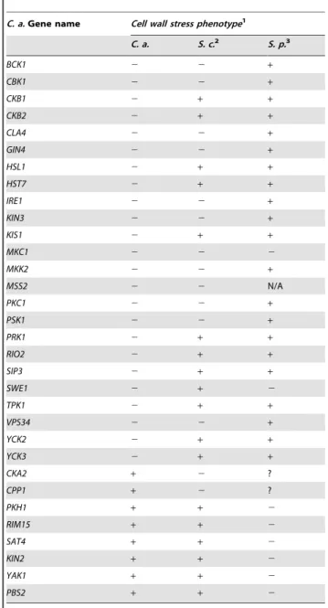
Table 1.Comparison ofC. albicans,S. cerevisiae, andS. pombe
mutant PK and PK-related cell wall stress sensitivity phenotypes.
C. a.Gene name Cell wall stress phenotype1
C. a. S. c.2 S. p.3
BCK1 2 2 +
CBK1 2 2 +
CKB1 2 + +
CKB2 2 + +
CLA4 2 2 +
GIN4 2 2 +
HSL1 2 + +
HST7 2 + +
IRE1 2 2 +
KIN3 2 2 +
KIS1 2 + +
MKC1 2 2 2
MKK2 2 2 +
MSS2 2 2 N/A
PKC1 2 2 +
PSK1 2 2 +
PRK1 2 + +
RIO2 2 + +
SIP3 2 + +
SWE1 2 + 2
TPK1 2 + +
VPS34 2 2 +
YCK2 2 + +
YCK3 2 + +
CKA2 + 2 ?
CPP1 + 2 ?
PKH1 + + 2
RIM15 + + 2
SAT4 + + 2
KIN2 + + 2
YAK1 + + 2
PBS2 + + 2
1
Genes listed are required for resistance to cell wall stress in at least one of the fungi represented:C. a. (C. albicans,) S. c. (S. cerevisiae), or S. p. (S. pombe). An entry of – means that a null mutation of this gene causes hypersensitivity to cell wall stress. An entry of+means that a null mutation of this gene does not alter sensitivity to cell wall stress. An entry of ? means the phenotype of the null mutation is unknown. Information for this table was gathered experimentally or from the Saccharomyces Genome Database (www. yeastgenome.org/), Biobase (portal.biobase-international.com/cgi-bin/portal/ login.cgi), reference [18], and literature searches.
2S. cerevisiaeortholog names do not always correspond to theC. albicansgene
name.HST7isScSTE7,MKC1isScSLT2, andCPP1isScMSG5.PSK1fromC. albicanshas two orthologs inS. cerevisiae,PSK1andPSK2. The cell wall stress hypersensitivity is only observed in the double mutant. The observation that theSckin3andScmss4mutant strains are hypersensitive to cell wall stress is first reported in this work.
3Many of the gene names are different between
C. albicansandS. pombe.MKC1is SpSPM1,SWE1isSpWEE1,PKH1isSpKSG1,RIM15isSpCEK1,SAT4isSpHAL4,KIN2 isSpKIN1,YAK1isSpPOM1,PBS2isSpWIS1,CKA2isSpCKA1, andCPP1isSpPMP1. Mutations inSpCKA1andSpPMP1have not been tested for sensitivity to cell wall stress and are marked ? for unknown. There is no obviousS. pombeortholog to MSS2and this cell is labeled N/A. The majority ofS. pombesensitivity assays were performed in SDS, which may impact more than just the cell wall.
doi:10.1371/journal.ppat.1000752.t001 Author Summary
Candida albicans is the major fungal commensal and
pathogen of humans. Like most microorganisms, C.
albicans is surrounded and protected by a cell wall. This cell wall has two purposes: to act as a rigid ‘‘exoskeleton’’ to prevent cells from bursting, and to provide a surface where a cell can interact with the outside environment while protecting the cell itself from this environment. Maintenance of this structure has been well studied in the
model fungus, Saccharomyces cerevisiae, but previous
S. cerevisiaemutants lacking the respective orthologs revealed new roles in CWR forS. cerevisiaeKin3 and Mss2 (data not shown), but not for the other orthologs. Complementation of each new C. albicanscaspofungin-hypersensitive mutant restored normal sensi-tivity to caspofungin (Fig. 1). Thus the defined mutation causes the CWR defect. Therefore, among the PKs and related proteins surveyed, we found 12 with conserved roles in CWR and 12 with apparentC. albicans-specific roles in CWR.
The cell wall PK network is unique
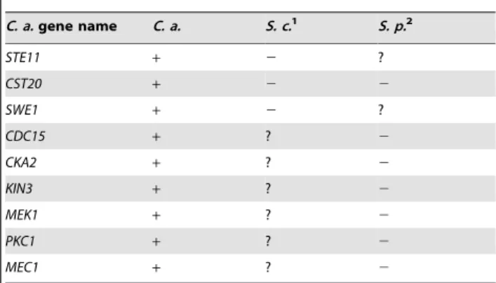
We also surveyed PK function in a panel of tests for traits related to survival and virulence. The mutants were tested for sensitivity to oxidative, osmotic, and pH stress, as well as for the ability to form filamentous cells and biofilms. The results of these screens are shown in Table 2 and Fig. S1. While these additional screens demonstrated both conserved and novel roles for PKs in these responses, only the oxidative stress assay uncovered a significant expansion of roles among conserved PKs. Five PK genes (PRK1,KIN4,VPS34,STE11, andGIN4) and two PK-related genes (CKB1 and KIS1) had previously undescribed roles in the Table 2.A comparison ofC. albicans,S. cerevisiae, andS.
pombePK and PK-related mutant phenotypes.
C. a.gene name C. a. S. c.1 S. p.2
Oxidative stress phenotype3
CHK1 2 + +
CKB1 2 + +
GIN4 2 + +
HOG1 2 2 2
KIN4 2 + +
KIS1 2 + +
PBS2 2 2 2
PRK1 2 + +
SLN1 2 + +
SSN3 2 2 +
SSN8 2 2 +
STE11 2 + +
VPS34 2 + +
MEC1 + 2 ?
RCK2 + 2 +
PKT2 + 2 ?
Osmotic stress phenotype3
GIN4 2 + +
HOG1 2 2 2
KIS1 2 + +
PBS2 2 2 2
SOK1 2 2 +
VPS34 2 2 2
CKA2 + 2 ?
CKB1 + 2 ?
CKB2 + 2 ?
HNT1 + 2 ?
IES1 + 2 N/A
KSP1 + 2 ?
MKC1 + 2 ?
SWE1 + 2 +
SAT4 + + 2
KIN2 + ? 2
BCK1 + + 2
TPK2 + ? 2
CPP1 + ? 2
VPS15 + ? 2
Filamentation/morphology phenotype3
CBK1 2 + 2
GIN4 2 + 2
IRE1 2 + +
MSS2 2 + N/A
RIO2 2 + +
SOK1 2 + +
TPK2 2 2 +
CLA4 + 2 2
KIS1 + 2 ?
HST7 + 2 ?
C. a.gene name C. a. S. c.1 S. p.2
STE11 + 2 ?
CST20 + 2 2
SWE1 + 2 ?
CDC15 + ? 2
CKA2 + ? 2
KIN3 + ? 2
MEK1 + ? 2
PKC1 + ? 2
MEC1 + ? 2
1S. cerevisiaeortholog names do not always correspond to theC. albicansgene
name.MKC1isScSLT2,CPP1isScMSG5,KIS1isScGAL83,HST7isSc STE7, and CST20isScSTE20. The observation thatScssn3andScssn8mutants are hypersensitive to oxidative stress is first reported in this work. For the morphology defect portion of the table, observed morphological defects ofS. cerevisiaefall into one of three categories: invasive growth defect, pseudohyphal growth defect, or both. TheS. cerevisiaeorthologs ofTPK1, CLA4,KIS1,HST7, andCST20are important for invasive growth. TheS. cerevisiae orthologs ofTPK2,HST7,STE11,CST20, andSWE1are important for pseudohyphal growth.
2Many of the gene names are different betweenC. albicansandS. pombe.HOG1
isSpSTY1,PBS2isSpWIS1,MEC1isSpRAD3,RCK2isSpSRK1,PKT2is most orthologous toSpSPCC70.05c,CKA2isSpCKA1,HNT1isSPCC1442.14c,MKC1is SpSPM1,SWE1isSpWEE1,SAT4isSpHAL4,KIN2isSpKIN1,BCK1isSpMKH1,TPK2 isPKA1,CPP1isSpPMP1,VPS15isSpPPK19,CBK1isSpORB6,GIN4isSpCDR2, CLA4isSpSHK2,KIS1isSpSPCC1919.03c,HST7isSpBYR1,STE11isSpBYR2,CDC15 isSpCDC7,CKB1isSpCKB2,KIN3isSpFIN1,CST20isSpPAK1, andPKC1isSpPCK2. N/A refers toC. albicansgenes that do not have known orthologs inS. pombe. Under the osmotic stress category, sensitivities of theSpkin2,Sptpk2and Spvps15mutants were noted using KCl instead of NaCl.
3Genes listed are required for resistance to the indicated stress in at least one of
the fungi represented:C. a. (C. albicans,) S. c. (S. cerevisiae),orS. p. (S. pombe). Oxidative stress generally refers to stress induced by H2O2, osmotic stress generally refers to stress induced by NaCl. Genes listed under filamentation/ morphology phenotype are required for appropriate morphology or are required for changes in morphology. An entry of – means that a null mutation of this gene causes hypersensitivity to cell wall stress or defective morphology. An entry of+means that a null mutation of this gene does not alter sensitivity to cell wall stress or have normal morphology. An entry of ? means the phenotype of the null mutation is unknown. Information for this table was gathered experimentally or from the Saccharomyces Genome Database (www. yeastgenome.org/), Biobase (portal.biobase-international.com/cgi-bin/portal/ login.cgi), reference [18], and literature searches.
doi:10.1371/journal.ppat.1000752.t002
oxidative stress response (Table 2 and Fig. S1). Analysis of S. cerevisiaemutants defective in their orthologs confirmed that their requirement for oxidative stress survival is unique to C. albicans (data not shown). This expanded role may reflect frequent encounters of C. albicans with host phagocytic cells that use oxidative attack during commensal growth. Rather than develop-ing entirely new signaldevelop-ing pathways to combat this frequent stress, it appears that conserved pathways were enlisted to improve this fungus’s chances for survivalin vivo. The identification of almost twice the number of PKs with novel roles in CWR suggests that cell wall damage is also an importantin vivostress.
PK mutants evoke a cell wall damage response in the absence of stress
We sought to determine whether the PK and PK-related genes with novel roles in CWR (Table 1) are required specifically for the response to cell wall damage, or have a role in cell wall biogenesis. Indeed, some of the conserved CWR PKs that appeared in our screens such as Cbk1, are important for cell wall biogenesis [20,21]. We hypothesized that such mutants would exhibit a cell wall damage response even in the absence of exogenous cell wall stress. We therefore monitored the transcription of six cell wall damage response genes in the absence of externally induced cell wall stress in PK mutant and complement strains (Fig. 2 and S2). Two of these cell wall damage response genes,DDR48andSOD5, exhibit the highest upregulation following cell wall stress in previous microarray studies [17] and are also upregulated in hyphal growth and in the presence of other stresses. The regulation ofRTA4is tied to both cell wall stress and to external pH [16,22]. ALS1, an adhesin, is upregulated during biofilm formation and cell wall stress whileSTP4andECM331appear to be more specific for caspofungin response [16,23,24].
Our first hypothesis, that strains mutant for genes involved in cell wall biogenesis will exhibit a damage profile in the absence of stress, is supported by the transcription profile from thecbk12/2 mutant (Fig. 2 and S2). We therefore tested the rest of the caspofungin-sensitive PK mutants for a damage response in the absence of cell wall stress by quantitative rtPCR (QrtPCR). Many of the PK mutant strains tested did display distinctive damage profiles compared to wild type and their respective complemented strains (data not shown). Hierarchical clustering of the QrtPCR data produced groupings of strains that may give us insight into shared functions between the PK and PK-related genes within the clusters. Cbk1 is important for cell wall biogenesis [20,21] and the cbk12/2 mutant is in Cluster I. This cluster also contains the ire12/2mutant, which had an even more pronounced damage profile than did thecbk12/2mutant, and thevps342/2mutant (Fig. 2). The magnitude of the damage response in Cluster I was significantly higher than other clusters (Fig. S2). We hypothesize that Cluster I represents PKs with intrinsic roles in cell wall biogenesis. Cluster II, representing mutant strains with very little difference in expression compared to the wild type strain, included thebck12/2mutant. Bck1 is a member of the Pkc1 MAP kinase cascade, which is involved in sensing cell wall stress and we hypothesize that other members of this cluster may also play roles in cell wall stress response. These PKs may govern induction of caspofungin-inducible genes that we have yet to test, or may have posttranslational roles in CWR. This QrtPCR analysis included only a small subset of genes downstream of cell wall stress, and more detailed analysis will likely be required to tease out their potential roles in cell wall stress response. Thus, our gene expression analysis not only suggests that many of the PK and PK-related genes identified in our study have some role in cell wall Figure 1. PKs play conserved and novel roles in CWR.A wild
type marker-matched strain (DAY286), a hypersensitive cas5D/cas5D
control, and the indicated prototrophic PK mutant strains and their complements were serially diluted onto YPD (2) or YPD+125 ng/ml
caspofungin (+) and grown for 2 days at 30uC. Data for PSK1were published in Rauceo, et al [17]. In most cases, complementation fully restored growth on the caspofungin plates, but it should be noted that complementation of thegin42/2strain with one copy ofGIN4was not sufficient to restore growth on caspofungin.
biogenesis, but also has separated these genes into clusters that may reflect function.
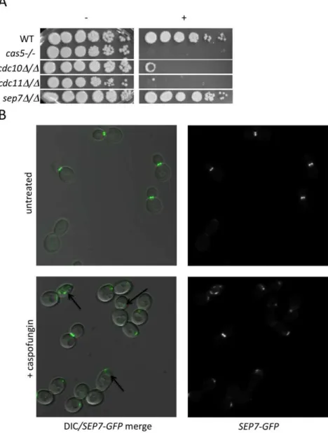
Relationship between septins and CWR
Cluster IV, identified in the previous analysis, contains several PKs with known connections to polar septin localization, including Cla4, Gin4, and Hsl1 [25,26]. We hypothesized that septin function itself may be related to CWR. Two further observations support this hypothesis. First, we found that mutants defective in the septin genescdc10andcdc11demonstrated increased sensitivity to caspofungin (Fig. 3A). These two septins are important for septin ring formation at high temperatures and chitin deposition at all temperatures [27]. In contrast, asep7deletion mutant, which causes no apparent defect in yeast-form septin ring formation or chitin deposition, does not have heightened sensitivity to caspofungin. Severity of sensitivity thus correlated with the severity of the septation defects (Fig. 3A), an indication that septin function is vital for C. albicans survival during cell wall stress. Second, we asked whether cell wall disruption might alter septin
localization. A Sep7-GFP fusion protein is concentrated at mother-bud necks in untreated budded cells, with two rings visible in separating mother-daughter cells (Fig. 3B), as previously reported [28]. Caspofungin treatment affected this localization dramatically: mislocalization was observed on one side of the mother-bud neck or in other regions of 46.7% of cells, while there were partial septa in 3.3% of budded cells (N = 92) (Fig. 3B). Caspofungin treatment ofCDC10-GFP,CDC12-GFP, and CDC3-GFP strains caused similar abnormalities (data not shown). Therefore, septin localization depends upon normal CWR.
We were led to the discovery of a role for septins in CWR by the sensitive phenotype of PK mutants with known roles in septin localization. We hypothesized that these PK mutants might display aberrant septin localization even in the absence of stress. Mutations causing a complete delocalization of septins at the mother-bud neck would probably be lethal to the cell and thus would not have been recovered in our library. To determine whether any of the caspofungin-sensitive PK and PK-related mutant strains displayed aberrant septin localization, a GFP tag Figure 2. PK and PK-related mutant strains show a damage response in the absence of cell wall stress.(A) The expression of six genes upregulated by caspofungin treatment,DDR48,SOD5,STP4,ALS1,RTA4, andECM331, was analyzed in PK and PK-related mutant strains in the absence of cell well stress. The expression ofTDH3, a gene involved in glycolysis, was used to normalize expression between strains and expression values were further normalized to wild type (DAY185) expression for comparison between experiments. Resultant values were log base 2 transformed (wild type expression for all six genes is therefore at 0). (B) A graphical representation of the expression data. Arrows point to targets upregulated in all (solid arrows), most (dashed lines), or half (dotted lines) of the mutants indicated in the clusters.
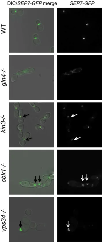
was inserted into the 39end of theSEP7gene at the endogenous SEP7locus. We were able to insert theSEP7-GFPtagged allele into all but two of the caspofungin-sensitive PK and PK-related mutant strains (hst72/2 and psk12/2). Cluster IV mutant strains, particularly gin42/2 and cla42/2 are the clearest candidates for delocalization based on phenotypes of the corresponding S. cerevisiaemutants [25,29]. Indeed, septin localization was disrupted in thegin42/2mutant strain (Fig. 4 and S3). Two cell phenotypes were observed: cells with normal buds had normal septin localization, but cells growing as pseudohyphae were either lacking septin rings or had septin rings that did not completely span the neck. Neither the cla42/2 nor the hsl12/2 had delocalized septins in the absence of stress (data not shown). This is consistent with the known roles of Cla4 in localization of septins in hyphal cells [30] and Hsl1 in signaling downstream of septin localization. However, one member of Cluster IV with no previous links to septin organization, Kin3, did demonstrate a septin organization defect, suggesting that this PK is linked to septin
localization as predicted by its inclusion in this cluster. Septins in this strain appear normal in buds until cytokinesis, where separation of septin rings is aberrant. In addition to these Cluster IV mutants, we also observed partial septin mislocalization in the vps342/2andcbk12/2mutant strains (Fig. 4 and S3). Whether septin disorganization contributes to the caspofungin sensitivity of these strains or it is a symptom of an intrinsic cell wall defect has not yet been determined. However, the disorganization of septins in these strains lends further support to the hypothesis that septin localization or regulation is linked to cell wall integrity.
Discussion
PKs occupy key positions between environmental sensors and responses. Our study ofC. albicans PKs emphasizes their pivotal roles: almost half of the PK mutants display a prominent phenotype. In contrast, only,5% of viableC. albicans transcrip-tion factor mutants have comparable phenotypes [16,17,31,32]. Figure 3. Septins play an integral role in CWR.(A) A wild type marker-matched strain (DAY185), acas5D/cas5Dnegative control, acdc10D/ cdc10D(YAW7), a cdc11Dcdc11/D(YAW11), and asep7D/sep7D(YAW41) mutant strain were serially diluted onto YPD (2) or YPD+125 ng/ml
caspofungin (+) and grown for 3 days at 30uC. (B)SEP7-GFPcells (JRB217) were grown to log phase and either treated with 125 ng/ml caspofungin (+
We observed thatC. albicansPK biological function is conserved with theirS. cerevisiaeandS. pombeorthologs in many cases (Tables 1 and 2). We also detected novel roles related to C. albicans
filamentation and biofilm formation, traits not examined system-atically with model organism mutant panels. However, our most striking result is thatC. albicansCWR relies upon conserved PKs with novel functions compared to their orthologs, representing an expansion of the circuitry involved in this process (Fig. 5). A few of these PKs have been linked to cell wall integrity previously, most notably Prk1 and Hst7 [33–35], but their importance in CWR seems much greater inC. albicansthan inS. cerevisiae, given that the orthologous S. cerevisiae mutants do not demonstrate heightened caspofungin sensitivity. CWR depends upon eight PKs with roles in this process that were not predicted from model yeast studies, plus a PK gene and a PK-related gene whose conserved CWR function is newly described in this report.
Two simple scenarios can account for the functional roles of the eight unpredicted CWR PKs: they may have a role in ongoing cell wall biogenesis, or they may only be required for response to cell wall stress. These possibilities are not mutually exclusive: a CWR PK may function in both capacities. Our analysis of PK mutant transcriptional responses strongly supports a role for Ire1 in normal cell wall biogenesis and suggests that several of the other novel PKs identified in this study may also have roles in cell wall biogenesis. In contrast, transcriptional responses of Cluster II PK mutants are not very distinct from wt and we suspect that these PKs may be important for cell wall stress response. Indeed,BCK1, a member of the cell wall stress response PKC1 MAPK pathway, andPSK1, which is also thought to have a role in cell wall stress response [17] are both in Cluster II. Microarray analysis of the other PKs in this cluster, Swe1 and Prk1, may identify a role for these PKs in stress response as well.
Our survey of gene expression alterations, though limited, revealed some qualitative differences among the mutants. Such distinctions may be useful in assembling the PKs into existing or novel pathways, a task that has generally proved challenging in organisms with limited genetic tractability. We were able to
Figure 4. Septins are mislocalized in some PK mutant strains. SEP7-GFP tagged wild type (JRB217), gin42/2 (JRB221), kin32/2 (JRB193), cbk12/2 (JRB224), and vps342/2 (JRB216) strains were grown to log phase and imaged at 1006on glass slides. An overlay of
DIC and GFP is on the left and GFP alone is on the right. Arrows point to aberrant septin localization.
doi:10.1371/journal.ppat.1000752.g004
Figure 5. A model of the role of PKs in septin morphology and cell wall biogenesis.Based on our observations, an intact septin ring is required for normal cell wall production and a normal cell wall is required for the formation of a septin ring. PK Cluster IV genes have known and predicted roles in septin morphology and signaling and may impact cell wall biogenesis indirectly via this role. We hypothesize PK Cluster I genes have direct roles in biogenesis of the cell wall, while PK Cluster III may indirectly effect cell wall biogenesis by regulating the flow of carbohydrates into cell wall biosynthesis pathways. We hypothesize that the genes in PK Cluster II are involved in cell wall stress response and the upregulation of cell wall integrity genes. PK Clusters I, III, and IV negatively regulate PK Cluster II, either directly or indirectly, based on the observation that cell wall integrity genes are upregulated in the absence of these PKs.
organize the data into distinct clusters that may reveal functional relationships. Indeed, mutants inGIN4,CLA4, and HSL1, genes important for the localization of septins (GIN4 and CLA4) or signaling downstream of septin localization (HSL1), fell into a cluster (Cluster IV, Fig. 3). Thekin32/2andckb22/2mutants were also in this cluster. Although only tentatively linked, close association of thekin32/2andckb22/2strains with mutants in genes involved in septin localization and signaling suggests Kin3 and Ckb2 may also play a role in this process (Fig. 5). Interestingly, thecbk12/2andire12/2strains also clustered together (Cluster I, Fig. 2), lending further support to the hypothesis thatIRE1has a role in cell wall biogenesis. Cbk1 is upstream of cell wall biogenesis genes, such as the chitin synthases [20], and along with the roles of Vps34 and Ire1 in cellular transport, this cluster seems to represent the production and delivery of cell wall building enzymes to the cell periphery (Fig. 5). Mutations in SWE1, PRK1, PSK1, and HST7, which fall into a cluster with thebck12/2mutant (Cluster II, Fig. 2), clustered together because the genes tested in the QrtPCR analysis were not differentially regulated compared to wild type. Perhaps, like bck12/2, they also have some role in responding to cell wall stress (Fig. 5). The third cluster includes both of the casein kinase I mutants (yck22/2 and yck32/2), tpk12/2, and the casein kinase II regulatory subunit mutant ckb12/2(Fig. 2). The genes included in this cluster have a variety of roles within cells, but one intriguing correlation between their functions is carbohydrate sensing (Yck2/3) and signaling (Tpk1). The cAMP pathway, of which Tpk1 is a key member, has a conserved role in glucose sensing (reviewed in [36]) and Yck1 and 2 have been shown to have a role in regulation of glucose transporters [37]. Glucose is the major building block of the b -glucans in the cell wall as well as an important source of energy for cells and perhaps these genes help cells balance the need for food versus protection (Fig. 5). While such inferences are clearly tentative, they make numerous testable predictions that will be useful in unraveling the complex relationships among cell wall regulators.
Ire1 showed the largest damage profiles in all three cell wall damage response genes. In all organisms where it has been studied, Ire1 is a component of the conserved unfolded protein response (UPR), which is activated by accumulation of unfolded proteins in the ER [38]. S. cerevisiae ire1 mutants do exhibit sensitivity to cell wall damage under certain conditions [39], but theC. albicans ire12/2mutant seems much more sensitive to cell wall damage, suggesting perhaps a more central role in this process. A simple model is that the UPR is critical forC. albicans cell wall damage response, perhaps because of the large number of cell wall protein genes that are induced by caspofungin [16]. We note that a relatively distal 39homozygous insertion mutation was created for this study (Table S1; insertion at bp 3520 of the 3675 bp ORF), and we have been unable to generate homozygous 59insertions or a deletion of this gene inC. albicans. Thus the UPR may be essential inC. albicanseven in the absence of exogenous stresses. We suggest that the UPR has a critical role in cell wall biogenesis and may be partially activated under our growth conditions.
Our findings also reveal a clear relationship between septin function and CWR. The PKs that govern CWR include Yck2, Cla4, and Gin4, which are involved in the assembly of septins at the mother-bud interface, and Swe1 and Hsl1, which are important for signaling that septins are appropriately localized [25,40]. Furthermore, we observed that septins are required for normal sensitivity to caspofungin, and that septins are delocalized in response to caspofungin treatment. This phenomenon appears to be conserved inS. cerevisiae(Briggs, Mitchell, and Blankenship
unpublished results), suggesting a conserved role for septin delocalization in cell wall integrity. Septins are connected to cell wall biogenesis through their known roles in determining sites of secretion and in directing the localization of chitin synthase. Septin delocalization may act positively in the cell wall damage response to permit delocalized secretion and cell wall synthesis when glucan synthase inhibition makes the bud neck an unsuitable site. A second possibility is that septin delocalization may function passively to prevent unbalanced production of cell wall compo-nents when glucan synthesis is blocked by caspofungin. A third possibility is that septin delocalization may protect cells by blocking cell cycle progression through Swe1 and Hsl1, thus reducing the need for new cell wall material. Thus, the analysis of C. albicans PKs has pointed toward a novel cell biological phenomenon, and several testable hypotheses may explain its functional consequences.
In some of our PK mutant strains, septins are mislocalized even in the absence of caspofungin treatment. This was expected for the gin42/2 mutant [29] and may contribute to the caspofungin sensitivity of this mutant strain, but we also identified several PKs with previously uncharacterized roles in septin organization. The kin32/2 strain clustered with mutants involved in septin organization and signaling (Class IV), and the septation defect of this mutant (Fig. 4) served as a validation of the clustering. Ckb2 was also a member of this cluster and although it did not have a septation defect, it may, like Hsl1, have a role in signaling downstream of septin organization. Septin mislocalization defects were also noted in thecbk12/2and vps342/2 mutant strains. Thecbk12/2 strain had septa at the mother-bud neck but also had aberrant septa in punctae throughout cells. Septa invps342/ 2mutant cells appeared normal in mature cells, but were wide in young buds, extending well into the bud, suggesting that septa in this mutant are disorganized at early stages of bud formation. Both of these mutants are in Cluster I, and perhaps the mislocalization of septins in these strains is due to the damaged walls present in these mutants. The cbk12/2 and vps342/2 phenotypes are different and the ire12/2 mutant does not have a septin localization defect at all, suggesting that the nature of the cell wall defect in these strains are distinct. We propose that Class I PKs are important for cell wall biogenesis and that defects in biogenesis or defects due to external stress inactivate Class IV PKs (perhaps via Class II PKs), which leads to septin mislocalization.
PK Snf1 [50,51], suggests that theC. albicans glucose repression system may be connected to cell wall biogenesis or integrity. Although aS. cerevisiae snf1mutant shows synthetic lethality with theb-1,3-glucan synthaseFKS1[52] we propose that this PK has a more prominent role in CWR inC. albicansand may explain why it is essential for viability in this fungus [53,54]. Caspofungin hypersensitivity of mutants defective in Rio2, Tpk1, and casein kinase 2 regulatory subunits Ckb1 and Ckb2 also suggests that PK rewiring has occurred during divergence of S. cerevisiae and C. albicans. These cases provide a foundation for further mechanistic inquiry into cell wall regulatory responses of this highly successful commensal and pathogen.
Materials and Methods
Strains and media
Strains were grown on yeast extract-peptone-dextrose (YPD) rich medium, defined synthetic dextrose medium, prepared as previously described [55], spider medium (10 g D-mannitol (Sigma), 10 g nutrient broth (BD Difco), 2 g K2HPO4(Sigma) in
1 l of H2O), and M199 cell culture medium (Roche) buffered with
150 mM Hepes (Gibco). NaCl (Sigma), H2O2 (Sigma),
Caspo-fungin (Merck), and sorbitol (Sigma) were added to media at the concentrations described. M199 medium was adjusted to the appropriate pH using NaOH and HCl, and pH was measured on a Corning pH Meter 240.
All PK mutants tested were made in the BWP17 [56] background. Construction of the PK mutant library followed the methods established in Davis et al [57]. Briefly, clones bearing genomic DNA from C. albicans strain CAI4 with an identified insertion of Tn7-UAU1 cassette were excised from the plasmid backbone by digestion with NotI. These linearized constructs were then transformed into BWP17. Putative heterozygote (Arg+) transformants were selected on SC-Arg+Uridine plates, and twelve independent colonies were grown in YPD liquid media overnight and patched onto SC-Arg-Ura plates. The Arg+Ura+ putative homozygotes were screened by colony PCR using primers,500 bp up and downstream of the insertion and within the transformant cassette (Arg4detect [32]) to ensure absence of the wild type allele and presence of the transformed alleles. A BWP17 colony was amplified with the same primer set as a control. These strains have been deposited and are available at the Fungal Genetics Stock Center http://www.fgsc.net/candida/ FGSCcandidaresources.htm (Kansas City, MO).
To complement specific deletions, a fragment of DNA,800 bp upstream and 200 bp downstream of the open reading frame was amplified from BWP17 genomic DNA. Primers for the comple-mentation were roughly 60–70 bp in length and the 3920–30 bp was gene specific. A 40mer sequence (upstream primer-TTCACAC-AGGAAACAGCTATGACCATGATTACGCCAAGCT, down-stream primer TCGACCATATGGGAGAGCTCCCAACGC-GTTGGATGCATAG) was tacked onto the 59end of each primer sequence to guidein vivorecombination into the pDDB78 plasmid [58]. The amplified complementation fragment was co-transformed into the S. cerevisiae BY4741 Dtrp1 strain with an EcoRI/NotI linearized pDDB78 plasmid, and the resulting complementation clone was amplified inE. coli. Each complement clone was digested with NruI to target insertion to theHIS1locus and transformed into the appropriate mutant strains, complementing the mutation and rendering the strains His+as well. Presence of a wild-type band was assayed by colony PCR using the complement primers. pDDB78, digested with NruI, was transformed into the same mutant strains to generate prototrophic, marker-matched strains for comparison with the complemented strains.
To insert GFP into theSEP7 locus in his12/2 PK mutant and wild type (DAY286) his12/2 strains, a fragment of the SEP7 gene was amplified from the YSM26-1 strain [28] containing the 39 end of the SEP7 gene and the N-terminal GFP tag. The same strategy used for creating the complemen-tation clones was utilized to make a HIS1-tagged SEP7-GFP integrating plasmid, generating pJRB103. This plasmid was digested with BclI (New England Biolabs), which cuts within the SEP7 sequence to direct integration at the native SEP7 locus. Integration of the construct into the native SEP7 locus was assayed by PCR using primers within the GFP sequence and upstream of theSEP7integration.
Identification of PK genes
A total of 108 PK were identified by scanningC. albicansgenome databases (CGD [59] and CandidaDB [60]) for genes annotated as PKs as well as searching forS. cerevisiaeandS. pombePK homologs by blastp analysis (Table S1). Identities between C. albicans PKs and putative S. cerevisiae orthologs ranged from 23%–83% and putative S. pombe orthologs from 22%–78% (although in four instances, the best matches with S. pombe were not PKs). An additional 13 genes, identified as PK-related, were also included in subsequent analyses.
Sensitivity assays
Osmotic, oxidative, pH, caspofungin sensitivity. Strains were grown overnight in YPD media at 30uC. Cell density was measured at OD600for each strain, which were then diluted to
an OD600 of 3 in H2O. Five-fold dilutions were made of the
OD600 3 stock and these were plated on the indicated media.
Plates were incubated at 30uC for 24–48 hours, and digitally photographed. When sensitivity was identified in C. albicans, orthologous a and a S. cerevisiae mutants from the S. cerevisiae mutant library [61] were also assayed for sensitivity in a similar manner.
Filamentation and biofilm assays
For filamentation assays, overnight cultures of strains were diluted to an OD600of 0.3 in M199 liquid medium. Cultures were
incubated at 37uC with shaking for 90 minutes and cells were examined under the microscope (see below).
Biofilm assays were performed as previously described [31]. Briefly, silicon disks were incubated at 37uC overnight in a 12-well plate with bovine serum (Sigma) and washed with 2 ml PBS. 2 ml of Spider medium was added to each well. Aliquots of overnight cultures were added to an OD600 of 0.5 to each well, and
incubated with the pre-treated silicon disks for 90 minutes at 37uC with light shaking. Following this adherence step, the silicon disks were carefully transferred to a new 12-well plate with 2 ml of PBS in each well and then to another 12-well plate with fresh spider medium. Cultures were incubated at 37uC with light shaking for 60 hours and then digitally imaged.
RNA collection and quantitative rtPCR
Overnight cultures of cells were diluted to an OD600of 0.2 in
harvesting. Strains carrying homozygous mutants ofYCK2,YCK3, RIO2, and PRK1 were generated after the completion of the treatment experiment and were thus not included in the final assay. Cells were kept frozen on filters at 280uC until RNA extraction. RNA was extracted using a RiboPure-Yeast kit (Ambion) following manufacturers instructions with the following modifications. Cells were resuspended from filters with 1.5 ml ice-cold dH2O followed by 10–15 seconds of vigorous vortexing.
Resuspended cells were transferred to a 1.5 ml tube and spun down following the manufacturers protocol. Furthermore, during the cell disruption step, cells were beaten with a Next Advance Bullet Blender for 3 min at 4uC to maximize cell lysis.
10mg of the resulting total RNA was treated with the DNA-free kit (Ambion) followed by first-strand cDNA synthesis from half of the DNA-free RNA using the AffinityScript multiple temperature cDNA synthesis kit (Stratagene). Absence of DNA contamination was confirmed using control sets for which reverse transcriptase was omitted from the cDNA reaction.
Primer3 software (http://frodo.wi.mit.edu/) was used to design primers for four genes, TDH3, DDR48, SOD5, and STP4. The primers were as follows: for TDH3, TDH3FOR, 59-ATCCCA-CAAGGACTGGAGA-39, and TDH3REV, 59 -GCAGAAGCTT-TAGCAACGTG-39; for DDR48, JRB212, 59 -TTTCGGTT-TCGGTAAAGACG-39, and JRB213, 59-CTGTTGGAGGAA-CCGTAGGA-39; for SOD5, JRB249, 59-TCCTGCTGCTCAT-GAAGTTG-39, and JRB250, 59 -TTGTGTTAGCATTGCC-GTGT-39; and forSTP4, JRB244, 59-TCCTTTCAAGAACATC-GATTCA-39, and JRB245, 59-TTATGCATCCAATCATC-GACA-39. 26iQ SYBR Green Supermix (Biorad), 1ml of first-strand cDNA reaction mixture, and 0.1mM of primers were mixed in a total volume of 50ml per reaction, and real-time PCR was performed in triplicate for each sample on an iCycler iQ real-time PCR detection system (Bio-Rad). The program for amplification had an initial denaturation step at 95uC for 5 min, followed by 40 cycles of 95uC for 45 s and 58uC for 30 s. Product amplification was detected using SYBR Green fluorescence during the 58uC step, and specificity of the reaction was monitored by melt-curve analysis following the real-time program.TDH3was used as a reference gene for normalization of gene expression, which was determined using Bio-Rad iQ5 software (DDCT method). Cluster analysis was performed with the Multiexperiment Viewer (MeV 4.3) from TIGR. Hierarchical clustering was performed with average linkage and a Manhattan distance metric.
Septin morphology and microscopy
SEP7-GFPcells were diluted and grown overnight to an OD600of
,1 in YPD. 200ml of this culture was pipeted onto glass bottom dishes (MatTek Corporation, Ashalnd, MA) coated with concanav-alin A or poly-D-lysine (for caspofungin treatment). The dishes were washed twice with PBS and resuspended with PBS for immediate visualization (untreated cells) or incubated with 125 ng/ml caspo-fungin in YPD for 30 minutes (treated cells). Following caspocaspo-fungin treatment, the dishes were washed again and PBS was added for visualization. Cells visualized with a Zeiss Axio Observer Z.1 fluorescence microscope and a 1006NA 1.4 objective. Fluorescent images were acquired with an exposure time of 1 s on a Coolsnap HQ2(Photometrics) camera using Axiovision (Zeiss) software.
Supporting Information
Figure S1 PK and PK-related genes play essential roles in survival of and response to commonin vivostresses. (A) A wild type marker-matched strain (DAY286) and strains mutant forhog1/hog1 (JMR115),pbs2/pbs2(JJH31),chk1/chk1(JJH33),sln1/sln1(SF040),
prk1/prk1(JJH50),kin4/kin4(SF021),ssn3/ssn3(JJH65),ste11/ste11 (SF041A),gin4/gin4(JJH87),ckb1/ckb1(SF039),kis1/kis1(JJH85), andssn8/ssn98(SF046) were serially diluted on YPD (2) or YPD+ 7mM H2O2and grown for 2 days at 30uC. (B) DAY286 (WT) and
strains mutant forhog1/hog1, pbs2/pbs2, sok1/sok1 (JJH106),gin4/ gin4, and kis1/kis1were serially diluted on YPD (2) or YPD + 1.5M H2O2and grown for 2 days at 30uC. (C) DAY286 (WT) and
a strain mutant for cbk1/cbk1 (JJH114) were serially diluted on M199 solid medium buffered at pH 7 or pH 4 and grown for 2 days at 30uC. For all assays, results shown are representative of 2 or more independently isolated strains where possible. (D) A wild type strain (DAY286) and strains mutant forire1/ire1 (SF008A), sok1/sok1 (JJH104), mss2/mss2 (JJH93), tpk2/tpk2 (SF026), cbk1/ cbk1, andrio2/rio2 (JJH243) were grown overnight in rich liquid medium (YPD), diluted, and then grown in either liquid YPD at 30uC with shaking, or in M199 cell culture medium at 37uC for 2 hours and then imaged. (E) DAY286 (WT), abcr1D/Dnegative control (CJN702), and strains mutant forcbk1/cbk1, ire1/ire1, and gin4/gin4 (JJH87) were tested for biofilm formation in spider medium. Biofilms were allowed to mature for 48 hours before imaging. In both assays, results are representative of 2 isolates where possible. Only those PK and PK-related mutant strains with distinct phenotypes from wt are shown in this figure.
Found at: doi:10.1371/journal.ppat.1000752.s001 (1.52 MB PDF)
Figure S2 Expression profiles of PK mutants in the absence of stress. The expression ofALS1, STP4, SOD5, DDR48, RTA4, and ECM331 were measured in caspofungin-sensitive PK and PK-related mutant strains and their respective complement strains. All strains were grown in rich media at 30uC in the absence of exogenous stressors. The expression of TDH3 was used to normalize expression between strains and all expression was compared to a marker-matched wild type strain (DAY185). The panels are divided into the clusters identified in Fig. 2. (A) Cluster I, (B) Cluster II, (C) Cluster III, and (C) Cluster IV. Error bars represent standard deviation from the mean.
Found at: doi:10.1371/journal.ppat.1000752.s002 (0.32 MB PDF)
Figure S3 Septins are mislocalized in a subset of PK mutants. The localization of SEP7-GFP was monitored in PK mutants sensitive to cell wall stress. Strains were grown in YPD without additional stress. Images shown are representative images from a panel of images taken of each strain. Strains are as follows:bck12/2(JRB194),cbk12/2 (JRB224), ckb12/2 (JRB198), ckb22/2 (JRB229), cla42/2 (JRB183), gin42/2 (JRB221), hsl12/2 (JRB188), ire12/2 (JRB227), kin32/2 (JRB193), kis12/2 (JRB167), mkc12/2 (JRB208), mkk22/2 (JRB205), mss22/2 (JRB177), pkc12/2 (JRB200), prk12/2 (JRB169), rio22/2 (JRB225), sip32/2 (JRB170), swe12/2 (JRB179), tpk12/2 (JRB174), vps342/2 (JRB216),yck22/2(JRB164),yck32/2(JRB212).
Found at: doi:10.1371/journal.ppat.1000752.s003 (4.38 MB PDF)
Table S1 Genetic and biological properties of C. albicans insertion mutants.
Found at: doi:10.1371/journal.ppat.1000752.s004 (0.07 MB XLS)
Acknowledgments
We thank members of our lab for their advice and discussions. We are grateful to Merck Research Labs for providing caspofungin and to the Konopka lab for the septin mutant strains.
Author Contributions
References
1. Walker LA, Munro CA, de Bruijn I, Lenardon MD, McKinnon A, et al. (2008) Stimulation of chitin synthesis rescuesCandida albicansfrom echinocandins. PLoS Pathog 4: e1000040. doi:10.1371/journal.ppat.1000040.
2. Wheeler RT, Fink GR (2006) A drug-sensitive genetic network masks fungi from the immune system. PLoS Pathogens 2: e35. doi:10.1371/journal.ppat.0020035. 3. Kumamoto CA (2008) Molecular mechanisms of mechanosensing and their
roles in fungal contact sensing. Nat Rev Microbiol 6: 667–673. 4. Sundstrom P (2002) Adhesion inCandida spp. Cell Microbiol 4: 461–469. 5. Chattaway FW, Holmes MR, Barlow AJ (1968) Cell wall composition of the
mycelial and blastospore forms ofCandida albicans. J Gen Microbiol 51: 367–376. 6. Chauhan N, Li D, Singh P, Calderone R, Kruppa M (2002) The cell wall of Candidaspp. In: Calderone R, ed (2002)Candidaand candidiasis. Washington, D. C.: ASM Press. pp 159–175.
7. Chaffin WL, Lopez-Ribot JL, Casanova M, Gozalbo D, Martinez JP (1998) Cell wall and secreted proteins of Candida albicans: identification, function, and expression. Microbiology & Molecular Biology Reviews 62: 130–180. 8. Levin DE (2005) Cell wall integrity signaling in Saccharomyces cerevisiae.
Microbiology & Molecular Biology Reviews 69: 262–291.
9. Gerik KJ, Donlin MJ, Soto CE, Banks AM, Banks IR, et al. (2005) Cell wall integrity is dependent on the PKC1 signal transduction pathway inCryptococcus neoformans. Mol Microbiol 58: 393–408.
10. Delley PA, Hall MN (1999) Cell wall stress depolarizes cell growth via hyperactivation of RHO1. J Cell Biol 147: 163–174.
11. Li Y, Moir RD, Sethy-Coraci IK, Warner JR, Willis IM (2000) Repression of ribosome and tRNA synthesis in secretion-defective cells is signaled by a novel branch of the cell integrity pathway. Mol Cell Biol 20: 3843–3851. 12. Nanduri J, Tartakoff AM (2001) Perturbation of the nucleus: a novel
Hog1p-independent, Pkc1p-dependent consequence of hypertonic shock in yeast. Mol Biol Cell 12: 1835–1841.
13. Imazu H, Sakurai H (2005)Saccharomyces cerevisiaeheat shock transcription factor regulates cell wall remodeling in response to heat shock. Eukaryot Cell 4: 1050–1056.
14. Paravicini G, Mendoza A, Antonsson B, Cooper M, Losberger C, et al. (1996) TheCandida albicans PKC1gene encodes a protein kinase C homolog necessary for cellular integrity but not dimorphism. Yeast 12: 741–756.
15. Navarro-Garcia F, Sanchez M, Pla J, Nombela C (1995) Functional characterization of the MKC1 gene of Candida albicans, which encodes a mitogen-activated protein kinase homolog related to cell integrity. Mol Cell Biol 15: 2197–2206.
16. Bruno VM, Kalachikov S, Subaran R, Nobile CJ, Kyratsous C, et al. (2006) Control of theC. albicanscell wall damage response by transcriptional regulator Cas5. PLoS Pathog 2: e21. doi:10.1371/journal.ppat.0020021.
17. Rauceo JM, Blankenship JR, Fanning S, Hamaker JJ, Deneault JS, et al. (2008) Regulation of theCandida albicanscell wall damage response by transcription factor Sko1 and PAS kinase Psk1. Mol Biol Cell 19: 2741–2751.
18. Bimbo A, Jia Y, Poh SL, Karuturi RKM, den Elzen N, et al. (2005) Systematic deletion analysis of fission yeast protein kinases. Eukaryot Cell 4: 799–813. 19. Eisman B, Alonso-Monge R, Roman E, Arana D, Nombela C, et al. (2006) The
Cek1 and Hog1 mitogen-activated protein kinases play complementary roles in cell wall biogenesis and chlamydospore formation in the fungal pathogenCandida albicans. Eukaryot Cell 5: 347–358.
20. Bidlingmaier S, Weiss EL, Seidel C, Drubin DG, Snyder M (2001) The Cbk1p pathway is important for polarized cell growth and cell separation in Saccharomyces cerevisiae. Mol Cell Biol 21: 2449–2462.
21. Song Y, Cheon SA, Lee KE, Lee SY, Lee BK, et al. (2008) Role of the RAM network in cell polarity and hyphal morphogenesis inCandida albicans. Mol Biol Cell 19: 5456–5477.
22. Bensen ES, Martin SJ, Li M, Berman J, Davis DA (2004) Transcriptional profiling inCandida albicansreveals new adaptive responses to extracellular pH and functions for Rim101p. Mol Microbiol 54: 1335–1351.
23. O’Connor L, Lahiff S, Casey F, Glennon M, Cormican M, et al. (2005) Quantification ofALS1gene expression inCandida albicansbiofilms by RT-PCR using hybridisation probes on the LightCycler. Mol Cell Probes 19: 153–162. 24. Liu TT, Lee RE, Barker KS, Lee RE, Wei L, et al. (2005) Genome-wide
expression profiling of the response to azole, polyene, echinocandin, and pyrimidine antifungal agents in Candida albicans. Antimicrobial Agents & Chemotherapy 49: 2226–2236.
25. Longtine MS, Theesfeld CL, McMillan JN, Weaver E, Pringle JR, et al. (2000) Septin-dependent assembly of a cell cycle-regulatory module inSaccharomyces cerevisiae. Molecular & Cellular Biology 20: 4049–4061.
26. Keaton MA, Lew DJ (2006) Eavesdropping on the cytoskeleton: progress and controversy in the yeast morphogenesis checkpoint. Current Opinion in Microbiology 9: 540–546.
27. Warenda A, Konopka J (2002) Septin function inCandida albicansmorphogenesis. Mol Biol Cell 13: 2732–2746.
28. Martin S, Konopka J (2004) SUMO modification of septin-interacting proteins inCandida albicans. J Biol Chem 279: 40861–40867.
29. Wightman R, Bates S, Amornrrattanapan P, Sudbery P (2004) In Candida albicans, the Nim1 kinases Gin4 and Hsl1 negatively regulate pseudohypha formation and Gin4 also controls septin organization. Journal of Cell Biology 164: 581–591.
30. Leberer E, Ziegelbauer K, Schmidt A, Harcus D, Dignard D, et al. (1997) Virulence and hyphal formation of Candida albicansrequire the Ste20p-like protein kinase CaCla4p. Curr Biol 7: 539–546.
31. Nobile CJ, Mitchell AP (2005) Regulation of cell-surface genes and biofilm formation by theC. albicanstranscription factor Bcr1. Curr Biol 15: 1150–1155. 32. Norice CT, Smith FJ, Solis N, Filler SG, Mitchell AP (2007) Requirement for Candida albicansSun41 in biofilm formation and virulence. Eukaryot Cell 6: 2046–2055.
33. Zeng G, Yu X, Cai M (2001) Regulation of yeast actin cytoskeleton-regulatory complex Pan1p/Sla1p/End3p by serine/threonine kinase Prk1p. Mol Biol Cell 12: 3759–3772.
34. Lee BN, Elion EA (1999) The MAPKKK Ste11 regulates vegetative growth through a kinase cascade of shared signaling components. Proc Natl Acad Sci U S A 96: 12679–12684.
35. Cullen PJ, Schultz J, Horecka J, Stevenson BJ, Jigami Y, et al. (2000) Defects in protein glycosylation causeSHO1-dependent activation of aSTE12signaling pathway in yeast. Genetics 155: 1005–1018.
36. Sabina J, Brown V (2009) Glucose sensing network inCandida albicans: a sweet spot for fungal morphogenesis. Eukaryot Cell 8: 1314–1320.
37. Moriya H, Johnston M (2004) Glucose sensing and signaling inSaccharomyces cerevisiaethrough the Rgt2 glucose sensor and casein kinase I. Proc Natl Acad Sci U S A 101: 1572–1577.
38. Patil C, Walter P (2001) Intracellular signaling from the endoplasmic reticulum to the nucleus: the unfolded protein response in yeast and mammals. Curr Opin Cell Bio 13: 349–356.
39. Scrimale T, Didone L, de Mesy Bentley KL, Krysan DJ (2009) The unfolded protein response is induced by the cell wall integrity mitogen-activated protein kinase signaling cascade and is required for cell wall integrity inSaccharomyces cerevisiae. Mol Biol Cell 20: 164–175.
40. Robinson LC, Bradley C, Bryan JD, Jerome A, Kweon Y, et al. (1999) The Yck2 yeast casein kinase 1 isoform shows cell cycle-specific localization to sites of polarized growth and is required for proper septin organization. Molecular Biology of the Cell 10: 1077–1092.
41. Ihmels J, Bergmann S, Gerami-Nejad M, Yanai I, McClellan M, et al. (2005) Rewiring of the yeast transcriptional network through the evolution of motif usage. Science 309: 938–940.
42. Tanay A, Regev A, Shamir R (2005) Conservation and evolvability in regulatory networks: the evolution of ribosomal regulation in yeast. Proc Natl Acad Sci U S A 102: 7203–7208.
43. Tsong AE, Tuch BB, Li H, Johnson AD (2006) Evolution of alternative transcriptional circuits with identical logic. Nature 443: 415–420.
44. Martchenko M, Levitin A, Hogues H, Nantel A, Whiteway M (2007) Transcriptional rewiring of fungal galactose-metabolism circuitry. Curr Biol 17: 1007–1013.
45. Tsong AE, Miller MG, Raisner RM, Johnson AD (2003) Evolution of a combinatorial transcriptional circuit: a case study in yeasts. Cell 115: 389–399. 46. Leberer E, Harcus D, Broadbent ID, Clark KL, Dignard D, et al. (1996) Signal transduction through homologs of the Ste20p and Ste7p protein kinases can trigger hyphal formation in the pathogenic fungusCandida albicans. Proc Natl Acad Sci U S A 93: 13217–13222.
47. Kohler JR, Fink GR (1996) Candida albicans strains heterozygous and homozygous for mutations in mitogen-activated protein kinase signaling components have defects in hyphal development. Proc Natl Acad Sci U S A 93: 13223–13228.
48. Chen J, Chen J, Lane S, Liu H (2002) A conserved mitogen-activated protein kinase pathway is required for mating inCandida albicans. Mol Microbiol 46. 49. Magee BB, Legrand M, Alarco AM, Raymond M, Magee PT (2002) Many of
the genes required for mating inSaccharomyces cerevisiaeare also required for mating inCandida albicans. Mol Microbiol 46: 1345–1351.
50. Yang X, Jiang R, Carlson M (1994) A family of proteins containing a conserved domain that mediates interaction with the yeast SNF1 protein kinase complex. Embo J 13: 5878–5886.
51. Lesage P, Yang X, Carlson M (1994) Analysis of the SIP3 protein identified in a two-hybrid screen for interaction with the SNF1 protein kinase. Nucleic Acids Res 22: 597–603.
52. Tong AH, Lesage G, Bader GD, Ding H, Xu H, et al. (2004) Global mapping of the yeast genetic interaction network. Science 303: 808–813.
53. Petter R, Chang YC, Kwon-Chung KJ (1997) A gene homologous to Saccharomyces cerevisiae SNF1appears to be essential for the viability ofCandida albicans. Infect Immun 65: 4909–4917.
54. Enloe B, Diamond A, Mitchell AP (2000) A single-transformation gene function test in diploidCandida albicans. J Bacteriol 182: 5730–5736.
55. Sherman F (1991) Getting started with yeast. In: Guthrie C, Fink GR, eds (1991) Methods in Enzymology. San DiegoCA: Academic Press, Inc. pp 3–21. 56. Wilson RB, Davis D, Mitchell AP (1999) Rapid hypothesis testing withCandida
albicansthrough gene disruption with short homology regions. J Bacteriol 181: 1868–1874.
58. Ma H, Kunes S, Schatz PJ, Botstein D (1987) Plasmid construction by homologous recombination in yeast. Gene 58: 201–216.
59. Arnaud MB, Costanzo MC, Skrzypek MS, Shah P, Binkley G, et al. (2007) Sequence resources at theCandidaGenome Database (CGD). Nucleic Acids Res 35: D452–456.
60. Rossignol T, Lechat P, Cuomo C, Zeng Q, Moszer I, et al. (2008) CandidaDB: a multi-genome database forCandidaspecies and relatedSaccharomycotina. Nucleic Acids Res 36: D557–561.