Improvement of
in vivo
anticancer and antiangiogenic potential
of thalidomide derivatives
Patrícia Marçal da Costa
a, Marcilia Pinheiro da Costa
b, Adriana Andrade Carvalho
c,
Suellen Melo Tibúrcio Cavalcanti
d, Marcos Veríssimo de Oliveira Cardoso
d,
Gevânio Bezerra de Oliveira Filho
d, Daniel de Araújo Viana
e, Francisco Vagnaldo Fechine-Jamacaru
a,
Ana Cristina Lima Leite
d, Manoel Odorico de Moraes
a, Claudia Pessoa
a,f, Paulo Michel Pinheiro Ferreira
g,⇑ aDepartment of Physiology and Pharmacology, School of Medicine, Federal University of Ceará, 60.430-270 Fortaleza, Ceará, BrazilbDepartment of Pharmacy, Federal University of Piauí, 64049-550 Teresina, Brazil
cDepartment of Pharmacy/Postgraduate Program in Health Sciences, Federal University of Sergipe, 49400-000 Lagarto, Sergipe, Brazil dDepartment of Pharmaceutical Sciences, Federal University of Pernambuco, 50670-901 Recife, Pernambuco, Brazil
eSchool of Veterinary Medicine, State University of Ceará, 60740-000 Fortaleza, Ceará, Brazil fFundação Oswaldo Cruz (FIOCRUZ), 60180-900 Fortaleza, Ceará, Brazil
gLaboratory of Experimental Cancerology, Department of Biophysics and Physiology, Postgraduate in Pharmaceutical Sciences, Federal University of Piauí, 64049-550 Teresina, Brazil
a r t i c l e
i n f o
Article history:
Received 15 March 2015
Received in revised form 22 June 2015 Accepted 26 June 2015
Available online 29 June 2015
Keywords:
Antitumor
Angiogenesis inhibition Sarcoma 180 Human cells
Phthalimide derivatives Toxicity
a b s t r a c t
The strategy of antiangiogenic drugs is based on inhibiting formation of new blood vessels as alternative to limit cancer progression. In this work, we investigated the antitumor and antiangiogenic potential of eight thalidomide derivatives. Most of the molecules was not cytotoxic but2a,2dand3drevealed weak antiproliferative activity on HL-60, Sarcoma 180 (S180) and normal peripheral blood mononuclear cells. Thalidomide,2aand2bwere able to inhibit tumor growth (53.5%, 67.9% and 67.4%, respectively) in S180-bearing mice and presented moderate and reversible toxicity on liver, kidneys and spleens. Both analogs (2a and 2b) inhibited cell migration of endothelial (HUVEC) and melanoma cells (MDA/MB-435) at 50lg/mL. Immunohistochemistry labeling assays with CD-31 (PECAM-1) antibody showed microvascular density (MVD) was significantly reduced in thalidomide, 2a and 2b groups (30 ± 4.9, 64.6 ± 1.8 and 46.5 ± 19.5%, respectively) (p< 0.05). Neovascularization evaluated by Chorioallantoic Membrane Assay (CAM) with compounds2aand2bshowed reduction of vessels’ number (12. 9 ± 2.3 and 14.8 ± 3.3%), neovascularization area (13.1 ± 1.7 and 14.3 ± 1.7%) and total length of ves-sels (9.2 ± 1.5 and 9.9 ± 1.9%). On the other hand, thalidomide did not alter vascularization parameters. Consequently, addition of thiosemicarbazone pharmacophore group into the phthalimidic ring improved thein vivoantitumor and antiangiogenic potential of the analogs2aand2b.
Published by Elsevier Ireland Ltd.
1. Introduction
Angiogenesis is a complex regulated biological process involved in the formation of new blood vessels in normal or pathological
conditions[1]. Various regulatory and signaling molecules manage
angiogenesis, including VEGF (vascular endothelial growth factor), PDGF (platelet-derived growth factor), FGF (fibroblast growth fac-tor), EGF (epidermal growth facfac-tor), HGF (hepatocyte growth factor), PlGF (placenta growth factor), receptor tyrosine kinases (RTKs), transcription factors, as well as molecules involved
in MAPK (Mitogen-activated protein kinases) and PI3K
(Phosphatidylinositol 3-kinase) signaling[1,2]. Angiogenic events
become activated in numerous illnesses, including ocular and
car-diovascular diseases, inflammation and cancer[3–5].
In cancer, tumor cells stimulate angiogenesis in an effort to maintain and allow tumor progression. Thus, neovascularization improves blood supply (oxygen and nutrient) to the neoplasic cells,
and consequently favors the tumor growth[1,3,4,6]. Therefore, the
angiogenesis has become an attractive target in therapeutic approaches due to its key role in tumor growth, invasion and
metastasis[2,3,6–8].
Thalidomide is the most known angiogenesis inhibitor. It is a synthetic derivative of glutamic acid with contains two imide rings: glutarimide and phthalimide. Due to its neurotoxicity, this
drug caused devastating teratogenic effects in the 1960s[9,10].
Nowadays, thalidomide is effective to treat different diseases, including leprosy, acquired immunodeficiency syndrome (AIDS),
http://dx.doi.org/10.1016/j.cbi.2015.06.037 0009-2797/Published by Elsevier Ireland Ltd.
⇑Corresponding author.
E-mail addresses:[email protected],[email protected](P.M.P. Ferreira).
Contents lists available atScienceDirect
Chemico-Biological Interactions
multiple myeloma, cancer and other angiogenesis-dependent
dis-orders[11,12]. Thalidomide is classified as an immunomodulatory
drug (IMiD) that inhibits the production of tumor necrosis factor
alpha (TNF-
a
) and may also affect the production ofinterleukin-1b (IL-1b), IL-2, IL-4, IL-5, IL-6, IL-10 and interferon-
c
(IFN-
c
). Moreover, secretion of both vascular endothelial growthfactor (VEGF) and beta fibroblast growth factor (bFGF) from tumor and bone marrow stromal cells is suppressed upon exposure to IMiDs, resulting in reduced endothelial cell migration and adhesion [10,11]. The idea of using thalidomide in the cancer treatment occurred after the discovery of its antiangiogenic properties in
the early 1990s[13]. Despite the thalidomide mechanism in cancer
is not fully understood, its derivatives are known by immunomod-ulatory and anti-inflammatory effects that likely contribute to
antiangiogenic effects [2,14]. In 2006, the Food and Drug
Administration (USA) approved it against multiple myeloma[13].
To overcome risks caused by thalidomide, such as teratogenic-ity, many analogs have been developed with anticancer proposal. Previous reports showed that structural variations of thalidomide could change the side-effect profile, potentially eliminating them, while still maintaining the desired anticancer activity and
demon-strating promising results in clinical trials [2,10]. Lenalidomide
(Revlimid™) was approved for use in patients with
myelodysplas-tic syndromes (MDS) and multiple myeloma[15]. Pomalidomide is
a potent second-generation IMiD.In vitrostudies have shown that
pomalidomide is 10-fold more potent than lenalidomide and up to
15,000 times more potent than thalidomide at inhibiting TNF-
a
.Pomalidomide has shown significant clinical activity in phase I and II trials on patients with relapsed and/or refractory multiple
myeloma[16].
In the current study, thalidomide and eight analogs were tested
usingin vitro andin vivoassays to assess their performance on
tumor growth inhibition and tumor angiogenesis. Based on the structure–activity relationship, these novel analogs were devel-oped with the phthalimide and thiosemicarbazone pharmacophore
groups[17] (Fig. 1). Data presented here add important findings
about antitumor effects of these analogs on solid tumors.
2. Materials and methods
2.1. Materials
Phthalimide derivatives (2a–fand 3a–b) were synthesized as
described by Pessoa et al.[17]. Purity of compounds was provided
by elemental analysis data.
Primary antibody CD-31 (Platelet Endothelial cell Adhesion Molecule-1, PECAM-1) and secondary antibody were purchased from Santa Cruz Biotechnology. Anti-VEGF monoclonal antibody (Bevacizumab – Avastin™) Genentech/Roche, CA, USA) was pur-chased from Santa Cruz Biotechnology. Endothelia basal medium (EBM), RPMI 1640 medium, fetal bovine serum (FBS), peni-cillin/streptomycin and glutamine were purchased from Gibco (USA). Alamar Blue™, 5-Fluorouracil (5-FU), hydrocortisone, gen-tamicin and mitomycin C were obtained from Sigma Aldrich (St. Louis, MO, USA). All the other chemicals were of analytical grade and used as indicated by manufacturer. Thalidomide tablets 100 mg were used (FUNED, MG, Brazil). All molecules were diluted in DMSO to a final concentration of 5 mg/mL (stock solution). Chemical names and purity data of all compounds tested are given inSupplementary material.
2.2. Animals
Adult female Swiss mice (Mus musculusLinnaeus, 1758) were
obtained from the animal facilities at Universidade Federal do
Ceará (UFC), Fortaleza, Ceará, Brazil. They were kept in well-ventilated cages under standard conditions of light (12 h with
alternate day and night cycles) and temperature (22 ± 2°C) and
were housed with free access to commercial rodent stock diet
(Nutrilabor, Campinas, Brazil). Ross chicken eggs of the Gallus
domesticus species weighing 50 g, 12–24 h after laid, were pur-chased from CIALNE™ Company (Fortaleza, Ceará, Brazil) and
stocked for 24 h at 19–20°C before incubation. All procedures
were approved by the Committee on Animal Research at UFC
(Process n° 106/2007) and they are in accordance with the
Brazilian (COBEA – Colégio Brasileiro de Experimentação Animal)
and International Standards on the care and use of experimental animals (Directive 2010/63/EU of the European Parliament and of the Council).
2.3. Cytotoxicity assays
Cytotoxic potential of the compounds (thalidomide,2a–2f,3a,
3b) was assessed after 72 h exposure using cancer cell lines
[HL-60 (leukemia), HCT-8 (colon), MDA/MB-435 (melanoma) and SF-295 (glioblastoma)], Sarcoma 180 cells (S180) and normal peripheral blood mononuclear cells (PBMC). Human cell lines were maintained in RPMI 1640 medium supplemented with fetal bovine
serum 10%, 100 U/mL penicillin/streptomycin at 37°C with 5% CO2
atmosphere. In similar conditions, S180 tumor cells and PBMC were cultured in RPMI 1640 medium supplemented with 20% fetal bovine serum. Quantification of cell proliferation was determined spectrophotometrically using a multiplate reader (DTX 880 Fig. 1.Phthalimide analogs derived from thalidomide used in this study. Analogs were obtained as demonstrate by Pessoa et al.[17].
Multimode Detector, Beckman Coulter). All wells were exposed to the same amount of dimethylsulfoxide (DMSO 0.1%).
2.3.1. Evaluation of the cytotoxicity on tumor cell lines
In vitroantiproliferative activity against four human tumor cell lines (HL-60, HCT-8, MDA-MB-435 and SF-295) was determined by MTT assay, a cell viability assay that detect reduction of a yellow
dye 3-(4,5-dimethyl-2-thiazolyl)-2,5-diphenyl-2H-tetrazolium
bromide (MTT) to a blue formazan product[18]. Briefly, cells were
plated in 96-well plates (0.1–0.3106cells/mL) to allow adhesion.
After 24 h, thalidomide and its analogs (0.78–100
l
g/mL) wereadded to each well. Doxorubicin (0.005–5
l
g/mL) was used aspos-itive control. After 69 h of incubation, plates were centrifuged and
the medium was replaced by fresh medium (150
l
L) containingMTT (0.5
l
g/mL). Three hours later, plates were centrifuged, andthe formazan was dissolved in DMSO. The effect of the molecules was quantified at 595 nm.
2.3.2. Evaluation on PBMC proliferation
Heparinized human blood (from healthy, non-smoker donors who had not taken any drug for at least 15 days prior to sampling, aged between 18 and 35 years) was collected and PBMC were iso-lated by a standard method of density-gradient centrifugation over Ficoll-Hypaque. PBMC were washed and resuspended in supple-mented RPMI 1640 medium and phytohemagglutinin (3%). Then,
PBMC were plated in 96-well plates (5105cells/well in 100
l
Lof medium). After 24 h, compounds (0.78–100
l
g/mL) were addedto each well and cells were incubated during 72 h. Twenty-four
hours before the late incubation, 10
l
L of stock solution(0.312 mg/mL) of Alamar Blue™ were added to each well [19].
Doxorubicin (0.005–5
l
g/mL) was used as a positive control. Theeffect was quantified as the percentage of control absorbance at 570 nm and 595 nm. These studies were performed in accordance to the Brazilian research guidelines (Law 466/2012, National Council of Health, Brazil) and the Declaration of Helsinki.
2.3.3. Evaluation on proliferation of Sarcoma 180 cells
Ascite-bearing mice between 7 and 9 days post inoculation were sacrificed by cervical dislocation and a suspension of S180 cells was harvested from the intraperitoneal cavity under aseptic
conditions. The suspension was centrifuged at 500gfor 5 min to
obtain a cell pellet and washed 3 times with RPMI medium. Cell
concentration was adjusted to 0.5106cells/mL in supplemented
RPMI 1640 medium, plated in a 96-well plate and incubated with
increasing concentrations of the compounds (0.4–100
l
g/mL). Cellproliferation was determined by the Alamar Blue assay after 72 h
[20]. Eight hours before the late incubation, 10
l
L of stock solution(0.312 mg/mL) of Alamar Blue™ were added to each well. The absorbance was measured at 570 nm and 595 nm and the antipro-liferative effect was quantified as percentage of the control.
2.4. In vivo antitumor assay
Female Swiss mice (20–25 g) obtained from the facilities of the Federal University of Ceará were used. Animals were kept in cages with free access to food and water under a 12/12 h light–dark cycle (lights on at 6:00 a.m.). Sarcoma 180 cells were maintained in the peritoneal cavities of the mice. Ten-day-old sarcoma 180 ascite tumor cells were removed from the peritoneal cavity, counted and subcutaneously implanted into the right hind axillary of the
animals (4106cells/mL). In the following day, thalidomide and
its analogs (50 mg/kg/day) dissolved in DMSO 10% were intraperi-toneally administered for 7 days. Negative and positive controls received DMSO 4% and 5-FU (25 mg/kg/day), respectively. On the 8th day, mice were sacrificed and tumors, livers, spleens and kid-neys were dissected out, weighed and fixed in 10% formaldehyde.
The inhibition ratio of tumor growth (%) was calculated as follows:
inhibition ratio (%) = [(A B)/A]100, where A is the average
tumor weight in the negative control, andBis the average in each
separately treated group.
2.4.1. Histopathology and morphological observations
Tumors, livers, spleens and kidneys fixed with formaldehyde were grossly examined for size or color changes and hemorrhage. Subsequently, these structures were cut into small pieces to
pre-pare histological sections (4–7
l
m) and stained with hematoxylinand eosin (H&E). Histological analyses were performed under light microscopy.
2.5. Antiangiogenic assays
2.5.1. Migration assay
The migration assay, also called Wound Healing Assay, was
per-formed as previously described by Bürk[21]with some
modifica-tions. HUVEC and MDA-MB-435 cells were used in this test. The human umbilical vascular endothelia cells (HUVEC-CC2517) was cultured in endothelia basal medium (EBM) supplemented with
20% FBS, human epidermal grow factor (hEGF) 10
l
g/mL,hydro-cortisone 1 mg/mL, gentamicin 50 mg/mL, bovine brain extract 3 mg/mL (BBE) and used at early passages. These cells were
incu-bated at 37°C with 5% CO2atmosphere. For this, HUVEC-CC2517
cells were cultured until a confluent monolayer formed (>90%)
and MDA/MB-435 cells (2105cells/mL) were both cultured in
12 well plates. After confluence, each well was treated with
mito-mycin C (5
l
g/mL) for 15 min. Wells were washed with PBS andappropriated medium was added. Using a sterile plastic pipette tip, a wound was made in the center of the plate by the removal
of the cell monolayer [22]. After washing, cells were incubated
with a nontoxic dose of thalidomide and analogs (2a and 2b,
50
l
g/mL). Negative control of both cells was treated only withculture medium and vehicle (DSMO). After 24 h of incubation, the wells were visualized and photographed using inverted
micro-scope (Nikon, UK, magnification of 200) to observe cellular
migration through the wound.
2.5.2. Assessment of angiogenesis by immunohistochemistry
In order to verify pathways involved on inhibiting
neovascular-ization, the samein vivomodel of the antitumor activity evaluation
was used to quantify intratumoral vessels staining with CD-31 (PECAM-1) antibody. To assess the antiangiogenic potential, the
treatment with thalidomide and its analogs (2a and 2b) were
started after 5 days of inoculation of Sarcoma 180 cells and extended for 10 consecutive days at 50 mg/kg/day. Negative and positive controls received 10% DMSO and 5-FU (25 mg/kg/day), respectively. Additionally, the monoclonal antibody bevacizumab (5 mg/kg/day) was used as standard drug. For each group of ani-mals, chosen tumors were those that apparently had a larger num-ber of peri- and intra-tumoral vessels.
Histological image samples were acquired under light micro-scope (BX4, Olympus Optical Co. Ltd., Japan) using a digital camera (C7070 Wide Zoom, Olympus Imaging America Inc., USA). The
pro-cedure included an initial scan of the tumor at 40magnification
in order to identify areas with increased vascular density called ‘hot spots’. Three hot spots selected for each tumor were
color-scanned at magnification of 200. Images were saved as
WindowsÒ
bitmaps (512384 pixels since 1 pixel = 24 bits, RGB
color scheme). Morphometric analyses of slides were processed by the System Quantification of Angiogenesis (SQAN), software
specifically developed for this purpose[23]. The system was
full images and user-defined regions. However, interactive
seg-mentation (making changes in segmentation parameters)
remained an option whenever the automatic mode was deemed inadequate. After the segmentation, the software provided the microvascular density of areas studied. The area density was obtained by dividing the area of microvasculature by the total area
analyzed[24].
2.5.3. In vivo chorioallantoic membrane (CAM) assay
To evaluate the activity of thalidomide and its synthetic analogs
(2a and2b) on the blood vessels’ formation, the chorioallantoic
membrane (CAM) assay was used[25,26]. Briefly, eggs were
incu-bated in a hatcher at 38.5°C under a humidified atmosphere (33%
relative humidity) and with rotary movement every 2 h. After 72 h of incubation (embryonic day – E3 stage 20-HH; Hamburger and
Hamilton[27]), a hole in the eggshell (approximately 100 mm2)
was done until reach the air camera, causing a displacement between the inner shell membrane and vitelline membrane. The albumin was withdrawn using a sterile syringe through a hole made on the opposite side. Subsequently, eggs were sealed and
re-incubated. At day 10 of incubation, eggs (n= 8) were treated
with filter paper disk (2 mm) impregnated with 20
l
L ofthalido-mide and analogs (1 e 5 mg/mL) and these disks were implanted over the chorioallantoic membrane. Negative control was treated with 10% DMSO. After treatment, the eggshell window was sealed
and eggs were re-incubated at 38.5°C. At 12th day of incubation,
eggs were removed from the incubator. A solution of white ink in distilled water (2:1) was injected within the allantoic sac and a white background was obtained underneath the treatment disks. Antiangiogenic response was determined by the number of blood vessels around the disk, and expressed as percent of vessels in the limit of the disk. For this, digital images of the CAM were stan-dardized in four quadrants (top, bottom, right and left) correspond-ing to the region around the filter paper disk containcorrespond-ing the tested compounds. Images acquired were analyzed by the System
Quantification of Angiogenesis (SQAN) as described above [24].
The parameters analyzed for angiogenesis quantification included area of neovascularization, vascular length and total number of blood vessels.
2.6. Statistical analysis
For cytotoxicity studies, IC50values and their 95% confidence
intervals were obtained by nonlinear regression. Allin vitrostudies
were carried out in triplicate represented by independent biologi-cal evaluations. In order to determine differences among treat-ments, data expressed as mean ± standard error of the mean
(S.E.M.) were compared by one-way analysis of variance
(ANOVA) followed by the Newman–Keuls test (p< 0.05) using the
Graphpad program (Intuitive Software for Science, San Diego, CA).
3. Results
3.1. In vitro antiproliferative action
Outcomes revealed that none of the studied molecules showed
potent cytotoxic action on tumor lines (HL-60, HCT-8,
MDA-MB-435 and SF 295).In vitroweak antiproliferative activity
was found in HL-60-treated cells with 2a [72.0 (39.6–
96.6)
l
g/mL], 2d [58.6 (52–65.3)l
g/mL] and thalidomide[40.3 (30.3–54)
l
g/mL] (Table 1).To evaluatein vitrocytotoxic activity on primary cell cultures,
the Alamar Blue™ assay was used. Once again, majority of the molecules showed weak action on sarcoma 180 and polymorphic
blood mononuclear cells, and compounds 2d and 3b exhibited
IC50 values of 11.3 (7.8–16.5)
l
g/mL and 26.6 (19.6–30.9)l
g/mLon sarcoma and PBMC, respectively.
3.2. Evaluation of the antitumor activity of thalidomide and its analogs in mice
The evaluation of the antitumor activity of thalidomide and its analogs was performed in Sarcoma 180-bearing mice. Thalidomide
(0.98 ± 0.15 g), 5-FU (0.34 ± 0.04 g), compound 2a (0.67 ± 0.12 g)
and compound 2b (0.69 ± 0.09 g) reduced tumor growth
(53.5 ± 7.2, 83.9 ± 2.3, 67.9 ± 6.0 and 67.4 ± 4.6%, respectively)
when compared to negative control (2.11 ± 0.18 g) (p< 0.05)
(Table 2).
3.2.1. Tumor morphological alterations
Analysis of the tumor tissue showed polygonal and mitotic cells, cellular pleomorphism and presence of anisokaryosis in all groups (Fig. 2). Areas of coagulative necrosis and hemorrhage were
espe-cially seen in 5-FU, 2a and 2b-treated groups (Fig. 2B–D).
Changes about relative weight of the livers and kidney were not
noticed (Table 2). On the other hand, 2band3b-treated groups
showed a significantly increasing in their spleen relative weights in comparison with the control group (0.73 ± 0.05, 0.82 ± 0.08
and 0.55 ± 0.05 g, respectively) (p< 0.05) while positive control
treated with 5-FU suffered from a reduction in the relative weight of this organ (0.22 ± 0.01 g). Kidneys showed glomerular and inter-stitial hemorrhage and presence of hyaline cylinders in all groups,
especially in2e-treated mice, which also showed intense cellular
Table 1
In vitrocytotoxic activity of thalidomide and its synthetic analogs on tumor cell lines evaluated by the MTT and on primary culture of Sarcoma 180 and peripheral blood mononuclear cells (PBMC) quantified by Alamar Blue assay after 72 h of incubation.
Compound CI50(lg/mL)
MDA-MB-435 HCT-8 SF-295 HL-60 Sarcoma 180 PBMC
2a >100 >100 74.3 (49.0–75.0) 72.0 (39.6–96.6) >100 >100
2b >100 >100 >100 >100 >100 >100
2c >100 >100 >100 >100 >100 >100
2d >100 >100 >100 58.6 (52–65.3) 11.3 (7.8–16.5) >100
2e >100 >100 >100 >100 >100 >100
3a >100 >100 >100 >100 >100 >100
2f >100 >100 >100 >100 >100 >100
3b >100 >100 >100 >100 >100 26.6 (19.6–30.9)
Thalidomide >100 >100 >100 40.3 (30.3–54) >100 90.7 (83.6–98.6)
Doxorubicin 0.8 (0.8–1.6) 0.8 (0.8–1.6) 1.9 (1.5–2.0) 1.6 (0.8–1.6) 0.3 (0.2–0.5) 0.9 (0.5–1.8)
Data are presented as IC50values and 95% confidence intervals for leukemia (HL-60), melanoma (MDA/MB-435), colon (HCT-8) and glioblastoma (SF-295) human tumor lines and for primary culture of Sarcoma 180 and peripheral blood mononuclear cells. Doxorubicin was used as positive control. Allin vitrostudies were carried out in triplicate represented by three independent biological evaluations.
swelling in the proximal tubules epithelium and hydropic degener-ation. Histological structure of the spleen did not reveal micro-scopic changes. In general, many megakaryocytes and preserved architecture of the white and red pulp were observed, even in
the2band3bgroups. In2eand2fgroups, light congestion and
disorganization in the red pulp were noted. Hemosiderin pigments were also found. Hyperplasia of the hepatic Kupffer cells, inflam-matory foci and focal microvesicular steatosis were exhibited in all treated and untreated animals. Intense cellular swelling and
necrosis areas were observed in the2egroup. However, all samples
Table 2
Effect of thalidomide and its synthetic analogs (50 mg/kg/day) on tumor growth in Swiss mice bearing Sarcoma 180 after 7 days of treatment.
Treatment Dose (mg/kg/day) Mice weight (g) Liver Kidney Spleen Tumor mass (g) Tumor inhibition (%)
g/100 g body weight
Negative control – 31.38 ± 1.25 5.26 ± 0.14 1.38 ± 0.03 0.55 ± 0.05 2.11 ± 0.18 – 5-FU 25 24.60 ± 0.40* 4.86 ± 0.27 1.28 ± 0.03 0.22 ± 0.01* 0.34 ± 0.04* 83.9 ± 2.3* Thalidomide 50 29.00 ± 0.94 4.93 ± 0.09 1.38 ± 0.02 0.46 ± 0.03 0.98 ± 0.15* 53.5 ± 7.2* 2a 50 29.38 ± 0.82 5.20 ± 0.16 1.50 ± 0.06 0.49 ± 0.03 0.67 ± 0.12* 67.9 ± 6.0* 2b 50 30.33 ± 0.76 5.13 ± 0.16 1.42 ± 0.04 0.73 ± 0.05* 0.69 ± 0.09* 67.4 ± 4.6* 2c 50 29.77 ± 1.83 5.47 ± 0.12 1.32 ± 0.04 0.54 ± 0.05 1.66 ± 0.35 28.9 ± 16.6 2d 50 29.67 ± 1.40 5.30 ± 0.16 1.40 ± 0.08 0.51 ± 0.02 1.74 ± 0.28 17.9 ± 12.5 2e 50 30.50 ± 0.89 5.67 ± 0.24 1.39 ± 0.06 0.69 ± 0.05 1.69 ± 0.22 20.1 ± 10.6
2f 50 30.00 ± 0.00 5.94 ± 0.21 1.43 ± 0.07 0.69 ± 0.05 1.90 ± 0.16 10.4 ± 7.6
3a 50 30.50 ± 1.16 6.03 ± 0.33 1.42 ± 0.09 0.71 ± 0.06 1.68 ± 0.19 20.6 ± 8.9
3b 50 31.43 ± 0.92 5.94 ± 0.26 1.39 ± 0.05 0.82 ± 0.08* 1.49 ± 0.28 29.6 ± 13.1
Values are means ± S.E.M.,n= 10 animals/group. Negative control was treated with the vehicle used to dilute the drug (10% DMSO). 5-Fluorouracil (5-FU) was used as positive control.
*p< 0.05 compared with the negative control by ANOVA followed by Newman–Keuls test.
analyzed suggested preservation of the hepatic parenchyma and glomerular architecture.
3.3. Antiangiogenic potential
The most active analogs againstin vivotumor (2aand2b) were
selected to execute detailed studies about the mechanism of action.
3.3.1. Cell migration inhibition
In order to assess the inhibitory effect of analogs,in vitroassays
were carried out to verify the ability to inhibit HUVEC and
MDA-MB-435 migration (Fig. 3). Both analogs (2aand2b)
inhib-ited cell migration at a concentration of 50
l
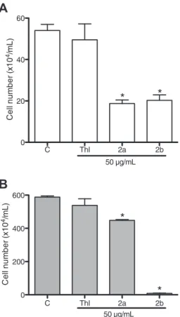
g/mL, leading to themigration inhibition of 65.3 ± 1.6 and 65.2 ± 2.4% on endothelial cells and 23.8 ± 0.8 and 98.5 ± 0.3% of inhibition on melanoma cells (Fig. 4). Thalidomide was not able to reduce cell migration.
3.3.2. Tumor angiogenesis inhibition analyzed by immunohistochemistry
Antitumor activity of thalidomide, 2a and2b (50 mg/kg/day)
was also performed to determine tumor vasculature related parameters. The treatment started at 5th day after the Sarcoma 180 tumor inoculation and was performed for 10 consecutive days. Subsequently, the animals were sacrificed on the 15th day and tumors were dissected out. Thalidomide (2.29 ± 0.13 g) and
ana-logs (2a: 2.73 ± 0.59 g; 2b: 3.09 ± 0.49 g) reduced tumor growth
(56.6 ± 6.5, 48.2 ± 11.2 and 41.4 ± 9.4%, respectively) in comparison
with negative control (5.28 ± 0.71 g) (p< 0.05) (Table 3). Only
thalidomide-treated animals presented weight loss in the posterior treatment (29.14 ± 1.16 g) compared to control (34.08 ± 1.25 g)
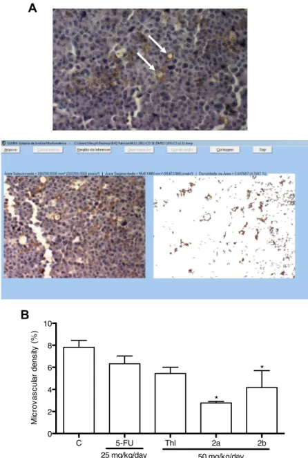
(p< 0.05). Fig. 5A shows a tumor section, indicating vessels and
cells immunostained with CD-31 antibody.
Immunohistochemistry assays showed that microvascular density
(MVD) was significantly reduced in thalidomide,2aand2bgroups
(30 ± 4.9, 64.6 ± 1.8 and 46.5 ± 19.5%, respectively) in comparison
with negative control group (Fig. 5B) (p< 0.05).
3.3.3. Angiogenesis assay in the chorioallantoic membrane (CAM) of chicken embryo
To evaluate thein vivoantitumor effect of thalidomide and
ana-logs, the chorioallantoic membrane assay, also known as in ovo
chicken embryo method, was performed in attempt to correlate the inhibition of vessel formation with antitumor potential.
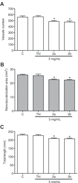
Compounds2aand2b(5 mg/mL) were able to reduce number of
vessels (12. 9 ± 2.3 and 14.8 ± 3.3%), neovascularization area (13.1 ± 1.7 and 14.3 ± 1.7%) and the total length of vessels in
mm2 (9.2 ± 1.5 and 9.9 ± 1.9%) as shown in Fig. 6. Thalidomide,
on the other hand, did not alter these vascularization parameters. Concentration dependent analyzes are in progress in attempt to observe pharmacological outcomes and possible toxic effects.
4. Discussion
In the fight against cancer, search for new chemical entities (NCEs) with chemotherapeutic properties is really worthy and numerous methods have been utilized to acquire compounds, including isolation from plants and animals and the use of syn-thetic and combinatorial chemistry and molecular modeling [19,28–31]. Thalidomide, for example, was the first drug with sig-nificant clinical outcomes against multiple myeloma (MM) and can be considered as alternative treatment for patients treated with
conventional therapy or after transplantation [32]. Benefits of
thalidomide for MM patients may be due to its ability to inhibit
Fig. 3.Effects of thalidomide and its analogs on the migration of human umbilical vein endothelial cells (HUVEC) and melanoma cells (MDA/MB-435) before and after 24 h of incubation. Each well was pretreated with mitomycin C (5lg/mL) for 15 min. (A) Negative control (DMSO 0.1%); (B) Thalidomide (50lg/mL); (C)2a(50lg/mL); (D)2b (50lg/mL). Magnification, 100. Studies were carried out in triplicate represented by three independent biological evaluations.
the growth of new blood vessels (angiogenesis); a phenomenon observed in different tumor types. Due to this potential, chemical modeling of the thalidomide is a source of novel precursors with therapeutic properties. Taking into consideration structure–activ-ity related studies (SAR), analogs and metabolites of thalidomide clearly showed that the presence of the pharmacophoric unit
phthalimide is essential for its pharmacological activity [33].
Thiosemicarbazone was another important pharmacophoric group used in this study. The anticancer effects of thiosemicarbazones
and its analogs (semicarbazones and N-acyl-hydrazones) have
been quite promising, acting on cell proliferation and combating
chemotherapy resistances[34].
Herein, the cytotoxic analyses of eight phthalimide and thiosemicarbazone analogs were performed by the MTT and
Alamar Blue™ assays, revealing weakin vitroantitumor activity
against tumor cells. Thus, the addition of the thiocarbonyl group to the phthalimide ring did not improve the cytotoxic action.
However, compound 2d showed promising action on sarcoma
180 cells. Previously, a series of 16 novel thalidomide sulfur ana-logs containing one and two sulfur atoms were screened for
in vitroantitumor activity against Ehrlich ascites carcinoma cell
line and also exhibited potent cytotoxic action[35]. A relationship
between the thiosemicarbazone subunit and antiproliferative effects on experimental tumors has been established, since this chemical structure inhibits ribonucleotide reductase processes, alters DNA structure, presents chelating capacity with potent and selective antineoplasic activity and generates iron complexes with
redox activity[34,36,37].
In vitroeffects can be not directly extrapolated forin vivo mod-els. Thus, evaluating the pharmacological actions of these com-pounds in complete biological systems becomes an obligatory
requirement. In order to assess thein vivoantitumoral action, the
compounds were evaluated using the Sarcoma 180 experimental model, a mouse-derived tumor widely exploited in antitumor
research[20,38–40]. In Sarcoma-180-bearing animal assays, only
thalidomide and compounds2aand2brevealed antitumor action.
Interestingly, the compound2d was not able to inhibit in vivo
tumor growth, though it has displayed moderatein vitro
cytotoxi-city against Sarcoma 180 cells. Probably,in vivometabolic
activa-tion is required to generate compounds with antiproliferative
effect on solid tumors as seen with thalidomide,2aand2b.
It has been demonstrated that inhibition of angiogenesis and tumor growth by thalidomide or analogs requires metabolic
acti-vation[12,41–43]. In a similar way, Pessoa et al.[17]also suggest
that a prior metabolic activation is necessary to produce one or
more active metabolites fromN-phthaloyl amino acids derivatives,
explaining their antiproliferative activity onin vivomodels only.
Therefore, it is also possible that the metabolizing of the molecules
also explains, at least in part, thein vivoantitumor action, since it is
very known that some molecules undergo hepatic enzymatic
reac-tions to generate active metabolites[44]. Zahran et al.[35], using
anin vivomodel of experimental carcinoma, also demonstrated
that thalidomide dithiocarbamate and dithioate analogs have activity against Ehrlich carcinoma-induced solid tumor in Swiss albino mice.
Histopathological analyses of livers in treated animals showed Kupffer cell hyperplasia, microvesicular steatosis and preserved hepatic parenchyma. The liver possesses a pronounced regenera-tive capacity: even when necrosis is found with conjuncregenera-tive tissue
preservation, there is often complete tissue restoration[44]. Thus,
hepatic alterations observed in2a and 2btreated groups were
reversible.
In spite of only compounds2aand2bhave exhibited antitumor
action, both2band3bcaused spleen enlargement, a finding
sug-gestive of immunostimulant action of these molecules. Indeed,
immunomodulatory drugs based on thalidomide, mainly
amino-substituted thalidomide analogs, have been reported to be
superior candidates as antitumor agents[45,46]. Despite
thalido-mide had not led to spleen morphological alterations in this work, it is known that some phthalimide analogs are co-stimulatory
sub-stances, increasing the response of T-lymphocytes to
T-cell-receptor-mediated stimulation, the production of
interleukin-2 and interferon-
c
as well as the number of naturalkiller cells[17,47]. On the other hand, most clinical chemotherapy
drugs are immunosuppressive and has negative side effects[48],
such as leucocyte suppression, hypoplasia of the splenic white pulp
and small lymphoid aggregates in 5-FU-treated mice[46]. These
findings display the importance about enhancement of host
C Thl 2a 2b 0
20 40 60
*
*
50 µg/mL
A
C
e
ll nu
m
b
e
r
(x
10
4/m
L
)
C Thl 2a 2b 0
200 400 600
50 µg/mL
*
*
B
C
e
ll nu
m
b
er
(
x
10
4/m
L
)
Fig. 4.Effects of thalidomide (Thl) and analogs (2aand2b) analyzed by the Wound Healing Assay measured after 24 h of incubation. (A) Human umbilical vein endothelial cells (HUVEC); (B) Melanoma cells (MDA-MB-435). Negative control (C) was treated with the vehicle used for diluting the tested substances (DMSO 0.1%). Results are mean ± S.E.M. of two independent experiments performed in triplicate obtained by cells counting that invaded the scar.*p< 0.05 compared to negative control by ANOVA test followed by Newman–Keuls test.
Table 3
Effect of thalidomide and synthetic analogs (50 mg/kg/day) on tumor growth in Swiss mice bearing Sarcoma 180. The treatment started after 5 days of Sarcoma 180 inoculation and extended for 10 consecutive days.
Treatment Dose (mg/kg/day)
Mice weight (g)
Tumor (g) Tumor inhibition (%)
Negative control – 34.08 ± 1.25 5.28 ± 0.71 – 5-FU 25 30.63 ± 1.21 3.09 ± 0.41* 41.5 ± 7.8* Thalidomide 50 29.14 ± 1.16* 2.29 ± 0.13* 56.6 ± 2.5* 2a 50 31.43 ± 0.75 2.73 ± 0.59* 48.2 ± 11.2* 2b 50 31.86 ± 1.24 3.09 ± 0.49* 41.4 ± 9.4*
Values are means ± S.E.M.,n= 10 animals/group. Negative control was treated with the vehicle used to dilute the drug (10% DMSO). 5-Fluorouracil (5-FU) was used as positive control.
*p< 0.05 compared with the negative control by ANOVA followed by Newman–
defenses as alternative to the traditional cancer cytotoxic chemotherapy since it involves slight side effects.
In general, the in vivoantitumor effect of thalidomide is not
commonly observed in the literature. Gutman et al.[49], testing
the efficacy of thalidomide daily administered by gavage (0.3– 1.0 mg until 10 days), reported no growth retardation in CT-26 bearing mice nor in mice with pulmonary or peritoneal metastases of B16-F10 melanoma. On the other hand, antitumor effects of thalidomide have been associated with its antiangiogenic proper-ties. It is well known that new analogs of thalidomide, including the second generation, have a great therapeutic potential. Therefore, these novel and active pharmacologically compounds were divided into two classes: SelCIDs (selective cytokine
inhibi-tory drugs) and IMiDs (immunomodulainhibi-tory drugs)[50].
In order to evaluate the antiangiogenic potential of the
mole-cules2aand2b, antiangiogenic assays were performed. Cell
migra-tion technique has been widely used as a screening assay for new
compounds with antiangiogenic activity[51,52]. Both molecules
(2aand2b) that showed promisingin vivo results inhibited cell
migration of endothelial (HUVEC) and melanoma cells
(MDA-MB-435) at 50
l
g/mL. On the other hand, thalidomide wasnot able to reduce cell migration, whose inefficacy can be explained by the fact that thalidomide requires metabolic activa-tion to produce one or more active metabolites after incubaactiva-tion
with microsomal proteins, as previously described[12,53]. These
data suggested that the absence of migration was not caused by cytotoxicity or inhibition of proliferation and other factors are probable involved in the antiangiogenic potential, a finding that needs to be investigated.
The continuous growth of tumors requires oxygen and nutrients
and these substances are provided by the blood flow. In vivo
antiangiogenic action of the compounds were performed in Sarcoma 180-bearing mice assay with a longer period of exposure (10 days) since effects of antiangiogenic therapy cannot be
A
C 5-FU Thl 2a 2b
0 2 4 6 8 10
M
ic
rov
a
s
c
u
lar
dens
ity
(%
)
50 mg/kg/day 25 mg/kg/day
*
*
B
Fig. 5.Effects on tumor angiogenesis evaluated by immunohistochemistry. (A) Images of a hot spot on the tumor obtained by inverted microscope and processed by the System Quantification of Angiogenesis (SQAN). White arrows indicate vessels and cells immunostained with CD-31 (PECAM-1) antibody (Magnification, 200); (B) Effects of thalidomide (Thl) and analogs (2aand2b) on microvascular density analyzed by immunohistochemistry in Sarcoma 180 tumors after 10 days of treatment started after on the 5th inoculation day. Negative control (C) was treated with the vehicle used to dilute the drug (DMSO10%). 5-Fluorouracil (5-FU) was used as positive control. Values are mean ± S.E.M. Microvascular density of areas was analyzed in 3 slides/group.*p< 0.05 compared with the negative control by ANOVA followed by Newman–Keuls test.
detected in vivoin shorter trials [54]. Thus, CD-31 or PECAM-1 labeling was determined. CD-31 or PECAM-1 is an adhesion mole-cule present in endothelial cells and platelets. It is a member of the immunoglobulin superfamily expressed on the surface of
endothe-lial cells, platelets, monocytes, neutrophils and T cells [55–58].
Thalidomide and analogs (2aand2b) inhibited tumor growth but
intratumor vessels stained with the CD-31 antibody revealed
reduction in microvascular density (MVD) only in 2a and 2b
groups.
In vivochorioallantoic membrane (CAM) assay demonstrated
effects of molecules on the neovascularization. The results
con-firmed that only compounds 2aand2bwere able to reduce the
number of vessels, neovascularization area and the total length of vessels at 5 mg/mL after 24 h exposure. These findings indicate that addition of the thiosemicarbazone pharmacophore group to
the phthalimidic ring in thalidomide increased its antitumor potential and angiogenesis activity. Thalidomide, on the other hand, did not change vascularization parameters probably because
in vivo hepatic biotransformation is necessary [12,53]. In fact, thalidomide antiangiogenic activity is significantly increased by human microsomal activation, but different findings are observed in rats. It is known that hydroxylation of thalidomide in positions
10and 50keep its antiangiogenic potential, but the hydroxylation
at 40position generates an inactive compound[59].
5. Conclusions
The addition of thiosemicarbazone pharmacophore group into
the phthalimidic ring improved thein vivoantitumor and
antian-giogenic potential of the analogs2aand2b. These molecules have
special interest for the development of new anticancer therapies and additional studies are in progress to elucidate the mechanism
of action of these bioactive phthalimides.
Conflict of Interest
The authors have declared no conflict of interest.
Transparency Document
TheTransparency documentassociated with this article can be found in the online version.
Acknowledgements
We wish to thank the Brazilian agencies Conselho Nacional de Desenvolvimento Científico e Tecnológico (CNPq), Fundação Cearense de Apoio ao Desenvolvimento Científico e Tecnológico (FUNCAP) and Fundação de Amparo à Pesquisa do Estado do Piauí (FAPEPI) for financial support. We are grateful to Silvana França dos Santos for technical assistance and Priscila Murolo for her help with English editing of the manuscript.
Appendix A. Supplementary data
Supplementary data associated with this article can be found, in
the online version, athttp://dx.doi.org/10.1016/j.cbi.2015.06.037.
References
[1]F. Shojaei, Anti-angiogenesis therapy in cancer: current challenges and future
perspectives, Cancer Lett. 320 (2012) 130–137.
[2]K.M. Cook, W.D. Figg, Angiogenesis inhibitors – current strategies and future
prospects, CA Cancer J. Clin. 60 (2010) 222–243.
[3]A.A. Carvalho, P.M. Costa, G.C. Vieira, F.V.F. Jamacaru, M.O. Moraes, B.C.
Cavalcanti, C. Pessoa, Natural products used as candidates for angiogenesis
inhibitors in cancer therapy, Trends Org. Chem. 15 (2011) 79–93.
[4]M. Potente, H. Gerhardt, P. Carmeliet, Basic and therapeutic aspects of
angiogenesis, Cell 146 (2011) 873–887.
[5]G.W. Prager, M. Poettler, Angiogenesis in cancer, Hämostaseologie 32 (2012)
105–114.
[6]S.M. Weis, D.A. Cheresh, Tumor angiogenesis: molecular pathways and
therapeutic targets, Nat. Med. 17 (2011) 1359–1370.
[7]A. Amini, S.M. Moghaddam, D.L. Morris, M.H. Pourgholami, The critical role of
vascular endothelial growth factor in tumor angiogenesis, Curr. Cancer Drug
Targets 12 (2012) 23–43.
[8]A.A. Carvalho, P.M. Costa, L.G.S. Souza, T.L.G. Lemos, A.P.N.N. Alves, C. Pessoa,
M.O. Moraes, Inhibition of metastatic potential of B16F10 melanoma cell line in vivoandin vitroby biflorin, Life Sci. 93 (2013) 201–207.
[9]J.H. Kim, A.R. Scialli, Thalidomide: the tragedy of birth defects and the effective
treatment of disease, Toxicol. Sci. 122 (2011) 1–6.
[10]M. Melchert, A. List, The thalidomide saga, Int. J. Biochem. Cell Biol. 39 (2007)
1489–1499.
C Thl 2a 2b 0
10 20 30
*
*
5 mg/mL
B
N
e
ov
a
s
c
ul
a
ri
z
a
tion ar
ea
(
mm
2)
C Thl 2a 2b 0
100 200 300 400 500 600 700
*
*
V
e
s
s
el
s
num
b
er
5 mg/mL
A
C Thl 2a 2b 0
50 100 150 200 250
*
*
T
o
ta
l le
n
g
th
(
mm
)
5 mg/mL
C
[11]M.V. Almeida, F.M. Teixeira, M.V.N. Souza, G.W. Amarante, C.C. Alves, S.H. Cardoso, A.M. Mattos, A.P. Ferreira, H.C. Teixeira, Thalidomide analogs from diamines: synthesis and evaluation as inhibitors of TNF-alpha production,
Chem. Pharm. Bull. 55 (2007) 223–236.
[12]T. Noguchi, H. Fujimoto, H. Sano, A. Miyajima, H. Miyachi, Y. Hashimoto,
Angiogenesis inhibitors derived from thalidomide, Bioorg. Med. Chem. 15
(2005) 5509–5513.
[13]T. Ito, H. Ando, H. Handa, Teratogenic effects of thalidomide: molecular
mechanisms, Cell. Mol. Life Sci. 68 (2011) 1569–1579.
[14]A.C.L. Leite, F.F. Barbosa, M.V.O. Cardoso, D.R.M. Moreira, L.C.D. Coêlho, E.B. da
Silva, G.B.O. Filho, V.M.O. de Souza, V.R.A. Pereira, L.C. Reis, P.M.P. Ferreira, C. Pessoa, A.G. Wanderley, F.V.B. Mota, T.G. da Silva, Phthaloyl amino acids as anti-inflammatory and immunomodulatory prototypes, Med. Chem. Res. 23
(2014) 1701–1708.
[15]C.I. Chen, Lenalidomide alone and in combination for chronic lymphocytic
leukemia, Curr. Hematol. Malig. Rep. 8 (2013) 7–13.
[16]P.G. Richardson, T.M. Mark, M.Q. Lacy, Pomalidomide: new
immunomodulatory agent with potent antiproliferative effects, Crit. Rev.
Oncol. Hematol. 88 (2013) S36–S44.
[17]C. Pessoa, P.M.P. Ferreira, L.V.C. Lotufo, M.O. Moraes, S.M.T. Cavalcanti, L.C.D.
Coelho, M.Z. Hernandes, A.C.L. Leite, C.A. Simone, V.M.A. Costa, V.M.O. Souza, Discovery of phthalimides as immunomodulatory and antitumor drug
prototypes, Chem. Med. Chem. 5 (2010) 523–528.
[18]T. Mosmann, Rapid colorimetric assay for cellular growth and survival –
application to proliferation and cytotoxicity assays, J. Immunol. Methods 65
(1983) 55–63.
[19]P.M.P. Ferreira, D.J.B. Lima, B.W. Debiasi, B.M. Soares, K.C. Machado, J.C.
Noronha, D.J. Rodrigues, A.P. Sinhorin, C. Pessoa, G.M. Vieira-Júnior,
Antiproliferative activity of Rhinella marina and Rhaebo guttatus venom
extracts from Southern Amazon, Toxicon 72 (2013) 43–51.
[20]P.M.P. Ferreira, D.F. Farias, M.P. Viana, T.M. Souza, I.M. Vasconcelos, B.M.
Soares, C. Pessoa, L.V. Costa-Lotufo, M.O. Moraes, A.F.U. Carvalho, Study of the antiproliferative potential of seed extracts from Northeastern Brazilian plants,
An. Acad. Bras. Cienc. 83 (2011) 1045–1058.
[21]R.R. Burk, A factor from a transformed cell line that affects cell migration, Proc.
Natl. Acad. Sci. U.S.A. 70 (1973) 368–372.
[22]K. Yanyong, W. Fengcai, F. Jing, Y. Dongling, Y. Xu, Y. Xiyun, Knockdown of
CD146 reduces the migration and proliferation of human endothelial cells, Cell
Res. 16 (2006) 313–318.
[23] F.V. Fechine-Jamacaru,In vivoquantification of corneal angiogenesis using digital image processing (Ph.D. Thesis in Surgery), Federal University of Ceará, Department of Surgery, Fortaleza, Ceará, Brazil, 2006.
[24]C.A. Dornelas, F.V. Fechine-Jamacaru, I.L. Albuquerque, H.I.F. Magalhães, T.A.
Dias, M.H.G. Faria, M.K.S. Alves, S.H.B. Rabenhorst, P.R.C. Almeida, T.L.G. Lemos, J.D.V. Castro, M.E.A. Moraes, M.O. Moraes, Angiogenesis inhibition by green
propolis and the angiogenic effect ofL-lysine on bladder cancer in rats, Acta
Cir. Bras. 27 (2012) 529–536.
[25]P.F. Dias, J.M. Siqueira-Junior, L.F. Vendrusco, T.J. Neiva, M. Maraschin, A.
Gagliardi, R.M. Ribeiro-Do-Valle, Antiangiogenic and antitumoral properties of polysaccharide isolated from the seaweed Sargassum stenophyllum, Cancer
Chemother. Pharmacol. 56 (2005) 436–446.
[26]M. Nguyen, Y. Shing, J. Folkman, Quantitation of angiogenesis and
antiangiogenesis in the chick-embryo chorioallantoic membrane, Microvasc.
Res. 47 (1994) 31–40.
[27]V. Hamburger, H. Hamilton, A series of normal stages in the development of
the chick embryo, J. Morphol. 88 (1951) 49–92.
[28]F.W.A. Barros, D.P. Bezerra, P.M.P. Ferreira, B.C. Cavalcanti, T.G. Silva, M.G.R.
Pitta, M.C.A. Lima, S.L. Galdino, I.R. Pitta, L.V. Costa-Lotufo, M.O. Moraes, R.R. Burbano, T.N. Guecheva, J.A.P. Henriques, C. Pessoa, Inhibition of DNA topoisomerase I activity and induction of apoptosis by thiazacridine
derivatives, Toxicol. Appl. Pharmacol. 268 (2013) 37–46.
[29]J.R.O. Ferreira, B.C. Cavalcanti, P.M. Costa, F.F.P. Arantes, E.S. Alvarenga, C.R.A.
Maltha, L.C.A. Barbosa, G.C.G. Militão, C. Pessoa, P.M.P. Ferreira, Induction of
G2/M arrest, caspase activation and apoptosis bya-santonin derivatives in
HL-60 cells, Toxicol. In Vitro 27 (2013) 1458–1466.
[30]A.C.L. Leite, L.M.F. Santos, D.R.M. Moreira, D.J. Brondani, Synthesis and
characterization of new amino acyl-4-thiazolidones, Quim. Nova 30 (2007)
284–286.
[31]V. Srivastava, A.S. Negi, J.K. Kumar, M. Gupta, S.P.S. Khanuja, Plant-based
anticancer molecules: a chemical and biological profile of some important
leads, Bioorg. Med. Chem. 13 (2005) 5892–5908.
[32]S.V. Rajkumar, R. Fonseca, A. Dispenzieri, M.Q. Lacy, J.A. Lust, T.E. Witzgi, R.A.
Kyle, M.A. Gertz, P.R. Greipp, Thalidomide in the treatment of relapsed
multiple myeloma, Mayo Clin. Proc. 75 (2000) 897–901.
[33]Y. Hashimoto, Thalidomide as a multi-template for development of
biologically active compounds, Arch. Pharm. 341 (2008) 536–547.
[34]H. Beraldo, Semicarbazones and thiosemicarbazones: their wide
pharmacological profile and clinical applications, Quím. Nova 27 (2004)
461–471.
[35]M.A.H. Zahran, T.A.R. Salem, R.M. Samaka, H.S. Agwa, A.R. Awad, Design
synthesis and antitumor evaluation of novel thalidomide dithiocarbamate and
dithioate analogs against Ehrlich ascites carcinoma-induced solid tumor in
Swiss albino mice, Bioorg. Med. Chem. 16 (2008) 9708–9718.
[36]D.R. Richardson, P.C. Sharpe, D.B. Lovejoy, D. Senaratne, D.S. Kalinowski, M.
Islam, P.V. Bernhardt, Dipyridyl thiosemicarbazone chelators with potent and selective antitumor activity form iron complexes with redox activity, J. Med.
Chem. 49 (2006) 6510–6521.
[37]Y. Yu, D.S. Kalinowski, K. Kovacevic, A.R. Siafakas, P.J. Jansson, C. Stefani, C.B.
Lovejoy, P.V. Bernhardt, D.R. Richardson, Thiosemicarbazones from the old to new: Iron chelators there are more than just ribonucleotide reductase
inhibitors, J. Med. Chem. 52 (2009) 5271–5294.
[38]A.A. Carvalho, D. Finger, C.S. Machado, E.M. Schmidt, P.M. Costa, A.P.N.N. Alves,
T.M.F. Morais, M.G.R. Queiroz, S.P. Quinaia, M.R. da Rosa, J.M.T. dos Santos, C. Pessoa, M.O. Moraes, L.V. Costa-Lotufo, A.C.H.F. Sawaya, M.N. Eberlin, Y.R.
Torres,In vivoantitumoural activity and composition of an oil extract of
Brazilian própolis, Food Chem. 126 (2011) 1239–1245.
[39]H.I. Magalhães, P.M.P. Ferreira, E.S. Moura, M.R. Torres, A.P. Alves, O.D. Pessoa,
L.V. Costa-Lotufo, M.O. Moraes, C. Pessoa,In vitroandin vivoantiproliferative
activity ofCalotropis procerastem extracts, An. Acad. Bras. Cienc. 82 (2010)
407–416.
[40] G.C.G. Militão, I.N.F. Dantas, P.M.P. Ferreira, A.P.N.N. Alves, D.C. Chaves, F.J.Q.
Monte, C. Pessoa, M.O. Moraes, L.V. Costa-Lotufo,In vitroandin vivoanticancer
properties of cucurbitacin isolated fromCayaponia racemosa, Pharm. Biol. 50
(2012) 1479–1487.
[41]B. Badamtseren, E. Odkhuu, N. Koide, A. Haque, Y. Naiki, S. Hashimoto, T.
Komatsu, T. Yoshida, T. Yokochi, Thalidomide inhibits interferon-c-mediated
nitric oxide production in mouse vascular endothelial cells, Cell. Immunol. 270
(2011) 19–24.
[42]K.S. Bauer, S.C. Dixon, W.D. Figg, Inhibition of angiogenesis by thalidomide
requires metabolic activation, which is species-dependent, Biochem.
Pharmacol. 55 (1998) 1827–1834.
[43]B.G. Pereira, S. Ligorio Fialho, C. Maria de Souza, G. Dantas-Cassali, A.
Silva-Cunha, Evaluation of the effects of thalidomide-loaded biodegradable devices
in solid Ehrlich tumor, Biomed. Pharmacother. 67 (2013) 129–132.
[44]V. Kumar, A.K. Abbas, N. Fausto, S.L. Robbins, R.S. Cotran, Pathologic Basis of
Disease, WB Saunders, Philadelphia, 2004.
[45]J.B. Bartlett, K. Dredge, A.G. Dalglish, The evolution of thalidomide and its IMiD
derivatives as anticancer agents, Nat. Rev. Cancer 4 (2004) 314–322.
[46]P.M.P. Ferreira, P.M. Costa, A.M. Costa, D.J.B. Lima, R.R. Drumond, D.R.M.
Moreira, G.O.B. Filho, J.F. Magalhães, M.G.R. Queiroz, A.C.L. Leite, C. Pessoa, Cytotoxic and toxicological effects of phthalimide derivatives on tumor and
normal murine cells, An. Acad. Bras. Cienc (2015) (in press).
[47]B. Pan, S. Lentzsch, The application and biology of immunomodulatory drugs
(IMiDs) in cancer, Pharmacol. Ther. 136 (2012) 56–68.
[48]N. Gonen, Y.G. Assaraf, Antifolates in cancer therapy: structure, activity and
mechanisms of drug resistance, Drug Resist. Updates 15 (2012) 183–210.
[49]M. Gutman, A. Szold, A. Ravid, T. Lazauskas, O. Merimsky, J.M. Klausner, Failure
of thalidomide to inhibit tumor growth and angiogenesisin vivo, Anticancer
Res. 16 (1996) 3673–3677.
[50] K. Dredge, J.B. Marriott, C.D. Macdonald, H.-W. Man, R. Chen, G.W. Muller, D.
Stirling, A.G. Dalgleish, Novel thalidomide analogues display anti-angiogenic activity independently of immunomodulatory effects, Br. J. Cancer 87 (2002)
1166–1172.
[51]I. Tsukamoto, N. Sakakibara, T. Maruyama, J. Igarashi, H. Kosaka, Y. Kubota, M.
Tokuda, H. Ashino, K. Hattori, S. Tanaka, M. Kawata, R. Konishi, A novel nucleic acid analogue shows strong angiogenic activity, Biochem. Biophys. Res.
Commun. 399 (2010) 699–704.
[52]K. Bala, K. Ambwani, N.K. Gohil, Effect of different mitogens and serum
concentration on HUVEC morphology and characteristics: implication on use
of higher passage cells, Tissue Cell 43 (2011) 216–222.
[53]Y. Li, Z. Jiang, Y. Xiao, L. Li, Y. Gao, Metabolism of thalidomide by human liver
microsome cytochrome CYP2C19 is required for its antimyeloma and
antiangiogenic activities in vitro, Hematol. Oncol. 30 (2011) 13–21.
[54]C.J. Bruns, M. Shrader, M.T. Harbison, C. Portera, C.C. Solorzano, K.-W. Jauch,
D.J. Hicklin, R. Radinsky, L.M. Ellis, Effect of the vascular endothelial growth factor receptor-2 antibody DC101 plus gemcitabine on growth, metastasis and angiogenesis of human pancreatic cancer growing orthotopically in nude mice,
Int. J. Cancer 102 (2002) 101–108.
[55]V.E. García, H.E. Chuluyan, SLAM and CD31: signaling molecules involved in
cytokine secretion during the development of innate and adaptive immune
responses, Cytokine Growth Factor Rev. 18 (2007) 85–96.
[56]P.J. Newman, The biology of PECAM-1, J. Clin. Invest. 100 (1997) 25–29.
[57]P.J. Newman, D.K. Newman, Signal transduction pathways mediated by
PECAM-1: new roles for an old molecule in platelet and vascular cell
biology, Arterioscler. Thromb. Vasc. Biol. 23 (2003) 953–964.
[58]C. Poncelet, P. Madelenat, G. Feldmann, F. Walker, E. Darai, Expression of von
Willebrand’s factor, CD34, CD31, and vascular endothelial growth factor in
uterine leiomyomas, Fertil. Steril. 78 (2002) 581–586.
[59]M.G. Marks, J. Shi, M.O. Fry, Z. Xiao, M. Trzyna, V. Pokala, M.A. Ihnat, P.K. Li,
Effects of putative hydroxylated thalidomide metabolites on blood vessel density in the chorioallantoic membrane (CAM) assay and on tumor and
endothelial cell proliferation, Biol. Pharm. Bull. 25 (2002) 597–604.