O
r i g i n a la
rt i c l e3 7 6 Arq Bras Oftalmol. 2016;79(6):376-9 http://dx.doi.org/10.5935/0004-2749.20160107
Atopic keratoconjunctivitis: long-term results of medical treatment
and penetrating keratoplasty
Ceratoconjuntivite atópica: resultados a longo prazo do tratamento clínico e da ceratoplastia penetrante
Yusuf KoçluK1, ZuleYha YalniZ-aKKaYa1, Ayşe Burcu1, Firdevs Örnek1
Submitted for publication: September 9, 2015 Accepted for publication: July 23, 2016
1 Ophthalmolgy Clinic, Ministry of Health Ankara Training and Research Hospital, Ankara, Turkey.
Funding: No specific financial support was received for this study.
Disclosure of potential conflicts of interest: None of the authors have any potential conflicts of interest to disclose.
Corresponding author: Yusuf Koçluk. Eye Department, Adana Numune Training and Research Hospital - Adana, 06520 - Turkey - E-mail: [email protected]
Approved by the following research ethics committee: Ankara Training and Research Hospital (# 0505/13).
ABSTRACT
Purpose: To evaluate the long-term outcomes of medically or surgically treated patients with atopic keratoconjunctivitis (AKC).
Methods: Charts of 16 patients with AKC (32 eyes) observed between 1996 and 2013 were reviewed retrospectively. Outcome measures included demographic features, follow-up duration, and biomicroscopic findings at the first and most recent visits. The corrected distance visual acuity (CDVA; in decimal units) was evaluated at the initial visit and the 1-, 6-, and 12-month follow-up visits. Results: In the medically treated group (25 eyes of 15 patients), the median follow-up duration was 3 (range, 1-9) years, and the median CDVA values were 0.01 (0.001-1.0) at the first visit and 0.01 (0.001-0.8) at the most recent visit (p=0.916). In the penetrating keratoplasty (PK) group (7 eyes of 6 patients), the median follow-up duration was 7 years (range, 1-11), and the median CDVA increased from 0.01 (0.001-0.01) to 0.2 (0.001-0.7) postoperatively (p=0.043).
Conclusion: Whereas most AKC patients maintained a useful CDVA with medical treatment, PK may be required in some cases. Despite the frequent occurrence of complications, PK can significantly improve the CDVA.
Keywords: Conjunctivitis, allergic/therapy; Conjunctivitis, allergic/surgery; Kerato-plasty, penetrating; Treatment outcome
RESUMO
Objetivo: Avaliar os resultados a longo prazo em ceratoconjuntivite atópica (AKC) pacientes que foram tratados clinicamente ou cirurgicamente.
Métodos: Os prontuários de 16 pacientes (32 olhos) com AKC, que foram acom-panhados entre 1996 e 2013 foram avaliados retrospectivamente. As medidas adotadas foram as características demográficas, tempos de seguimento, e resultados biomicroscópicos da visita inicial e da visita mais recente. A acuidade visual corrigida para distância (CDVA), apresentada em unidades decimais, foi avaliada na visita inicial e nas visitas do 1o mês, 6o mês e 1o ano de seguimento.
Resultados: No grupo tratado clinicamente (25 olhos de 15 pacientes), a mediana do tempo de seguimento foi de 3 anos (variação, 1-9) e a CDVA média foi de 0,01 (0,001-1,0) na visita inicial e 0,01 (0,001-0,8) na visita mais recente (p=0,916). No grupo de cera-toplastia penetrante (PK) (7 olhos de 6 pacientes), a mediana de tempo de seguimento foi de 7 anos (variação, 1-11) e a CDVA média aumentou de 0,01 (0,001-0,01) para 0,2 (0,001-0,7) (p=0,043) no pós-operatório.
Conclusões: Embora a maioria dos pacientes AKC mantém a CDVA útil com o trata-mento clínico, alguns necessitam de PK a fim de obter CDVA útil. Embora as compli-cações pós-PK ocorrem com freqüência, a CDVA pode melhorar significativamente.
Descritores: Conjuntivite alérgica/terapia;Conjuntivite alérgica/cirurgia; Cerato plas -tia penetrante; Resultado do tratamento
INTRODUCTION
Atopic keratoconjunctivitis (AKC) is a bilateral chronic inlamma-tory disease of the ocular surface and eyelids. The pathomechanism of AKC involves both chronic immunoglobulin (Ig) E-mediated mast cell degranulation and immune reactions mediated by T helper 1 (Th1)- and T helper 2 (Th2)-lymphocyte derived cytokines, as well as other inlammatory cells(1,2). Notably, eosinophils, which are never observed in normal tissues, are present in the substantia propria of patients with AKC and express increased surface levels of activation markers(3). The onset of disease usually occurs from the second through the ifth decades(4).
Itching is the most characteristic symptom and may be accompa-nied by watering, mucous discharge, redness, blurred vision, photo-phobia, and pain. Itching and other symptoms may be continuous or more pronounced in certain seasons. Clinical signs include hy pe-remia of the conjunctiva and episcleral vessels, papillae in the tarsal conjunctiva, and the presence of concomitant blepharitis. Conjunc-tival scarring with subepithelial ibrosis, fornix foreshortening,
sym-blepharon, and corneal ulceration and neovascularization may occur in the most severe cases(5).
For patients with AKC, the topical application of a vasoconstric-tor-antihistamine combination might provide transient symptom relief but is unlikely to afect the immunopathologic process or its se-quelae. In contrast, the topical administration of steroids, such as pred-nisolone acetate, may provide some control of symptoms and signs. However, patients must be warned of the potential risks of cataract formation and glaucoma with steroid therapy. To address this risk, previous studies have found that both the oral and topical forms of cyclosporin A (CSA) can efectively treat AKC and reduce the usage of topical steroids(6,7), and steroid-sparing medications, including the mast cell stabilizer sodium cromolyn 4%, have been shown to efecti-vely reduce symptoms(8).
Ko ç l u K Y, e t a l.
3 7 7 Arq Bras Oftalmol. 2016;79(6):376-9
causes of loss of vision. Although penetrating keratoplasty (PK) typi-cally results in similar surface problems (corneal scarring), this proce-dure has been shown to improve vision in some patients(9). However, no studies comparing the outcomes of medical treatment with those of PK for AKC were identiied in the current body of literature. Accordingly, in this study we aimed to compare the long-term visual and other clinical outcomes of AKC patients who received medical or surgical treatment.
METHODS
This retrospective case-control study, which compared the results of patients with AKC who underwent surgical transplantation with the results of those who received medical treatment, was conducted according to the tenets of the Declaration of Helsinki after receiving institutional board approval. All medical charts of patients with AKC who were followed for at least 1 year between 1996 and 2013 at an ophthalmology clinic of a tertiary care center were reviewed. Patients who were treated only for AKC and its complications were included; those who received other treatments (surgical or medical) for other ocular conditions were excluded. The characteristics of 16 patients (32 eyes) who met the criteria were evaluated in this study.
The outcome measures were demographic features, follow-up duration, corrected distance visual acuity (CDVA; preoperative and 1-, 6-, and 12-month postoperatively), biomicroscopic indings of the irst and last visits, and treatment modality (medical or surgical). Signs and results were compared between patients who did and did not undergo PK. The evaluated clinical indings included eczema, blepharitis, meibomianitis, tarsal margin keratinization, trichiasis, ma darosis, ectropion, entropion, conjunctival subepithelial ibrosis, fornix foreshortening, symblepharon, giant papillae, follicles, punctate keratitis, neovascularization or conjunctivalization, persistent epi the-lial defects, ilamentary keratitis, stromal scarring, and graft clarity. Visual acuities, which are expressed in decimals throughout the text, were measured using a Snellen chart. The visual acuity of counting ingers was converted to 0.01 decimal values; hand motion was con-verted to 0.001 decimal values(10).
The medical treatment administered to patients who did not un-dergo PK comprised topical CSA 4 times daily and preservative-free artiicial tears 8 times daily. CSA 0.3% (prepared by mixing a Sandim-mun 50 mg ampule [Novartis AG, Basel, Switzerland] + 15 ml artiicial tears) was used prior to 2004; CSA 0.05% (Restasis, Allergan Inc., Waco, TX, USA) was used after 2004. Treatment was switched from CSA 0.05% to CSA 0.3% if needed. During exacerbation periods (2-6 weeks), topical dexamethasone sodium phosphate 0.1% was applied 4 times daily (Maxidex; Alcon Laboratories, Inc., Fort Worth, TX, USA), and topical anti-histamines and mast cell stabilizers were added to the treatment regimen. Decreasing doses of topical dexamethasone were administered for 2-6 weeks. Systemic anti-histamines were used in intractable cases. Patients with accompanying rhinitis, asthma, and eczema consulted with the relevant clinical department.
All PK procedures were performed for optic purposes and in pa tients with reduced visual acuity and greater corneal AKC involve-ment. Patients were made as comfortable as possible via symptom treatment. Gentamicin 40 mg/0.5 ml and dexamethasone 4 mg/1 ml were injected subconjunctivally at the end of each PK procedure. Postoperatively, all patients received topical oloxacin (Exocin, Allergan Pharmaceuticals LTD., Mayo, Ireland), topical 0.1% dexamethasone 8 times daily, topical CSA 0.05% four times daily, and preservative-free artiicial tears hourly. Topical oloxacin was discontinued after 1 month. Topical dexamethasone was tapered and maintained according to the needs of each patient. Topical CSA and artiicial tears were conti-nued throughout the follow-up period.
SPSS for Windows 16.0 (SPSS Inc. Chicago, IL, USA) was used for the statistical analysis. The Kolmogorov-Smirnov test was used to eva-luate the normal distributions of variables (ages, follow-up time, sex,
clinical indings, symptoms, irst and inal CDVA, intraocular pressure, complications, and graft clarity). Numerical variables with abnor mal distributions were compared using the Mann-Whitney U test, and descriptive statistics are expressed as medians (minimum-maximum). Qualitative variables were compared using the chi-square test, and descriptive statistics are expressed as percentages (%) and frequencies. A P value <0.05 was considered statistically signiicant.
RESULTS
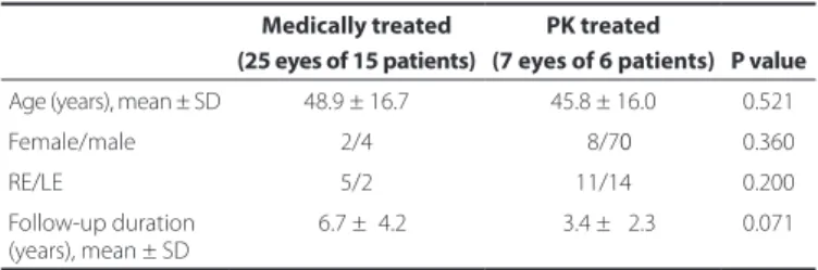
The median age of the 16 patients with AKC (9 women, 7 men) was 47.50 (range: 16-68) years. The median ages of patients who underwent PK and those who received only medical treatment were 45.0 (20-68) years and 48 (16-68) years, respectively (p=0.521). The patient groups did not difer signiicantly with regard to follow-up time (p=0.071). Ten (62.5%) patients had eczema of the eyelids and periorbital skin, 2 (12.5%) had asthma, and 6 (37.5%) had rhinitis. All patients had bilateral signs of AKC. The characteristic features of the included patients are presented in table 1.
The most common modes of eyelid involvement were blepha-ritis (62.5%) and meibomianitis (62.5%). The most common corneal inding was corneal opacity (75%). Elevated intraocular pressure was observed in only 3 (18.7%) eyes, and all events were post-PK. Associa-ted keratoconus was present in 1 (6.2%) patient. Other clinical indings in our cohort included punctate keratitis (62.5%), neovascularization (62.5%), conjunctival subepithelial ibrosis (43.7%), cataract (37.5%), persistent epithelial defects (25%), ilamentary keratitis (18.7%), giant papillae (18.7%), tarsal margin keratinization (12.5%), trichiasis (12.5%), fornix foreshortening (12.5%), symblepharon (12.5%), follicles (12.5%), entropion (6.2%), and ptosis (6.2%).
For the 25 eyes that received only medical treatment, the median follow-up duration was 3 (1-9) years, and the median CDVAs at the irst and last visits were 0.01 (0.001-1.0) and 0.01 (0.001-0.8), respecti-vely, a non-signiicant diference (p=0.916). Similarly the intraocular pressure did not difer statistically between the irst and last visits (p=0.675). Patients exhibited greater symptomatic (e.g., itching, eye redness, irritation) stability during medical treatment.
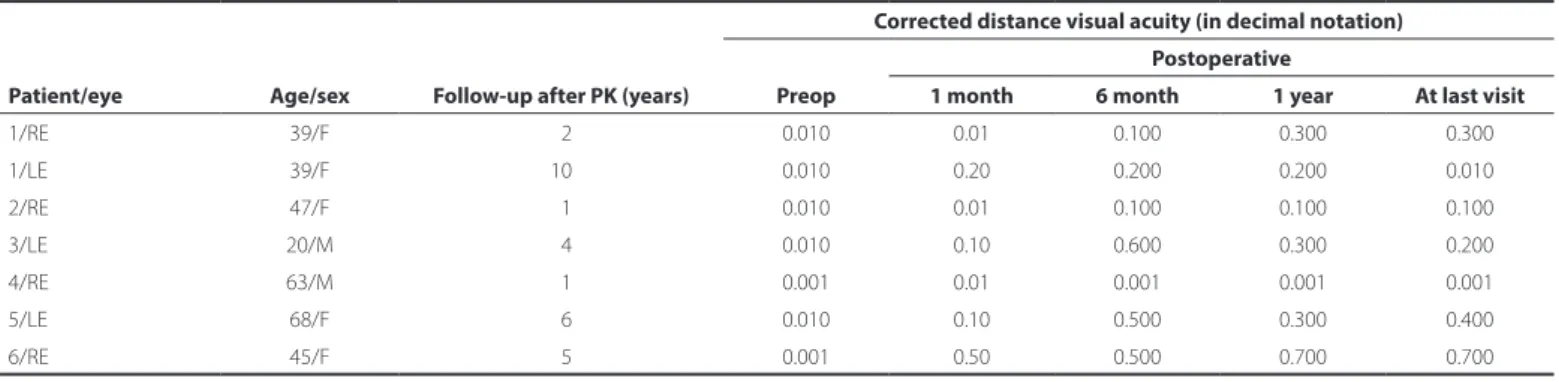
For the 7 eyes (6 patients) subjected to PK, the median follow-up duration was 7 (1-11) years. The median preoperative and postope-rative CDVAs were 0.01 (0.001-0.01) and 0.2 (0.001-0.7), respectively, a signiicant diference (p=0.043; Table 2). However, only 1 patient did not experience postoperative complications. The complications associated with PK and patients’ graft statuses are listed in table 3.
Among all patients, 3 eyes had both initial and inal CDVAs ≥0.5, whereas 10 and 15 eyes respectively had initial and inal CDVAs ≥0.1. Although only 7 patients initially had a CDVA ≥0.1 in at least 1 eye, 11 patients had achieved this level of vision by the inal examination. Regarding treatment modalities, the median initial CDVAs were 0.01 (range, 0.001-0.01) and 0.01 (range, 0.001-1.0) among patients who did and did not undergo PK, respectively, whereas the corresponding median inal CDVAs were 0.2 (range, 0.005-0.7) and 0.04 (range, 0.001-0.8), respectively. The diferences between the examinations were not statistically signiicant in either group (p=0.175, p=0.253, respectively). Similarly, the groups did not signiicantly difer in terms of changes
Table 1. Characteristic features of study patients
Medically treated PK treated
P value (25 eyes of 15 patients) (7 eyes of 6 patients) Age (years), mean ± SD 48.9 ± 16.7 45.8 ± 16.0 0.521
Female/male 2/4 08/70 0.360
RE/LE 5/2 11/14 0.200
Follow-up duration
(years), mean ± SD 06.7 ±04.2 03.4 ± 02.3 0.071
At o p i ck e r At o c o n j u n c t i v i t i s: l o n g-t e r mr e s u lt so fm e d i c A lt r e At m e n tA n dp e n e t r At i n gk e r At o p l A s t y
3 7 8 Arq Bras Oftalmol. 2016;79(6):376-9
in intraocular pressure between the initial and final examinations (p=0.853, p=0.067, respectively). However, patients who underwent PK were signiicantly more likely to present with cataracts, anterior sinechias, and glaucoma at the inal examination (6 (85.7%) eyes that underwent PKP vs. 4 (16.0%) eyes that did not undergo PK; p<0.001).
The most common complication was cataracts (71.5%), followed by intraocular pressure elevation; the latter occurred in half of the patients and was controlled with topical medications. Conjunctival and corneal complications at the last visit were statistically more frequent among patients who did not undergo PK (p<0.001); however, this group was larger than the PK group, which might present a statistical limitation.
Graft rejections occurred in 2 patients (1 within the irst year and 1 after the irst year). The graft remained clear in 1 such patients but became opaque in the other. Although 6 (85.7%) grafts remained clear after the irst postoperative year, 1 (14.3%) graft had become semi-opaque. At the inal examination, 4 (57.1%) grafts remained clear, 2 (28.6%) were semi-opaque due to the recurrence of AKC-related corneal scarring, and 1 (14.3%) graft was opaque consequent to graft rejection. In our series, 57.1% of grafts were clear at the inal visit, and complication rates were high (5/7 cataracts and 3/7 glaucoma).
Amniotic membrane transplantation (AMT) was performed in 6 (18.8%) eyes (2 for persistent epithelial defects following PK, 4 for pri mary persistent epithelial defects). Although 1 of those grafts re-mained clear after AMT, the others healed with varying degrees of subepithelial and stromal opacity. At the last visit, visual acuity was not found to difer according to sex. Patients with a CDVA of ≥0.1 had signiicantly higher inal visual acuities (p=0.036).
DISCUSSION
AKC is a chronic allergic inlammatory disease with bilateral invol-vement of the cornea and conjunctiva. This disease has an onset during adolescence but primarily afects adults, with a peak incidence between 30 and 50 years of age(11). Accordingly, the median age of our cohort was compatible with the literature. Patients with AKC typi-cally describe severe, persistent, periocular itching associated with dermatitis. There is usually a family history of atopic disease in 1 or both parents, and patients often have another atopic manifesta tion such as asthma (65%) or allergic rhinitis (65%)(12). Inversely, the re-ported incidence of ocular involvement among patients with atopic dermatitis ranges from 25% to 42%(4,13-15). In our patients, 62.5% of patients had eczema of the eyelids and periorbital skin, 12.5% had asthma, and 37.5% had rhinitis. The discrepancies between the literature and our results might be related to the small number of patients in our study.
AKC is among the most debilitating allergic conjunctival diseases because of its ability to induce visual losses consequent to corneal complications(16). These complications may be mild or severe and include supericial punctate keratitis, exfoliated supericial punctate keratitis, corneal erosions, and shield ulcers that progress to ulceration and neovascularization, conjunctival subepithelial ibrosis, fornix shortening, and symblepharon formation(16,17). In our study, corneal opacity, corneal neovascularization, punctate keratitis, and persistent epithelial defects were observed in 75%, 62.5%, 62.5%, and 25% of patients, respectively.
The immunophilin CSA inhibits T lymphocyte activation by inhi-biting the expression of the IL-2 receptor, as well as the activation of eosinophils and mast cells, thereby preventing the release of in-lammatory mediators(18,19). A large prospective observational study of patients with vernal keratoconjunctivitis or AKC (n=594) evaluated the eicacy of a CSA 0.1% aqueous ophthalmic solution. Over a 6-month period, 30% of patients were able to discontinue topical steroid use. The most common adverse complaint was eye irritation (12%), and >1% of patients (n=5) reported infectious complications (e.g., bacte rial corneal ulcer or herpetic keratitis)(20). In our study, topical CSA was used as a primary treatment for the long-term prevention of AKC exacerbations and complications. Despite the limited number of cases in our series, 16 patients experienced symptom improvement with CSA treatment, and CDVA remained stable (p=0.916).
Takano et al.(21) observed dramatic healing of a persistent (dura-tion: 6 months) allergic corneal ulcer following amniotic membrane patching in a patient with AKC. In another study, signiicant decreases in symptoms and complete corneal ulcer re-epithelialization were observed in all cases of chronic AKC within 7 days of treatment(22). In our study, 6 eyes of 6 patients were subjected to AMT for persistent epi thelial defects and corneal ulcers, with successful corneal
epithe-Table 2. Preoperative and postoperative corrected distance visual acuities of patients who underwent penetrating keratoplasty (PK)
Patient/eye Age/sex Follow-up after PK (years)
Corrected distance visual acuity (in decimal notation) Postoperative
Preop 1 month 6 month 1 year At last visit
1/RE 39/F 02 0.010 0.01 0.100 0.300 0.300
1/LE 39/F 10 0.010 0.20 0.200 0.200 0.010
2/RE 47/F 01 0.010 0.01 0.100 0.100 0.100
3/LE 20/M 04 0.010 0.10 0.600 0.300 0.200
4/RE 63/M 01 0.001 0.01 0.001 0.001 0.001
5/LE 68/F 06 0.010 0.10 0.500 0.300 0.400
6/RE 45/F 05 0.001 0.50 0.500 0.700 0.700
M= male; F= female; RE= right eye; LE= left eye.
Table 3. Postoperative complications and graft statuses in patients treated wtih penetrating keratoplasty
Patients/eye Graft status* Graft rejection Complications Graft status#
1/RE Clear - Cataract Clear
1/LE Clear Yes, irreversible Glaucoma and cataract
Opaque
2/RE Clear - Glaucoma Clear
3/LE Clear - Cataract Semi-opaque
4/RE Semi-opaque Yes, reversible Glaucoma and
cataract Semi-opaque
5/LE Clear - Cataract Clear
6/RE Clear - - Clear
Ko ç l u K Y, e t a l.
3 7 9 Arq Bras Oftalmol. 2016;79(6):376-9
lium recovery in all cases. We believe that AMT should be considered a beneicial adjunctive treatment for cases in which epithelization must be achieved quickly.
Patients with AKC may require corneal transplantation to treat corneal scarring, vascularization, or perforation(9). Although such pa-tients are considered to have a high risk of corneal graft failure, espe-cially in the context of high serum IgE levels(9), some reports in the literature have discussed the prognosis of PK in patients with AKC. In one series of patients with keratoconus who underwent keratoplas-ty, the outcomes of corneal grafting in the 30 atopic patients were comparable to the outcomes of non-atopic patients(23). Ghoraishi et al.(9) studied 11 eyes in 9 patients with AKC who required PK and evaluated the visual outcomes and prognostic factors. The authors observed a inal visual acuity of ≥0.5 in 46% of the eyes; in addition, the grafts remained clear in 10 eyes, and an average improvement of 4.5 Snellen acuity lines was recorded. Despite the frequent posto-perative complications observed in our patients, the CDVA improved signiicantly after PK. We note, however, that our study was limited by the imbalance in patient numbers between the patient groups and the small number of total patients.
In conclusion, most patients with AKC are able to maintain a useful CDVA with medical treatment and follow-up, as shown by the obser-vation of similar long-term CDVAs in patients with AKC who were treated medically or surgically. However, in some cases PK may be required to achieve a useful CDVA. Although PK is associated with frequent postoperative complications, it has also been shown to yield signiicant postoperative improvements in CDVA.
REFERENCES
1. Bonini S. Atopic keratoconjunctivitis. Allergy.2004;59(78):71-3.
2. Leonardi A, De Dominicis C, Motterle L. Immunopathogenesis of ocular allergy: a schematic approach to diferent clinical entities.Curr Opin Allergy Clin Immunol.2007; 7(5):429-35.
3. Hingorani M, Calder V, Jolly G, Buckley RJ, Lightman SL. Eosinophil surface antigen expression and cytokine production vary in diferent ocular allergic diseases. J Allergy Clin Immunol. 1998;102(5):821-30.
4. Chen JJ, Applebaum DS, Sun GS, Plugfelder SC. Atopicc Keratoconjunctivitis: A review. J Am Acad Dermatol. 2014;70(3):569-75.
5. Heustein Sy, Leonard Bielory. Atopic keratoconjunctivitis Allergy Asthma Proc. 2013; 34(1):33-41.
6. Hoang-Xuan T, Prisant O, Hannouche D, Robin H. Systemic cyclosporin A in severe atopic keratoconjunctivitis. Ophthalmology. 1997;104(8):1715-20.
7. Hingorani M, Moodaley L, Calder VL, Buckley RJ, Lightman S. A randomized, pla ce bo-controlled trial of topical cyclosporin A in steroid-dependent atopic kerato conjunctivitis. Ophthalmology. 1998;105(9):1715-20.
8. Avunduk AM, Avunduk AC, Kapıcıoglu Z, Akyol N, Tavli L. Mechanisms and compari-son of anti-allergic eicacy of topical lodoxamide and cromolyn sodium treatment in vernal keratoconjunctivitis. Ophthalmology. 2000;107(7):1333-37.
9. Ghoraishi M, Akova YA, Tugal-Tutkun I, Foster CS. Penetrating keratoplasty in atopic keratoconjunctivitis. Cornea. 1995;14(6):610-3.
10. Holladay JT. Visual acuity measurements. J Cataract Refract Surg. 2004;30(2):287-90. 11. O’Brien TP. Allergic conjunctivitis: an uptade on diagnosis and management. Curr Opin
Allergy Clin İmmunol. 2013;13(5):543-9.
12. Power WJ, Tugal-Tutkun I, Foster CS. Long-term follow-up of patients with atopic ke ratoconjunctivitis Ophthalmology. 1998;105(2):637-42.
13. Kaujalgi R, Handa S, Jain A, Kanwar AJ. Ocular abnormalities in atopic dermatitis in İndian patients. İndian J Dermatol venereol Leprol. 2009;75(2):148-51.
14. Moscovici BK, Cesar AS, Nishiwaki-Dantas MC, Mayor SA, Marques JC. Atopic kerato-conjunctivitis in patients of the pediatric dermatology ambulatory in a reference center. Arc Bras Oftalmol. 2009;72(6):805-10.
15. Ebihara N, Funaki T, Matsuda H, Okumura K, Muurakami A, Ra C. Corneal abnormalities in the NC/Nga mouse: an atopic dermatitis model. Cornea. 2008:2(8):923-9. 16. Wakamatsu TH, Tanaka M, Satake Y, Dogru M, Fukagava K, Igarashi A, et al. Eosinophil
cationic protein as a marker for assessing the eicacy of tacrolimus ophthalmic so lu-tion in the treatment of atopic keratoconjunctivitis. Mol Vis. 2011;13(17):932-8. 17. Takamura E, Uchio E, Ebihara N, Ohno S, Ohashi Y, Okamoto S, et al. Japanese guideline
for allergic conjunctival diseases. Alergol Int. 2011;60(2):191-203.
18. Daniell M, Constantinou M, Vu HT, Taylor HR. Randomised controlled trial of topical ciclosporin A in steroid dependent allergic conjunctivitis. Br J Ophthalmol. 2006;90(4): 461-4.
19. Akpek EK, Dart JK, Watson S, Christen S, Dursun D, Yoo S, et al. A randomized trial of topical cyclosporin 0.05% in topical steroid-resistant atopic keratoconjunctivitis. Oph-thalmology. 2004;111(3):476-82.
20. Ebihara N, Ohashi Y, Uchio E, Okamoto S, Kumagai N, Shoji J, et al. A large prospective observational study of novel cyclosporine 0.1% aqueous ophthalmic solution in the treatment of severe allergic conjunctivitis. J Ocular Pharmacol Ther. 2009;25(4):365-72. 21. Takano Y, Fukagawa K, Miyake-Kashima M. Dramatic healing of an allergic corneal ulcer
persistent for 6 months by amniotic membrane patching in a patient with atopic ke-ratoconjunctivitis: a case report. Cornea. 2004;23(7):723-5.
22. Rouher N, Pilon F, Dalens H. Implantation of preserved human amniotic membrane for the treatment of shield ulcers and persistent corneal epithelial defects in chronic allergic keratoconjunctivitis. J Fr Ophtalmol. 2004;27(10):1091-7.