Echocardiography and 6-Minute Walk Test in Left Ventricular Systolic
Dysfunction
Venera Berisha
1, Gani Bajraktari
1, Dritero Dobra
2, Edmond Haliti
1, Reshat Bajrami
1, Shpend Elezi
2Second Division of Cardiology, Internal Medicine Clinic, University Clinical Center of Kosova1; Medical School, University of Prishtina2, Prishtina - Kosovo
Summary
Background: Chronic heart failure is a major cardiovascular disorder.
Objective: The aim of this study was to prospectively examine whether a 6-min walk test (6-MWT) result correlates with echocardiographic variables in patients with left ventricular systolic dysfunction and stable chronic heart failure.
Methods: We prospectively studied 52 patients (65% male; mean age 60±11 years) who had chronic heart failure secondary to ischemic heart disease or idiopathic cardiomyopathy. All patients had left ventricular systolic dysfunction
(ejection fraction ≤0.45), and were in stable NYHA class II or III heart failure. An echo-Doppler study and a 6-MWT
were performed on the same day.
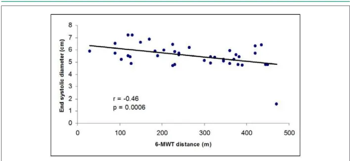
Results: 6-MWT had moderate, but statistically significant correlation with end-systolic diameter (ESD) (r=-0.46;
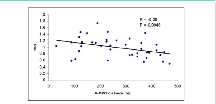
p=0.0006), with shortening fraction-SF (r=0.52; p=0.0001), and with ejection fraction-EF (r=0.5; p=0.0001), whereas it had poor, but statistically significant correlation with myocardial performance index-MPI (r=-0.39; p=0.0046), E/ A(tricuspid) ratio (r=-0.333; p=0.016), pulmonary acceleration time (r=0.328; p=0.018), and lateral long axis amplitude (r=0.283; p=0.04). Linear regression model demonstrated that age (c2=-0.59, p=<0.001), restrictive transmitral filling pattern (c2=-0.44, p=0.004) and left ventricular end-systolic dimension (c2=-0.34, p=0.012) were independent factors
that influenced the 6-MWT.
Conclusion: In patients with heart failure due to left ventricular systolic dysfunction, the 6-MWT as a clinical assessment tool of the functional capacity has a significant correlation with the most important global LV systolic function parameters, as well as with LV MPI. In patients with LV systolic dysfunction, age, restrictive transmitral filling pattern, and left ventricular systolic dimension, were independently associated with the 6-MWT. (Arq Bras Cardiol 2009;92(2):121-127)
Key words: Heart failure; echocardiography; walking, quality of life.
Mailing address: Gani Bajraktari •
Bregu i Diellit, Z.Q. 7/19, 10000, Prishtina - Kosovo E-mail: [email protected]
Manuscript received January 21, 2008; revised manuscript received February 18, 2008; accepted February 29, 2008.
an important prognostic role in the diagnosis of heart failure5
and in the prediction of cardiac events in patients with acute myocardial infarction6. The severity of congestive heart failure
is usually graded according to patients’ symptoms, in particular to the physical activity that induces dyspnea or fatigue. The NYHA functional heart failure classification meets only some of these criteria. However, the NYHA classification is based on the patient’s self-assessment and its reliability is still a matter of debate7. The ideal instrument for risk stratification and optimal
clinical management of congestive heart failure patients should be objective, as well as simple, inexpensive and safe. The peak oxygen uptake, measured during cardiopulmonary exercise testing is a more objective parameter to assess functional capacity and to predict survival in patients with heart failure8-10.
However, this method is limited only to specialized centers, because it requires sophisticated equipment and specially trained personnel.
The 6-min walk test (6-MWT) has been proposed as a simple and accurate alternative technique for measurement of submaximal exercise capacity in congestive heart failure
Introduction
Chronic heart failure is a major cardiovascular disorder with increasing incidence and prevalence1. Two-dimensional
and Doppler echocardiography is a sensitive and noninvasive method of detecting cardiac abnormalities and systolic and/ or diastolic dysfunction2. Variables of diastolic filling allow
further identification of subgroups with divergent long-term prognoses3. Recently, myocardial performance index (MPI)
has been suggested for the evaluation of ventricular function4.
patients11. It is considered to be realistically related to daily
physical activity. Consequently, it has been incorporated into studies of assessment of exercise capacity12,13, of quality of
life14, of efficacy of new therapeutic agents15-17, including
beta-blockers18-22, of physical training programs23 and of
prognostic stratification24-29.
The aim of the present study was to asses the relationship between 2-dimensional and Doppler echocardiographic parameters and 6-MWT in patients with heart failure and left ventricular systolic dysfunction. 6-MWT is used as a proxy measure of the quality of life; therefore, echocardiographic parameters that have independent association with 6-MWT would facilitate prediction of the quality of life.
Methods
Study population
The study group included 52 patients with stable chronic heart failure secondary to ischemic heart disease or idiopathic cardiomyopathy, referred to the Second Division of Cardiology at the Internal Medicine Clinic of the University Clinical Center of Kosova, in Prishtina, Kosovo, between December 2005 and April 2007.
The mean age was 60±11 years. The cause of heart failure was idiopathic dilated cardiomyopathy in 19 patients and ischemic cardiomyopathy in 33 patients. All patients had an enlarged left ventricle (left ventricular [LV] diastolic dimension >5.7cm) and global left ventricular systolic dysfunction (left
ventricular ejection fraction [LVEF] <0.45), and all were in
stable New York Heart Association (NYHA) functional class II or III heart failure. The functional capacity of 27 patients was class II and that of the remaining 25 was class III. Table 1 summarizes the patients’ clinical and laboratory characteristics.
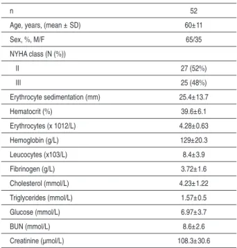
Table 1 - Clinical and laboratory data of patients enrolled in the study
n 52
Age, years, (mean ± SD) 60±11
Sex, %, M/F 65/35
NYHA class (N (%))
II 27 (52%)
III 25 (48%)
Erythrocyte sedimentation (mm) 25.4±13.7
Hematocrit (%) 39.6±6.1
Erythrocytes (x 1012/L) 4.28±0.63
Hemoglobin (g/L) 129±20.3
Leucocytes (x103/L) 8.4±3.9
Fibrinogen (g/L) 3.72±1.6
Cholesterol (mmol/L) 4.23±1.22
Triglycerides (mmol/L) 1.57±0.5
Glucose (mmol/L) 6.97±3.7
BUN (mmol/L) 8.6±2.6
Creatinine (μmol/L) 108.3±30.6
Patients with rhythm disturbances (atrial fibrillation, serious ventricular arrhythmias), patients who had decompensated heart failure, limitation of physical activity due to factors other than exertional dyspnea and fatigue (such as arthritis), and those with moderate to severe renal failure, with chronic obstructive pulmonary disease were excluded from the study. We also excluded patients who had experienced unstable angina, acute myocardial infarction or stroke, anemia, and any febrile condition or infectious disease.
The study was approved by the Institutional Ethics Committee and all patients gave their written informed consent. Standard medical therapy with e.g. ACE-inhibitors, beta-blockers, digoxin and diuretics was individually optimized based on symptoms and renal function at least 2 weeks prior to exercise testing. All patients continued to take their routine medication when the echocardiography and 6-MWT were performed.
Data collection
The history and physical examination was performed in all subjects. In all patients the routine biochemical measurements were performed: the measurements of erythrocytes, leucocytes, hemoglobin, hematocrit, erythrocyte sedimentation, BUN, creatinine, blood glucose, cholesterol, triglycerides and inflammatory blood analysis (fibrinogen, CRP). Measurements of weight, height, waist and hip were also performed.
Echocardiographic examinations
Echocardiographic examination was performed using a Philips Intelligent E-33 echocardiograph with a multi-frequency transducer, and harmonic imaging as appropriate. All echocardiographic studies were performed by the same investigator. The echocardiographic study was performed in the left lateral decubitus in the parasternal long-axis and 4-chamber views.
The cross-sectional, two-dimensionally guided M mode recordings of LV short axis were performed using the left parasternal long axis view with the cursor at the tips of the mitral valve leaflets. Fractional shortening was calculated as the percentage fall in LV cavity dimension during systole in relation to that at end-diastole. Left ventricular ejection fraction was estimated using Simpson’s biplane method. Left atrial dimension was measured from M-mode recordings with the cursor crossing the aortic root at the level of the aortic valve leaflets. LV long axis M-mode recordings were obtained with the cursor positioned at the lateral and septal angles of the mitral ring and right ventricular long axis recording with the cursor positioned at the lateral angle of the tricuspid ring. All M-mode recordings were zoomed to obtain better images for accurate measurements.
Diastolic function of the left and right ventricles was assessed by pulsed Doppler. The pulsed Doppler sample volume was positioned at the tips of the mitral and tricuspid leaflets, respectively. We registered the E wave, A wave, from which the E/A ratio was calculated, and the deceleration time of E wave (DT-E). We also registered Er wave, Ar wave and Er/Ar ratio.
measuredfrom the cessation to the onset of mitral inflow is equal to the sum of isovolumetric contraction time (ICT), ejection time (ET), and isovolumetric relaxation time (IRT). Left ventricular ejection time b is the duration of left ventricular outflow velocity profile. Thus the sum of ICT and IRT was obtained by subtracting b from a. The index of combined LV systolic and diastolic function (the sum of ICT and IRT divided by ejection time) was calculated as (a - b) / b (Fig.1). The IRT can be measured by subtracting the d interval, between the R wave and the cessation of LV outflow, from the c interval, between the R wave and the onset of mitral inflow. ICT was calculated by subtracting the IRT from a-b.
Mitral regurgitation severity was assessed by color and continuous wave Doppler and was graded as mild, moderate, or severe according to the jet distance from the valve orifice and flow velocity profile, respectively, in line with the recommendations of the American Society of Echocardiography31. Likewise, tricuspid regurgitation was
assessed by color flow and continuous-wave Doppler from the apical four-chamber view. Retrograde transtricuspid pressure drop > 35 mmHg was taken as a sign of pulmonary hypertension when the right atrium was not dilated.
6-min walk test
Within 24 h of echocardiographic examination, a 6-MWT was performed on a level hallway surface; the test was administered by a registered nurse blinded to the results of echocardiography. According to the method of Gyatt et al11,12
all patients were informed of the purpose, methods and use of the 6-min walk test results. The 6-MWT was conducted following a standardized protocol, between 10 am and 4 pm after usual medication11. A 15 m flat, obstacle-free
corridor, with chairs placed at either end was used. Patients were instructed to walk as far as possible, turning 180º every 15 m in the allotted time of 6 minutes. Patients walked unaccompanied so as not to influence walking speed, and they were asked to walk as far as possible within 6-minutes.
Figure 1 -Schematic drawing of the myocardial performance index (MPI). The index (ICT+IRT/ET) is derived as (a - b / b).
Patients were permitted to rest, if needed, then they were told of the remaining time at every two minutes. At the end of the 6 min, the physician measured the total distance walked by the patient.
Statistical analysis
Data are presented as mean ± SD or proportions (% of
patients). Continuous data are compared with two-tailed unpaired Student’s t test. Discrete data are compared with chi- square test. Correlation between variables was performed using
Pearson’s correlation test. A p value < 0.05 was considered
to indicate statistical significance. Multivariate analysis was performed using linear regression model. Factors included in this model were age, restrictive transmitral filling pattern, left ventricular systolic dimension, myocardial performance index and diabetes mellitus.
Results
The clinical and laboratory characteristics of the 52 patients enrolled in the study are shown in Table 1, and the echocardiographic and Doppler measurements are shown in Table 2. There was a moderate, but statistically significant correlation of 6-MWT with LV posterior wall thickness ( r=
0.49; p<0.001), left ventricular end-systolic dimension (r=-left ventricular end-systolic dimension
(r=-0.46, p<0.001, Fig.2), LV fractional shortening (r = 0.517,
p<0.001, Fig. 3), and LVEF (r = 0.50, p<0.001), whereas
there was a poor, but statistically significant correlation with myocardial performance index-MPI (r=-0.39; p=0.0046, Fig . 4), E/A (tricuspid) ratio (r=-0.333; p=0.016), pulmonary acceleration time (r=0.328; p=0.018), and lateral long axis amplitude (r=0.283; p=0.04) (Table 3). The dimensions of aorta, left atrium, interventricular septal thickness, E wave, A wave, E/A ratio, deceleration time of E wave, E (tricuspid) wave, A (tricuspid) wave, E (tricuspid) /A (tricuspid) ratio, deceleration time of E (tricuspid) and LV septal long axis amplitude had no significant correlation with 6-MWT (Table 3).
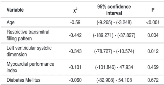
Multivariate analysis was performed using linear regression model. Results of this model (Table 4) demonstrated that age, restrictive transmitral filling pattern and left ventricular end-systolic dimension were independent factors that influence the 6-MWT. All these parameters were inversely related to the 6-MWT. Age of the patients emerged as the strongest independent predictive factor, followed by restrictive filling pattern and left ventricular end- systolic dimension.
Discussion
To our knowledge, this is the first study to correlate 6-MWT performance in patients with chronic heart failure. The main finding of our study is that the 6-MWT has a moderate, but statistically significant correlation with LV systolic dimension, global LV systolic function parameters, LV MPI, as well as with long axis systolic function of left and right ventricles in patients with chronic stable heart failure due to LV systolic dysfunction.
Figure 2 -Correlation between 6-minute walk test (6-MWT) distance (in meters) and end systolic diameter (in centimeters).
Figure 3 -Correlation between 6-minute walk test (6-MWT) distance (in meters) and left ventricular shortening fraction (in %).
dimension are independently related to the 6-MWT in stable chronic heart failure patients due to left ventricular systolic dysfunction, without rheumatic valvular heart disease.
Heart failure is an epidemiological entity that is increasing worldwide, and now represents a major health problem in most developed countries1. The measurement of quality of
life in heart failure patients has proved more elusive than what was anticipated. It is still unclear which one is better for the assessment of grading of heart failure: if the judgment of the physician, the use of the NYHA classification or the use of many sophisticated questionnaires.
The 6-MWT was originally used by physicians to assess patients with lung disease32,33, and later was introduced as
an alternative objective measure of submaximal exercise capacity of congestive heart failure patients11. Previous studies
show controversial results about the correlation of 6-MWT with functional exercise capacity in chronic heart failure patients. The 6-min walk test was shown to be correlated with peak oxygen uptake in dilated cardiomyopathy and to have a predictive value in these patients34-36. The 6-MWT
was also shown to be a useful prognostic marker in these patients26,27,37. However, Opasich et al38 demonstrated that
in moderate to severe chronic heart failure patients, the 6-min walk test is not related to cardiac function and only moderately related to exercise capacity.
Figure. 4 -Correlation between the 6-minute walk test (6-MWT) distance (in meters) and myocardial performance index (MPI).
Table 2 - Echocardiographic and Doppler measurements in patients enrolled in the study
Parameter Patients
(n = 52)
Left ventricular end-diastolic diameter (cm) 6.66±0.74
Left ventricular end-systolic diameter (cm) 5.52±0.93
Left ventricular fractional shortening (%) 16.03±5.03
Left ventricular ejection fraction (%) 32.7±9.5
Interventricular septal thickeness (cm) 1.2±0.12
Posterior wall thickeness (cm) 0.99±0.12
Left atrial dimension (cm) 4.66±0.57
Aorta (cm) 3.54±0.54
Early diastolic low velocity in mitral valve (cm/s) 70.8±27 Late diastolic low velocity in mitral valve (cm/s) 58.15±27.7 Early diastolic low velocity in mitral valve /
late diastolic low velocity in mitral valve ratio 1.54±0.94 Deceleration time of early diastolic low velocity in mitral
valve (ms) 133±43.4
Isovolumetric relaxation time (ms) 112.4±34.4
Pulmonary acceleration time (ms) 90.5±28.8
Myocardial performance index 0.996±0.58 Early diastolic low velocity in tricuspid valve (cm/s) 39.9±5.96 Late diastolic low velocity in tricuspid valve (cm/s) 40.7±9.1 Early diastolic low velocity in tricuspid valve /
Late diastolic low velocity in tricuspid valve ratio 1.02±0.29
Lateral long axis amplitude (cm) 0.91±0.32
Septal long axis amplitude (cm) 0.72±0.24
Right ventricular long axis amplitude (cm) 1.99±0.69
Table 3 - Relationship of Echocardiographic and Doppler measurements with the 6-minute walk test in patients enrolled in the study
6-minute walk test
R value P value
Aorta 0.063 0.66
Left atrial dimension -0.032 0.82
Interventricular septal thickness 0.249 0.075
Posterior wall thickness 0.490 <0.001
Left ventricular end-diastolic dimension -0.214 0.135
Left ventricular end-systolic dimension -0.46 <0.001
Left ventricular fractional shortening 0.517 <0.001
Left ventricular ejection fraction 0.504 <0.001
Pulmonary acceleration time 0.328 0.018 Early diastolic low velocity in mitral valve -0.0219 0.155 Late diastolic low velocity in mitral valve 0.2227 0.112 Early diastolic low velocity in mitral valve /
Late diastolic low velocity in mitral valve ratio -0.138 0.329 Deceleration time of early diastolic low velocity
in mitral valve 0.227 0.106
Isovolumetric relaxation time -0.269 0.054
Myocardial performance index -0.388 0.045 Early diastolic low velocity in tricuspid valve -0.119 0.4008 Late diastolic low velocity in tricuspid valve 0.2005 0.155 Early diastolic low velocity in tricuspid valve /
Late diastolic low velocity in tricuspid valve ratio -0.333 0.016 Deceleration time of early diastolic low velocity
in tricuspid valve 0.192 0.177
Lateral long axis amplitude (cm) 0.283 0.04
Septal long axis amplitude (cm) 0.108 0.446
Table 4 - Results of Multivariate Linear Regression Model for the 6-Minute Walk Test in Patients with Heart Failure
Variable χ2 95% conidence
interval P
Age -0.59 (-9.265) - (-3.248) <0.001
Restrictive transmitral
illing pattern -0.442 (-189.271) - (-37.827) 0.004 Left ventricular systolic
dimension -0.343 (-78.727) - (-10.574) 0.012
Myocardial performance
index -0.101 (-101.846) - 47.934 0.469
Diabetes Mellitus -0.060 (-82.908) - 54.108 0.672
factors associated with aging.
The small number of patients could be mentioned as a limitation of this study, especially for the comprehensive multivariate analysis.
In conclusion, in patients with heart failure due to left ventricular systolic dysfunction, the 6-MWT as a clinical assessment tool of the functional capacity has a significant correlation with the most important global LV systolic function parameters, as well as with LV MPI. In patients with LV systolic dysfunction, age, restrictive transmitral filling pattern, and left ventricular systolic dimension, were independently associated with the 6-MWT.
Potential Conflict of Interest
No potential conflict of interest relevant to this article was reported.
Sources of Funding
There were no external funding sources for this study.
Study Association
This study is not associated with any post-graduation program.
after a follow-up in patients with LV systolic dysfunction that received ACE-inhibitors39,40, new-generation beta-blockers18-22,
and underwent exercise training23.
There are several clinical implications that could be suggested from this study. Several important echocardiographic parameters are independently related to 6-MWT and therefore can be used to predict the quality of life of the patients. Age emerged as an independent factor inversely related to 6-MWT. Therefore, independently from echocardiographic results, the quality of life deteriorates with age probably due to complex
References
1. Hoes AW, Mosterd A, Grobbee DE. An epidemic of heart failure? Recent evidence from Europe. Eur Heart J. 1998; 19 (Suppl 1): I 2-I 9.
2. DeMaria AN, Wiswnbaugh TW, Smith MD, Harrison MR, Berk MR. Doppler echocardiographic evaluation of diastolic dysfunction. Circulation. 1991; 84: 288-95.
3. Rihal CS, Nishimura RA, Hatle LK, Bailey KR, Tajik AJ. Systolic and diastolic dysfunction in patients with clinical diagnosis of dilated cardiomyopathy: relation to symptoms and prognosis. Circulation. 1994; 90: 2772-9.
4. Tei C, Ling LH, Hodge DO, Bailey KR, Oh JK, Rodeheffer RJ, et al. New index of combined systolic and diastolic myocardial performance: a simple and reproducible measure of cardiac function- a study in normal and dilated cardiomyopathy. J Cardiol. 1995; 26 (6): 357-66.
5. St John Sutton M, Wiegers SE. The Tei index- a role in the diagnosis of heart failure? Eur Heart J. 2000; 21 (22): 1822-4.
6. Moller JE, Sondergaard E, Poulsen SH, Egstrup K. The Doppler echocardiographic myocardial performance index predicts left-ventricular dilation and cardiac death after myocardial infarction. Cardiology. 2001; 95: 105-11.
7. New York Heart Association (NYHA). Nomenclarute and criteria for diagnosis of disease of the heart and great vessels. 7th ed. Boston: Little and Brown Company; 1973. p. 286.
8. Weber KT, Kinasewitz GT, Janicki JS, Fishman AP. Oxygen utilization and ventilation during exercise in patients with chronic cardiac failure. Circulation. 1982; 65: 1213-23.
9. Mancini DM, Eisen H, Kussmaul W, Mull R, Edmunds LH Jr, Wilson JR. Value of peak exercise oxygen consumption for optimal timing of cardiac transplantation in ambulatory patients with heart failure. Circulation. 1991; 83: 778-86.
10. Riley M, McFarland J, Stanford CF, Nicholls DP. Oxygen consumption during corridor walk testing in chronic heart failure. Eur Heart J. 1992; 13: 789-93.
11. Guyatt GH, Sullivan MJ, Thompson PJ, Fallen EL, Pugsley SO, Taylor DW, et al. The 6-minute walk test: a new measure of exercise capacity in patients with chronic heart failure. Can Med Assoc J. 1985; 132: 919-23.
12. Guyatt GH, Thompson PJ, Berman LB, Sullivan MJ, Townsend M, Jones NL, et al. How should we measyre function in patients with chronic heart and lung disease? J Chronic Dis. 1985; 28: 517-24.
13. Lipkin DP, Scriven AJ, Crake T, Poole-Wilson PA. Six minute walking test for assessing exercise capacity in chronic heart failure. Br Med J (Clin Res Ed.). 1986; 292: 653-5.
14. Dracup K, Walden JA, Stevenson LW, Brecht ML. Quality of life in patients with advanced heart failure. J Heart and Lung Transplant. 1992; 11: 273-9.
15. Packer M, Gheorghiade M, Young JB, Constantini PJ, Adams KF, Cody RJ, et al. for the RADIANCE Study. Withdrawal of digoxin from patients with chronic heart failure treated with angiotensin converting enzyme inhibitors. N Engl J Med. 1993; 329: 1-7.
16. Uretsky BF, Young JB, Shahidi E, Yellen LG, Harrison MC, Jolly MK on behalf of the PROVED Investigative Group. Randomized study assessing the effect of digoxin withdrawal in patients with mild to moderate chronic congestive heart failure: results of the PROVED Trial. J Am Coll Cardiol. 1993; 22: 955-62.
17. Califf RM, Adams KF, McKenna WJ, Gheorghiade M, Uretsky BF, McNulty SE, etal. A randomized trial of epoprostenol therapy for severe congestive heart failure: The Flolan International Randomized Survival Trial (FIRST). Am Heart J. 1997; 134: 44-54.
Failure Research. Circulation. 1995; 92: 212-8.
19. Packer M, Bristow ML, Cohn JN, Colucci WS, Fowler MB, Gilbert EM, et al. The effect of carvedilol on morbidity and mortality in patients with chronic heart failure. US Carvedilol Heart Failure Study Groups. N Engl J Med. 1996; 334: 1349-55.
20. Metra M, Nardi M, Giubbini R, Dei Cas L. Effects of short and long-term carvedilol administration on rest and exercise hemodynamic variables, exercise capacity and clinical conditions in patients with idiopathic dilated cardiomyopathy. J Am Coll Cardiol. 1994; 24: 1678-87.
21. Krum H, Sackner-Bernstein JD, Goldsmith RL, Kurkin ML, Schuwartz B, Penn J, et al. Doubleblind, placebo-controlled study of the long-term efficacy of carvedilol in severe chronic heart failure. Circulation. 1995; 92: 1499-506.
22. Olsen SL, Gilbert EM, Renlund DG, Taylor DO, Yanowitz FD, Bristow MR. Carvedilol improves left ventricular function and symptoms in chronic heart failure: a double-blind randomized study. J Am Coll Cardiol. 1995; 25: 1225-31.
23. Meyer K, Scwaibold M, Westbrooh S, Beneke R, Hajric R, Lehmann M, et al. Effects of exercise training and activity restriction on 6-minute walking test performance in patients with chronic heart failure. Am Heart J. 1997; 133: 447-53.
24. Bittner V, Weiner D, Yusuf S, Rogers WJ, McIntyre KM, Bangdiwala SI, et al. for the SOLVD Investigators. Prediction of mortality and morbidity with a 6-minute walk test in patients with left ventricular dysfunction. JAMA. 1993; 270: 1702-7.
25. Cahalin L, Mathier M, Semigran M, Dec W, DiSalvo T. The six-minute walk test predicts peak oxygen uptake and survival in patients with advanced heart failure. Chest. 1996; 110: 325-32.
26. Roul G, Germain P, Bareiss P. Does the 6-minute walk test predict the prognosis in patients with NYHA class II or III chronic heart failure? Am Heart J. 1998; 136: 449-57.
27. Swedberg K, Califf RM, Adams K. FIRST Investigators. Six minute walk test gives prognostic information in severe heart failure. J Am Coll Cardiol. 1995; 25: 329A.
28. Zugck C, Kruger C, Durr S, Gerber SH. Is the 6-minute walking test a reliable substitute for peak oxygen uptake in patients with dilated cardiomyopathy? Eur Heart J. 2000; 21: 540-9.
29. Lucas C, Stevenson LW, Johnson W, Hartley H, Hamilton MA, Walden J, et al. The 6-min walk and peak oxygen consumption in advanced heart failure:
aerobic capacity and survival. Am Heart J. 1999; 138: 618-24.
30. Tei C, Ling LH, Hodge DO, Bailey KR, Oh JK, Rodeheffer RJ, et al. New index of combined systolic and diastolic myocardial performance: a simple and reproducible measure of cardiac function- a study in normal and dilated cardiomyopathy. J Cardiol. 1995; 26 (6): 357-66.
31. Zoghbi WA, Enriquez-Sarano M, Foster E, Grayburn PA, Kraft CD, Levine RA, et al. American Society of Echocardiography. Recommendations for evaluation of the severity of native valvular regurgitation with two-dimensional and Doppler echocardiography. J Am Soc Echocardiogr. 2003; 16: 777-802.
32. McGavin CR, Gupta SP, McHardy GJ. Twelve-minute walking test for assessing disability in chronic bronchitis. Br Med J. 1976; 1: 822-3.
33. Butland RJ, Pang J, Gross ER, Woodcock AA, Geddes DM. Two-, six-, and 12-minute walking tests in respiratory disease. Br Med J. 1982; 284: 1607-8.
34. Zugck C, Kruger C, Durr S, Gerber SH, Haunstetter A, Hornig K, et al. Is the 6-minute walk test a reliable substitute for peak oxygen uptake in patients with dilated cardiomyopathy? Eur Heart J. 2000; 21: 540-9.
35. Ingle L, Rigby AS, Nabb S, Jones PK, Clark AL, Cleland JG. Clinical determinants of poor six-minute walk test performance in patients with left ventricular systolic dysfunction and no major structural heart disease. Eur J Heart Fail. 2006; 8 (3): 321-5.
36. Ingle L, Goode K, Rigby AS, Cleland JG, Clark AL. Predicting peak oxygen uptake from 6-min walk test performance in male patients with left ventricular systolic dysfunction. Eur J Heart Fail. 2006; 8 (2): 198-202.
37. Arslan S, Erol MK, Gundogdu F, Sevimli S, Aksakal E, Senocak H, et al. Prognostic value of 6-minute walk test in stable outpatients with heart failure. Tex Heart Inst J. 2007; 34 (2): 166-9.
38. Opasich C, Pinna GD, Mazza A, Febo O, Riccardi R, Riccardi PG, et al. Six-minute walking performance in patients with moderate-to-severe heart failure; is it a useful indicator in clinical practice? Eur Heart J. 2001; 22 (6): 488-96.
39. Hutcheon SD, Gillespie ND, Crombie IK, Struthers AD, McMurdo MET. Perindopril improves six minute walking distance in controlled trial dysfunction: a randomised double blind placebo older patients with left ventricular systolic. Heart. 2002; 88: 373-7.