1
Original Article
Funct ion of t he Lef t At rium in t he Chagas’
Cardiom yopat hy
M aria do Carmo Pereira Nunes, M árcia de M elo Barbosa, Édson Siqueira da Rocha,
M anoel Ot ávio da Cost a Rocha
Hospit al Socor Serviço de Ecocardiograf ia Ecocent er e Universidade Federal de M inas Gerais
-Belo Horizont e, M G
Mailing address: Maria do Carmo Pereira Nunes - Rua Ludgero Dolabela, 8 0 1 /6 0 1 - 3 0 4 3 0 -1 3 0 - Belo Horizonte, MG - Brazil E-m a il-m ca rm o@ wa ym a il. com . b r
Sent for publishing on 0 4 /1 3 /2 0 0 4 Accepted on 1 1 /0 8 /2 0 0 4
Objective
To study thefunction of the left atrium patients with of dilated cardiomyopathy of chagasic etiology and relate it to the diastolic function and to the functional class.
M ethods
We studied 75 chagasic with cardiomyopathy from July to 1999 to May to 2001, submitted to clinical exams, electrocar-diogram and transesophageal echocarelectrocar-diogram. The left atrium function was assessed by means of the velocities in the left atrial appendix and the atrial reverse in the pulmonary vein. The control group consisted of 20 normal patients.
Results
The age was 48± 13 years old and 69% were men. Most of patients (88%) were in functional classes I and II, under a conven-tional treatment for cardiac insufficiency. The fraction of ejection of the left ventricle was 39± 13%. The indicators of diastolic function associate to those of systolic function and the functional class. The carriers of pseudonormal or restrictive pattern of diastolic dysfunction presented a larger diameter of the left atrium, lower flow velocities in the left atrial appendix and a longer duration of the atrial reverse. There was no difference among the patients with normal pattern and abnormal diastolic relaxation in relation to the control group.
Conclusion
The left atrial function is an important parameter in the as-sessment of patients with chagasic myocardiopathy and it is related to the systolic and diastolic functions of the left ventricle.
Key w ords
chagas’ disease, cardiomyopathy, left atrium and transeso-phageal echocardiogram
The pattern of filling of the left ventricle provides important clinical and prognostic information in patients with cardiac
insuf-ficiency1 -4. However, the diastolic function comprises a complex
sequence of interrelated events and depends on several factors,
such as age, heart rate and ventricular function1 ,5 -7. The left atrial
function has had lower attention as a possible determinant of the dynamics of the ventricular filling, presumably due to technical difficulties to assess the size and function of such chamber.
The Doppler echocardiogram constitutes the main instrument for the non-invasive assessment of the left ventricular diastolic
func-tion6 -8. Multiple parameters used are influenced by the ventricular
load and complacence conditions, in addition to the filling pressures. The left atrium function, directly influenced by the diastolic pressure of the left ventricle, represents a stable marker of the duration and
severity of the diastolic dysfunction, by showing a prognostic value4.
In Chagas’ disease, the diastolic dysfunction can be premature,
preceding the systolic compromising9 -1 3. However, its pattern of
ventricular filling has not been systematically studied and related to the left atrial function, at the advanced stage of chagasic car-diopathy. The present study aims at assessing the function of the left atrium in patients with chagasic myocardiopathy and check its correlation with the diastolic function of the left ventricle and functional class.
M ethods
Seventy-five patients with chagasic dilated cardiomyopathy were studied. They came from the Reference Ambulatory in Cha-gas’ Disease of Hospital das Clínicas of UFMG, consecutively en-roled from July ’9 9 to May ’0 1 . The echocardiographic exams took place at the Ecocenter - Hospital Socor, in Belo Horizonte. Patients with a diagnostic of Chagas’ disease and cardiac onset defined in the echocardiogram for the presence of dilatation of
the left ventricle (diastolic diameter ≥ 5 5 mm or 2 7 mm/m2)
measured at M mode and ejection fraction ≤ 5 5 %, through the
method of Teichholz1 4 ,1 5 were included. Those with other associated
cardiopathies, hypertension, mellitus diabetes, thyroid dysfunction, pulmonary disease and alcoholism, as well as the cases with atrial fibrillation or pacemaker were excluded.
All patients were submitted to a clinical exam, aiming at
deter-mine the functional class of New York Heart Association (NYHA)
and 1 2 -derivation electrocardiogram.
2
Function of the Left Atrium in the Chagas’ Cardiomyopathy
Fig. 1 - Flow of an upper left pulmonary vein, obtained at the transesophageal Doppler echocardiogram, highlighting two anterograde peaks (systolic and dias-tolic), followed by the atrial reverse. ULPV - upper left pulmonary vein; S - systolic peak; D - diastolic peak; AR - atrial reverse.
Fig. 2 - Left atrial appendix and the record of its velocities (biphasic pattern) at the transesophageal echocardiogram.
echocardiogram were included in our service. The clinical indica-tions of the exam were varied, as patients with cerebral ischemic event for the research of cardiac source of em bolus were not included. Despite the regular values of the velocities in the appendix were established, the objective was to select a control group to identify regular values of the velocities in our service, gauged by a single examiner, and compare those measurements to those in the literature. Those patients presented or not several diseases, but they did not follow the changes of the left atrium. All of them were in a sinus rhythm in relation to the electrocardiographic m onitoring during the tra nsesopha gea l electroca rdiogra m , a l-though the electrocardiogram had not been obtained.
The echocardiograms were performed by a single examiner soon after the inclusion in the study, by using a Hewlett-Packard 5 5 0 0 , with 2 .5 and 3 .5 MHz transducers and the measurements
were taken in accordance to the established technique1 6 ,1 7.
For the study of the diastolic function of the left ventricle, the velocities of the mitral flow and pulmonary veins were analyzed,
in addition to the measuring the time of isovolumetric relaxation1 8.
According to those measurements, the left ventricular filling pattern was classified as: normal, abnormal diastolic relaxation,
pseudo-normal and restrictive, according to the literature5 -7.
The transesophageal echocardiogram was performed using a Hewlett-Packard 5 MHz multiplan transducer, with standardized
se-quential images following the service routine1 9. The analysis of the
flow of the pulmonary veins was made with the use of a color Dop-pler, by placing the volume sample 0 .5 cm far from the inlet opening
of the upper left pulmonary vein, with a velocity of 1 0 0 m/s2 0 (fig. 1 ).
The velocities in the left atrial appendix were obtained with the pulsing Doppler, by positioning the volume sample in its inlet,
1 cm from the left atrial cavity2 1. The velocity of dissection of the
left atrial appendix was obtained through the measurement of the positive flow tha t precedes the QRS of the electroca rdiogra m (after the atrial contraction). The filling velocity was measured through the maximum velocity of the followed negative flow (fig. 2 ). Both measurements were made in three consecutive cardiac cycles, using the average values from three measurements.
The function of the left atrium was assessed through the velo-cities of filling (V1 ) and ejection (V2 ) of the left atrial appendix (LAA) and the peak velocity of the atrial reverse flow in the pulmonary vein. The patients with atrial fibrillation, pacemaker or important mitral regurgitation, which is subjectively defined by the assessment of the area of the regurgitant jet in relation to the left atrium area using the color Doppler, were excluded.
The continuing variables were analyzed through their descriptive statistics and the differences among the means were compared
by the t test of Student, regarding the samples as independent.
The analysis of variance (ANOVA) was employed whenever ap-propriate. The discreet and categorical variables were tabulated by their absolute and relative frequency. The chi-square test of Pearson was applied to test association and/or homogeneity. The relation between the clinical and Doppler echocardiographic va-riables was analyzed through the simple linear regression method.
Results
The average age of the patients with chagasic cardiomyopathy was 4 8 ± 1 3 years old (2 6 -7 3 ), without any difference in relation
to the control group (5 2 ± 1 5 years old). The proportion of men was 6 9 % among the chagasic and 6 0 % in control. Most of cha-gasic patients were in functional class I and II (tab. I).
History of cerebral thromboembolism took place in 1 4 (1 9 %) patients. There were 7 0 patients under the use of ECA inhibitor, 2 9 of amiodarone, 2 4 of diuretics, 1 3 of anticoagulants, 1 0 of
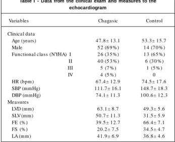
Table I - Data from the clinical exam and measures to the echocardiogram
Variables Chagasic Control Clinical data
Age (years) 47.8± 13.1 53.3± 15.7 Male 5 2 (6 9 %) 1 4 (7 0 %) Functional class (NYHA) I 2 6 (3 5 %) 1 3 (6 5 %) II 4 0 (5 3 %) 6 (3 0 %) III 5 (7 %) 1 (5 %) IV 4 (5 %) 0 HR (bpm) 67.4± 12.9 74.5± 17.6 SBP (mmHg) 111.7± 16.1 148.7± 18.3 DBP (mmHg) 74.1± 11.3 100.6± 12.3 Measures
3
S
p
e
e
d
o
f
e
je
c
ti
o
n
o
f
th
e
L
A
A
(
c
m
/s
)
Normal ADR PN Restrictive Non-conclusive Distribution of patients according to the dyastolic function pattern Fig. 3 - Association between the LAA ejection velocity and left ventricular diastolic function in the patients with chagasic cardiomyopathy. Normal (2 1 patients); ADR - Abnormal diastolic relaxation (1 8 ); PN - pseudonormal pattern (1 3 ); Restrictive (1 0 ) and non-conclusive (1 3 ).
digital and only 3 of beta-blockers. Thirteen (1 8 %) patients were using oral anticoagulant.
The m ost frequent electroca rdiogra phic cha nges were the blocking of the right branch (5 4 %) and ventricular extra-systoles (5 3 %). The left branch blocking happened in 1 8 % of the patients and atrial fibrillation rhythm in 5 %.
The mean of the ejection fraction of the left ventricle was 3 9 ± 1 3 %. The M mode measurement can be found on table I.
The classification of patients in accordance to the diastolic function is in figure 3 . The parameters used for the assessment of the dias-tolic function were associated to those of the sysdias-tolic function and the functional class. Except for the maximum velocity of the atrial reverse flow, all other parameters employed for the assessment of the left atrial function were different in the patients with pseudo-normal and restrictive pattern in relation to the others (tab. II).
The maximum velocities of the atrial reverse in the patients with pseudonormal and restrictive pattern were similar, but different in their duration.
There was no difference of V1 and V2 among the patients with normal pattern and ARR and the control group (p= 0 .2 1 and p= 0 .4 6 ). The atrial reverse velocity was also similar among the groups.
The velocities in the LAA correlated with the diastolic dysfunc-tion pattern. Changes of the ventricular filling pressure, as occurred in the cases with pseudonormal and restrictive pattern, resulted in lower velocities in the LAA (fig. 3 ). The diameter of the left atrium was associated to the diastolic function pattern (fig. 4 ).
Thrombus inside the LAA was found in four (5 %) patients, and it did not associate with cerebral thromboembolism or the flow velocities, although low velocities (V1 = 3 2 .0 cm/s) tended to be associated to thrombus (p= 0 .0 7 ). The flow velocities in the LAA correlated negatively (r= -0 .7 ) with the LA diameter (fig. 5 ).
Discussion
Many methods have been developed to estimate the contractile function of the left atrium. Generally, they are difficult and time-consuming and they are not used in clinical practice. The velocities in the left atrial appendix appear as a clinically applicable method to
estimate the atrial function1. Our objective was to estimate the left
atrial function through such technique and correlate that function with the diastolic dysfunction in the chagasic dilated myocardiopathy. Unlike it happens in the rheumatic mitral stenosis, in patients with sinus rhythm, there is not a clear association between the d ia m e te r of th e le ft a triu m a n d th e ve locitie s in th e a tria l
appendix2 2 ,2 3. In the patients with atrial fibrillation, the appendix
flow is irregular as demonstrated in the literature. Therefore, its
values were not measured in the present study2 4 -2 9.
The values of the velocities in the LAA, which were obtained in our study, in the patients with normal diastolic function or abnormal diastolic relaxation, are within the limits of normality, when compa-red to the control group. The size of the atrium was correlated with those velocities. The atrial dilatation was more frequent in
Table II - Association between the assessment parameters of the left atrium function and the diastole pattern
Parameters Controlδ Normal/ADR PN/Restrictive p * LA (mm) 36.8± 4.6 37.8± 3.8 45.9± 66.4 p< 0.00001 V1 (cm/s) 63.7± 13.9 72.4± 18.7 44.1± 25.1 p< 0.00001 V2 (cm/s) 58.0± 10.9 60.6± 14.4 42.6± 20.3 p< 0.00010
Atrial reverse
Vmax. (cm/s) 24.7± 4.2 22.7± 10.5 27.2± 13.2 p= 0.16000
Duration(ms) 121.3± 23.1 120.9± 36.7 167.1± 34.9 p< 0.00010
*Norm al group/ADR in relation to the PN/Restrictive; δ - No difference with the norm al group/ADR; ADR abnorm al diastolic relaxation; PN -pseudonorm al; Vm ax - m axim um velocity of the atrial reverse flow.
Fig. 4 - Association between the left atrium diameter and the left ventricular diastolic function in the patients with chagasic cardiomyopathy. Normal (2 1 patients); ADR - Abnormal diastolic relaxation (1 8 ); PN - pseudonormal pattern (1 3 ); Restrictive (1 0 ) and non-conclusive (1 3 ).
D
ia
m
e
te
r
o
f
th
e
L
A
(
m
m
)
Normal ADR PN Restrictive Non-conclusive Distribution of patients according to the dyastolic function pattern
Fig. 5 - Correlation between the LA diameter and ejection velocity of the LAA.
S
p
e
e
d
o
f
e
je
c
ti
o
n
o
f
th
e
L
A
A
(
c
m
/s
)
Diameter of the LA (mm)
4
Function of the Left Atrium in the Chagas’ Cardiomyopathy
1. Ito T, Suwa M, Otake Y et al. Left ventricular Doppler filling pattern in dilated car-diom yopathy: relation to hem odynam ics and left atrial function. J Am Soc Echo-cardiogr 1 9 9 7 ;1 0 :1 8 -2 5 .
2. Hansen A, Haass M, Zugck C et al. Prognostic value of doppler echocardiographic m itral inflow patterns: im plications for risk stratification in patients with chronic congestive heart failure. J Am Coll Cardiol 2 0 0 1 ;3 7 :1 0 4 9 -5 5 .
3. Kamel CS, Siqueira-Filho AG, Barreto LFM et al. Insuficiência cardíaca congestiva. Correlação entre a classe funcional e as funções sistólica e diastólica avaliadas pela ecocardiografia com Doppler. Arq Bras Cardiol 2 0 0 1 ; 7 6 :1 2 7 -3 1 . 4. Moller JE, Graham SH, Oh JK et al. left atrial volume a powerful predictor of survival
after acute myocardial infarction. Circulation 2 0 0 3 ; 1 0 7 : 2 2 0 7 -1 2 .
5. Nishim ura RA, Tajik J. Evaluation of diastolic filling of left ventricle in health and disease:doppler echocardiography is the clinician’s rosetta stone. J Am Coll Cardiol 1 9 9 7 ; 3 0 : 8 -1 8 .
6. Coh en GI, Pietrolungo J F, Th om a s J et a l. A pra ctica l guide to a ssessm ent of ventricular diastolic function using Doppler echocardiography. J Am Coll Cardiol 1 9 9 6 ; 2 7 : 1 7 5 3 -6 0 .
7. Garcia MJ, Thomas JD, Klein AL. New Doppler echocardiographic applications for the study of diastolic function. J Am Coll Cardiol 1 9 9 8 ;3 2 :8 6 5 -7 5 .
8. Appleton CP, Jensen JL, Hatle LK et al. Doppler evaluation of left and right ventri-cular diastolic function: a technical guide for obtaining optimal flow velocity recor-dings. J Am Soc Echocardiogr 1 9 9 7 ;1 0 : 2 7 1 -9 1 .
9. Martínez OR, Guerra CH, Molina CA et al. Estudio de la función diastólica ventricular izquierda en pacientes com enfermedad de Chagas. Arq Bras Cardiol 1 9 8 6 ; 4 7 : 3 1 -6 . 10. Sousa ACS, Marin-Neto JA, Maciel BC et al. Disfunção sistólica e diastólica nas form as indeterm inada, digestiva e cardíaca crônica da m oléstia de Chagas. Arq Bras Cardiol 1 9 8 8 ; 5 0 : 2 9 3 -9 9 .
11. Maciel BC, Almeida Filho OC, Schmidt A et al. Função ventricular na moléstia de Chagas. Rev Soc Cardiol Estado de São Paulo 1 9 9 4 ; 4 :1 4 4 -5 1 .
12. Mady C, Ianni BM, Arteaga E et al. Capacidade funcional m áxim a e função dias-tólica em portadores de cardiomiopatia chagásica sem insuficiência cardíaca con-gestiva. Arq Bras Cardiol 1 9 9 7 ; 6 9 :2 3 7 -4 1 .
13. Ba rros MVL, Rocha MOC, Ribeiro ALP et a l. Tissue Doppler im a ging in the eva -lua tion of the regiona l dia stolic function in Cha ga s’disea se. Eur J Echoca rdiogr 2 0 0 1 ;2 : 9 4 -9 .
14. Teichholz LE, KreulenT, Herman MV, et at. Problems in echocardiographic volume determinations: echocardiographic-angiocardiographic correlations in the presen-ce or absenpresen-ce of asynergy. Am J Cardiol 1 9 7 6 ; 3 7 :7 -1 5 .
15. Baker BJ, Wilen MM, Boyd CM et al. Relation of right ventricular ejection fraction to exercise capacity in chronic left ventricular failure. Am J Cardiol 1 9 8 4 ;5 4 :5 9 6 -9 9 . 16. Tajik AJ, Seward JB, Hagler DJ et al. Two-dimensional real-time ultrasonic imaging
of the heart and great vessels. Technique, image orientation, structure, identifica-tion and validaidentifica-tion. Mayo Clin Proc 1 9 7 8 ;5 3 :2 7 1 -3 0 3 .
17. Sahn DJ, De Maria A Kisslo J et al. Recom endation regarding quantitation in M-mode echocardiography:results of a survey of echocardiographic measurements. Circulation 1 9 7 8 ; 5 8 : 1 0 7 2 -8 2 .
18. Rakowski H, Aplleton C, Chan KL et al. Canadian consensus recommendations for de measurement and reporting of diastolic dysfunction by echocardiography. J Am Soc Echocardiogr 1 9 9 6 ; 9 :7 3 6 -6 0 .
19. Seward JB, Khandheria BK, Freeman WK et al. Multiplane transesophageal echo-cardiography: image orientation, examination technique, anatomic correlations, and clinical applications. Mayo Clin Proc 1 9 9 3 ; 6 8 :5 2 3 -5 1 .
20. Castello R, Pearson AC, Lenzen P et al. Evaluation of pulm onary venous flow by
References
of restrictive pattern pf diastolic dysfunction can cause atrial me-chanic failure, which leads to the decrease of the velocity of the atrial reverse, as it occurred in the present study, without any difference
among the patients with milder ways of onset of diastole7.
The increase of the size of the left atrium is associated to a cardiovascular disease and represents a risk factor for atrial
fibril-lation, cerebrovascular accident and death3 4 ,3 5. Moller et al.4
de-monstrated that the increase of the left atrial volume was a strong mortality predictor after acute myocardial infarction. The prognostic value persisted after stratification for clinical predictors of events and for conventional Doppler echocardiographic indexes of systolic and diastolic function of the left ventricle. Those authors also verified the association between the volume and size of the left atrium (p< 0 .0 0 1 ). The volume of the left atrium was not analyzed in the present study.
In another study, Tsang et al.3 6 showed that the size of the
left atrium was a predictor of development of a nonvalvular atrial fibrillation. The chronic diastolic function reflects the extent of the changes in the atrial substratum, which predisposes electro-physiological abnormalities and development of arrhythmias. That mechanism can also contribute for thromboembolic complications, which are classically associated with the cardiac insufficiency of chagasic etiology. So, patients with a restrictive pattern of diastolic dysfunction show a greater predisposition for the occurrence of atrial fibrillation and throm boem bolic events. However, in the present study, the patients with restrictive pattern were not ho-mogenous concerning the atrial function. The atrial reverse velocity in those patients varied from increased, suggesting atrial function preserved and diastolic dysfunction, to normal or reduced, in the presence of atrial dysfunction.
Concluding, the left atrial function constitutes an important p a ra m eter in th e a s s es s m ent of th e p a tients with ch a ga s ic cardiom yopathy, and it is related to the systolic and diastolic functions of the left ventricle.
the patients with an intense compromising of the left ventricular contra ctile function, showing a dva nced dia stolic function a nd depressed ejection fraction, with the changes associated to the
dysfunction of the LAA. Agmon et al.considered that the contractility
of the appendix is affected by the systolic and/or diastolic dysfunction,
primarily due to the rising of the ventricular filling pressure2 2.
Tri-poskiadis et al.3 0, on the other hand, suggested that there is a left
atrial myopathy in the dilated myocardiopathy, which contributes to the depression of the atrial function. That can precede, develop simultaneously or follow the ventricular the myopathic process.
Ito et a l.3 1 ha ve m a de clea r tha t the function of the LAA
improves after the treatment of the cardiac insufficiency and de-monstrated a correlation between the fraction of ejection of the left ventricle and the velocity of dissection of the atrial appendix (r= 0 .8 , p< 0 .0 0 0 5 ).
The outline of the pulmonary venous flow is another strategy currently employed for the study of the left atrium function. The analysis of the record of the atrial reverse can be limited to the
transthoracic, even with the evolution of the transducers3 2. The
transesophageal stays as a useful strategy, allowing for a clear tra cing a nd a ccura te m ea s urem ents of th e velocities of th e
pulmonary venous flow. Hoit and Gabel3 3, in an experimental study,
demonstrated that the reverse flow, during the atrial systole, was absent in the cases of isolated or combined atrial dysfunction, and showed increased velocity after isolated left ventricular dysfunction. In the chagasic myocardiopathy, the loss of the effective atrial systole can cause the decrease of the cardiac output, which can explain the differences in the clinical evolution of the patients and an unfavorable diagnostic.
The left atrium is directly exposed to the diastolic pressure of the left ventricle. Therefore, its dimension is determined by the same factors that influence the diastolic filling. It is regarded as a more stable indicator, which reflects the duration and severity of the diastolic
5
transesophageal echocardiography in subjects with a norm al heart: com parison with transthoracic echocardiography. J Am Coll Cardiol 1 9 9 1 ;1 8 :6 5 -7 1 . 21. Özer N, Tokgözoglu L, Övünç K et a l. Left a tria l a ppenda ge function in pa tients
with cardioembolic stroke in sinus rhythm and atrial fibrillation. J Am Soc Echocar-diogr 2 0 0 0 ;1 3 :6 6 1 -6 5 .
22. Agmon Y, Khandheria B K, Gentile F et al. Echocardiographic assessment of the left atrial appendage. J Am Coll Cardiol 1 9 9 9 ;3 4 :1 8 6 7 -7 7 .
23. AgmonY, Khandheria BK, Meissner I et al. Age-associated changes in left atrial ap-pendage function: a population-based transesophageal echocardiographic study. J Am Coll Cardiol 1 9 9 8 ; 1 1 7 :1 6 3 A (abstract).
24. Pozzolli M, Febo O, Torbicki A et al. Left atrial appendage dysfunction: a cause of throm bosis? Evidence by transesophageal echocardiography-Doppler studies. J Am Soc Echocardiogr 1 9 9 1 ; 4 : 4 3 5 -4 1 .
25. Fernández MAG, Torrecila EG, Román DS et al. Left atrial appendage Doppler flow patterns: implications on thrombus formation. Am Heart J 1 9 9 2 ; 1 2 4 : 9 5 5 -6 1 . 26. Chim owitz MI, DeGeorgia MA, Poole M et al. Left atrial spontaneous echo
con-trast is highly associated with previous stroke in patients with atrial fibrillation or mitral stenosis. Stroke 1 9 9 3 ; 2 4 : 1 0 1 5 -1 9 .
27. Li YH, Lai LP, Shyu KG et al. Clinical implications of left atrial appendage function: its influence on throm bus form ation. Int J Cardiol 1 9 9 4 ; 4 3 : 6 1 -6 .
28. Yao S, Meisner JS, Factor SM et al. Assessm ent of left atrial appendage structure
and function by transesophageal echocardiography: A Review. Echocardiography 1 9 9 8 ; 1 5 :2 4 3 -5 3 .
29. Klein AL, Murray D, Grim m RA et al. Role of transesophageal echocardiography-gu id e d c a rd iove rs ion of p a tie n ts with a tria l fib rilla tion . J Am Coll Ca rd iol 2001;37:691-704.
30. Triposkiadis F, Pitsavos C, Boudoulas H et al. Left atrial myopathy in idiopathic di-lated cardiomyopathy. Am Heart J 1 9 9 4 ; 1 2 8 :3 0 8 -1 5 .
31. Ito T, Suwa M, Kobashi A, Yagi H et al. Influence of altered loading conditions on left atrial appendage function in vivo. Am J Cardiol 1 9 9 8 ;8 1 :1 0 5 6 -5 9 . 32. Tabaca T, Thom as JD, Klein AL. Pulm onary venous flow by doppler
echocardio-graphy: revisited 1 2 years later. J Am Coll Cardiol 2 0 0 3 ; 4 1 :1 2 4 3 -5 0 . 33. Hoit BD, Gabel M. Influence of left ventricular dysfunction on the role of atrial
con-tra ction. a n echoca rdiogra phic-hem odyna m ic study in dogs. J Am Coll Ca rdiol 2 0 0 0 ; 3 6 : 1 7 1 3 -9 .
34. Benjamin EJ, Agostino RBD, Belanger AJ et al. Left atrial size and the risk of stroke and death. the framingham heart study. Circulation 1 9 9 5 ; 9 2 :8 3 5 -4 1 . 35. Pritchett AM, Ja cobsen SJ, Ma honey DW et a l. Left a tria l volum e a s a n index of