Acoustic and hearing-perceptual
voice analysis in individuals with
idiopathic Parkinson’s disease
in “on” and “of” stages
Luiza Lara M. Santos1, Larissa Oliveira dos Reis2, Iara Bassi2, Clara Guzella2, Francisco Cardoso3, César Reis4, Ana Cristina Côrtes Gama5
ABSTRACT
Objective: To evaluate the voice quality of patients with idiopathic Parkinson’s disease, at the “on” and “off” moments of the disease. Method: Five individuals with Parkinson’s disease and five of the control group were assessed. All of them underwent the recording of voice and speech. The acoustic parameters analyzed were: fundamental frequency, jitter, shimmer, harmonic noise proportion and index of tremor, besides performing the hearing-perceptual analysis by means of GRBASI scale. The findings were analyzed using statistics through t test and the level of significance adopted was p<0.05. Results: There was no difference in the acoustic parameters in the three analyzed groups. In the hearing-perceptual analysis, patients with idiopathic Parkinson’s disease showed altered voice quality and the ones from the control group, neutral vocal quality. Conclusion: Patients with idiopathic Parkinson’s disease present rough, breathy and unstable vocal quality in both stages. In the acoustic analysis, there are no differences in the studied parameters. Key words: Parkinson’s disease, voice, speech acoustics, hearing perception.
Análise acústica e perceptivo-auditiva da voz em indivíduos com doença de Parkinson idiopática nos estágios “on” e “off”
RESUMO
Objetivo: Avaliar a qualidade vocal de pacientes portadores da doença de Parkinson idiopática, nos momentos “on” e “off” da doença. Método: Foram avaliados 5 indivíduos com doença de Parkinson idiopática e 5 do grupo controle. Todos foram submetidos à gravação da voz e fala. Os parâmetros acústicos analisados foram: frequência fundamental, jitter, shimmer, PHR e índice de tremor, além da realização da análise perceptivo-auditiva por meio da escala GRBASI. Os achados foram analisados pelo teste t e o nível de significância adotado foi p<0,05. Resultados: Não foi encontrada diferença nos parâmetros acústicos nos três grupos estudados. Na análise perceptivo-auditiva, os pacientes com doença de Parkinson idiopática apresentaram qualidade vocal alterada e os do grupo controle qualidade vocal neutra. Conclusão: Os indivíduos com doença de Parkinson idiopática apresentam qualidade vocal rugosa, soprosa e instável, em ambos estágios. Na análise acústica, não há diferenças nos parâmetros estudados.
Palavras-chave: doença de Parkinson, voz, acústica da fala, percepção auditiva.
Correspondence
Ana Cristina Côrtes Gama Universidade Federal de Minas Gerais Faculdade de Medicina
Av. Alfredo Balena 190 / 3005 30130-100 Belo Horizonte MG - Brasil E-mail: [email protected]
Support
Conselho Nacional de Desenvolvimento Científico e Tecnológico (CNPq)
Received 1 September 2009 Received in final form 15 April 2010 Accepted 23 April 2010
1Graduation Student in Speech and Language Therapy of the Federal University of Minas Gerais (UFMG), Belo Horizonte
MG, Brazil; 2Speech and Language Therapist Voluntary; 3Neurologist, Neurology Service Full Professor, Clinical of Movement
Disturbances, Neurology Service, UFMG; 4Phonetics Laboratory Coordinating Teacher Associate, Letters-UFMG Faculty; 5Speech and Language Therapist. Adjunctive Teacher of the Department of Speech and Language herapy of UFMG, Doctor
Parkinson’s disease (PD) can be deined as a neurode-generative disorder mainly characterized by the degrada-tion of dopaminergic neurons of the basal ganglia, often associated with other etiologies1. he combination of two
or more clinical symptoms (among them, tremor, rigidi-ty, slowness, reduction or loss of movement and postural changes), along with low concentration of dopamine in the basal ganglia, is the parkinsonism2.
Due to the disorder in this neuromuscular control in individuals with Parkinson’s disease, the alterations that may afect oral communication are noted, due to lack of coordination of muscle movements that control the re-sponsible organs for speech, causing vocal and articula-tion disorders and swallowing diiculties, which interfere negatively in communicative expression. Studies describe that vocal alterations, such as imprecise articulation, de-creased speech speed, reduced vocal intensity and low-er variation of fundamental frequency, are commonly al-terations presented by people sufering from Parkinson3.
According to scholars, the entire phonatory mechanism will be afected in Parkinson’s disease, as neuromuscular functions are necessary for the production of an intelli-gible speech4.
Other alterations that may be associated with Parkin-son’s disease are autonomic disorders. Some examples are: cardiovascular and pupillary changes, gastrointestinal and kidney changes, and appetite and mood alterations5.
Among the therapeutic strategies used for the treat-ment of the disease, symptomatic therapy aims to reduce the signs and symptoms of Parkinsonism. he drug most commonly used today is levodopa, an aromatic amino acid that is converted to dopamine in the central ner-vous system. When the patient is under the inluence of the drug (“on” stage), the symptoms disappear or reduce noticeably, and they reappear after ceasing the action of levodopa (“off ” stage).
Despite the advantages brought by the drug, patients with Parkinson’s disease and using levodopa tend to de-velop, after some time, a series of motor complications - such as luctuations and dyskinesias - and non-motor complications such as gastrointestinal and sleeping disor-ders6. During treatment, the motor luctuations become
more and more abrupt and at random, culminating in the “on-off effect”, deined as sudden and unpredictable luc-tuations in rates of motor deiciencies, not related to the time of levodopa ingestion7. It is estimated that after ive
years of levodopa use around about 50% of patients will have luctuations, while 30% have hyperkinesias in ad-dition, most of the time coinciding with the peak of the medication efect (levodopa-induced dyskinesias)8.
To study the likely vocal alterations present in indi-viduals with Parkinson’s disease, a speech assessment is essential because it is considered an efective tool for the
analysis of voice disorders. Currently there are two forms of voice assessment: a hearing-perceptual analysis of vo-cal quality and acoustic analysis of the voice.
The hearing-perceptual assessment is the classical evaluation of voice quality, sovereign and traditional in clinical practice. It is a subjective test based especially in the impression of the speech therapist about the voice. hrough the hearing impression about the voice, the praiser classiies the voice quality of the individual as ap-propriate (neutral) or changed (rough, breathy, whispered, tense, asthenic etc.). here are a number of scales for the hearing assessing the voice, through the use of various tasks for the assessment of the vocal quality9.
he acoustic analysis, in turn, is a complementary tool that allows a more objective assessment of the human voice. hrough this analysis it is possible to obtain con-crete values of acoustic characteristics of the voice, as the fundamental frequency, perturbation measures, as jitter and shimmer, noise measurements and the obtaining of a sound wave trace. In clinical practice, the acoustic analy-sis acts mainly as an auxiliary tool of the hearing percep-tual assessment, in order to increase the diagnostic accu-racy of vocal alterations9.
he purpose of this study was to evaluate the voice of patients with idiopathic Parkinson’s disease diagnosis at the “on” and “off ” moments, objectively and subjectively, by means of acoustic and hearing perceptual analysis.
METHOD
his is an experimental longitudinal study conduct-ed with two groups pairconduct-ed by sex and age. he experi-mental group consisted of ive patients with idiopathic Parkinson’s disease, recruited from the Outpatient Clinic for Movement Disorders of the Neurology Hospital Bias Fortes, UFMG. hey were three males (aged between 58 and 72 years, mean of 63.6 years) and two females (aged between 48 and 72 years, mean of 60 years) and dura-tion of the disease from 1 to 18 years, with an average of 8 years. According to the Hoehn-Yahr Scale, four patients were on the scale 2 and one patient on the scale 3 of the development of Parkinson’s disease.
he control group was composed of ive individuals without neurological disorders, three males (aged from 54 to 71 years, mean of 58.6 years) and two females (aged from 54 to 65 years, mean of 59.5 years).
his-tory of laryngeal surgery or alteration and do not present another neurological disease.
he criteria for inclusion of individuals selected for the control group were to be literate and able to read, have complete dentition or use dental prosthesis, have adequate visual acuity to allow the reading, do not have a history of laryngeal surgery or alteration and do not pres-ent another neurological disease.
Participants signed a free and clear consent form and were informed about the voluntary and conidential char-acter of the research.
The voices of the individuals were recorded in an acoustically treated booth belonging to the Department of Speech Therapy of São Geraldo Hospital, in a Dell® computer, model Optiplex GX260, with Direct Sound® professional sound board. he acoustic analysis software used was the CSL, MDVP Kay Elemetrics MDVP® mod-ule. Professional condenser unidirectional Shure® micro-phone was used, installed on a pedestal, laterally posi-tioned at a distance of 5 cm from the mouth of the infor-mants, who remained standing during the recording.
he control group was subjected to a voice record-ing on a unique moment, while the group of informants with idiopathic Parkinson’s disease was submitted for re-cording in two moments: initially after the abstention of levodopa for a period of 12 hours (“off ” stage) and then after a period of 30 minutes to 1 hour of levodopa ad-ministration (“on” stage). Thus, three groups were an-alyzed: control group (CG), the experimental group in “off ” stage (EG1) and the experimental group in “on” stage (EG2). All people performed the recording of voice and speech through the prolonged vowel /a/ and the read-ing of a text.
he voice samples were analyzed in a hearing-percep-tual way by three speech therapists, voice specialists, using the GRBASI scale. he GRBASI scale aims at the dyspho-nia global assessment (G-Grade) by identifying the fol-lowing factors: roughness (R), breathness (B-breathness), asthenia (A) and strain (S-strain)10,11. he instability
fac-tor (I) was added to the scale later10. For these aspects the
grade 0 (absent), 1 (mild), 2 (moderate) and 3 (severe) can be assigned. he GRBASI scale is the most used proto-col of the hearing-perceptual analysis in clinical practice. To perform the acoustic analysis, the sustained vow-el /a/ was transferred and analyzed by the acoustic anal-ysis software. he investigated parameters in the acous-tic analysis were: fundamental frequency (F0), which
cor-responds to the number of vibration cycles per time unit; jitter (%): an index of fundamental frequency variabili-ty in the short term; shimmer (%): an index of variabilivariabili-ty of the sound wave amplitude in the short-term; noise to harmonic noise proportion (HNP), which computes the noise in a series of pulses produced by the oscillation of
the vocal folds; voice turbulence index (VTI): calculates the energy level of high frequency noise.
he statistical analysis used was the T test and the ad-opted level of signiicance was p<0.05.
his work was reviewed by the Research Ethics Com-mittee of the Federal University of Minas Gerais and ap-proved on the report number ETIC 676/07.
RESULTS
The mean values of acoustic parameters for each group studied were obtained. It is observed that the fun-damental frequency (F0) presented higher values for both
experimental groups (EG1 and EG2), compared to the control group (CG), and even higher for individuals with Parkinson’s disease in “on” stage (GE2); the parameter shimmer (%) was higher for the group of individuals in “off ’’ stage (GE1) and smaller for the ones in ““off ” stage; jitter (%) had also elevated values for individuals in the group with Parkinson’s disease in “off ” stage and reduced in “on” stage; the HNP and VTI parameters showed high-er values for the control group individuals, and lowhigh-er for the “on” and “off ” experimental groups, respectively.
Table 1 sets out the following results.
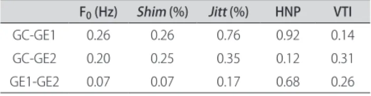
After obtaining the average values of each acoustic pa-rameter, the values of signiicance were calculated for the acoustic measures comparing the three studied groups. here was no statistically signiicant diference for the an-alyzed acoustic parameters among the three groups.
Table 2 presents the values of signiicance that were found.
Table 1. Average values of the acoustic parameters in the three studied groups.
F0 (Hz) Shim (%) Jitt (%) HNP VTI
GC 144.34 4.80 1.50 0.20 0.73 GE1 187.30 8.53 1.95 0.19 0.04 GE2 203.07 3.07 0.62 0.13 0.05
GC: group control; GE1: experimental group in the stage on; GE2: experimental group in the of stage; FO: fundamental frequency in Hz; Shim (%): shimmer (vibration amplitude variation cycle by cycle); Jitt (%): jitter (vibration frequency variation cycle by cycle); HNP: harmonic-noise proportion; VTI: index of vocal turbulence.
Table 2. Values of signiicance for the acoustic measures comparing the three studied groups.
F0 (Hz) Shim (%) Jitt (%) HNP VTI
GC-GE1 0.26 0.26 0.76 0.92 0.14 GC-GE2 0.20 0.25 0.35 0.12 0.31 GE1-GE2 0.07 0.07 0.17 0.68 0.26
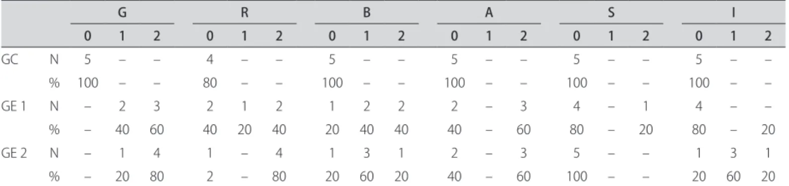
After the hearing-perceptual assessment, values for each parameter of the GRBASI scale, in the three groups, were extracted. It was found that 100% of the individuals in the control group present neutral vocal quality, 60% of individuals in the “off ” stage of the disease have moder-ate dysphonia and 40% had mild dysphonia; 80% of indi-viduals in the “on” stage of the disease present moderate dysphonia and 20% a mild one.
Table 3 sets out the values assigned to the voices of each group, analyzed in a hearing-perceptual way through GRBASI scale.
DISCUSSION
he acoustic and hearing-perceptual analysis used for vocal assessment of the individuals who composed the study sample enabled the veriication of the vocal features of the experimental and control groups, with subsequent comparison among the obtained values.
Although higher values for acoustic parameters of fun-damental frequency (F0) were identiied, shimmer (%) and
jitter (%) and lower values for HNP and VTI in the exper-imental groups (“on” and “off ” stages), the study in ques-tion did not show statistically signiicant diference for any of these parameters when compared with the three studied groups (Tables 1 and 2). he results of this study agree in part with the researched literature and, during their interpretation, one should consider the methodolog-ical diferences, especially regarding the quantity of sam-ple used in the studies that were found and the acoustic analysis software which was used.
Scholars held voice acoustic analysis of 20 patients with Parkinson’s disease in the “on” and “off ” stages of the disease, in comparison with a control group, and identi-ied a signiicant increase of fundamental frequency in the “on” stage and a also signiicant reduction in the jit-ter, HNP and VTI values, after medication12, unlike the
results from our study, whose F0 value, despite increased
for the “on” stage group, was not signiicantly higher, and the jitter, HNP and VTI values were not signiicantly re-duced in the group in the “on” stage (Table 2). Possibly the
diference in the results is due to the signiicantly smaller sample in this study, besides the methodological difer-ences of the type of extraction of acoustic parameters.
In a study that quantiied the acoustic measures of voice and speech of 41 patients with Parkinson’s disease under drug treatment (“on” stage), compared with a con-trol group paired by sex and age, the groups with Parkin-son’s disease showed increased measures of F0 and jitter
and reducing of vocal intensity, of the HNP values and of the variability of frequency and intensity in relation to the group control13. Our results agree with the literature in
relation to the values of F0, which were elevated in groups
with Parkinson’s disease in the “on” stage. However, there was disagreement in relation to the values of jitter, which in this study showed reduced values on the “on” group compared with the control group (Table 1). Nevertheless, these values were not statistically signiicant (Table 2).
In a study evaluating the voice and speech of patients with Parkinson’s disease before and after pallidotomy in the “on” and “off ” stages of the disease, by means of acoustic analysis, it was not found a statistically signif-icant diference among the values of F0, jitter, shimmer,
PPQ (pitch perturbation quotient), APQ (amplitude per-turbation quotient) and NHR (noise-harmonic ratio), pre-and post-pallidotomy, both in the “on” pre-and “off ” stages of the disease. However, except for the F0, which increased
with drug intake, other values were reduced during the “on” stage before and after the surgery14. Considering that
pallidotomy did not interfere signiicantly in the acoustic measurements analyzed and that the diferences found are due to the drugs, the results agree with this study, which found increased values for F0 and reduced values for
jit-ter, shimmer and HNP in the “on” stage of the disease. In order to identify changes in the values of funda-mental frequency in the “on” and “off ” stages of Parkinson’s disease, a study analyzed the speech of patients immedi-ately before and immediimmedi-ately after the producing of a con-sonant and found increased values of F0 for individuals in
the “on” stage. he study linked the increase of F0 to an
in-crease in tension caused by the use of the antiparkinsonian
Table 3. Values of the hearing-perceptual analysis in the three studied groups.
G R B A S I
0 1 2 0 1 2 0 1 2 0 1 2 0 1 2 0 1 2
GC N 5 – – 4 – – 5 – – 5 – – 5 – – 5 – –
% 100 – – 80 – – 100 – – 100 – – 100 – – 100 – –
GE 1 N – 2 3 2 1 2 1 2 2 2 – 3 4 – 1 4 – –
% – 40 60 40 20 40 20 40 40 40 – 60 80 – 20 80 – 20
GE 2 N – 1 4 1 – 4 1 3 1 2 – 3 5 – – 1 3 1
% – 20 80 2 – 80 20 60 20 40 – 60 100 – – 20 60 20
drug15. he present study found F
0 values extracted from
the sustained vowel /a/, increased for both experimental groups, especially for the group in the “on” stage. How-ever, there is no statistically signiicant diference when compared to the control group and the “off ” stage group (Tables 1 and 2). he discrepant results may be justiied by the methodological diference between the two surveys.
A survey on the hearing-perceptual and acoustic anal-ysis in neurological dysphonias revealed that in all the types of dysarthrias, including the hypokinetic dysarthria present in patients with idiopathic Parkinson’s disease, the jitter and shimmer measures are altered and elevated16.
In agreement with these results, Table 1 of our study de-scribes an increase of the same measures for the group in the “off ” stage of the disease.
A study examined the efect of levodopa on the speech of patients with Parkinson’s disease. It was observed that the drug efect on the acoustic features of the speech will depend on the proile of the individual’s speech at the time of assessment, as levodopa promotes articulation, sound, rhythm, vocal amplitude and speech intelligibili-ty of speech in a general form17.
As for the hearing-perceptual evaluations carried out by GRBASI scale and described in Table 3, changes in vo-cal quality for all patients with Parkinson’s disease were observed, showing, preferably, rough, breathy and unsta-ble voices. hese hearing-perceptual features are usually associated with hypokinesia and rigidity of the muscles involved in respiration, phonation and articulação18,19. he
incomplete glottic closing prevents the increase of sub-glottic pressure and justiies the perception of a breathy voice20.
he literature is in favor of the results found in this re-search. Experts say that the altered voice quality is com-monly found in patients with Parkinson’s disease assessed in a hearing-perceptual form14.
Studies classify the voice quality of individuals with Parkinson’s disease as hoarse-harsh-breathy, with the presence of phonation instability20-22. In this study, most
of the voices in the experimental groups were classiied as rough, breathy and unstable, while the neutral vocal qual-ity was predominant in the group without Parkinson’s dis-ease. hese results are consistent with the data found in the literature and are described in Table 3.
A study conducted the survey of vocal complaints of 118 patients with Parkinson’s disease, with subsequent hearing-perceptual assessment of the voices, through GR-BASI scale and the vocal attack. Weak voice was the most frequent complaint, which justiies the mild to moderate degree of deviation of the “asthenia” parameter, obtained in the hearing-perceptual analysis. he vocal instability was as diverted as the asthenia, which gives the negative impact of a deteriorated speech19.In this study, the
“asthe-nia” and “instability” parameters are present in most pa-tients with Parkinson’s disease, both in the “on” and “off ” stages (Table 3).
In order to evaluate the efect of levodopa on respi-ration and speech intelligibility of patients with Parkin-son’s disease, 25 patients were assessed using a dysarthria protocol. It was found that both respiratory standard and speech intelligibility improved with the drug. However it can not be stated that these results are related to an im-provement in vocal quality of these individuals. hus, the study ponders over the need for research that assesses the changes of vocal characteristics of individuals with Par-kinson’s disease under levodopa treatment23. his study
found no statistically signiicant diferences in vocal qual-ity of individuals with neurological disease under dop-aminergic medication, as it is represented in Table 3.
Considering the indings of this study and all the re-searched literature, one sees that the efect of levodopa does not carry out signiicantly improvement in the voice patterns of the patient with Parkinson’s disease, objective-ly and subjectiveobjective-ly assessed.
he literature identiies and describes several advan-tages brought by the administration of levodopa, which especially promotes an increase of life quality of patients under medication treatment. Among the beneits of the drug, it can be highlighted: better performance of muscu-lar behavior, increase of the activities of the olfactory and blood system, increase of cognition, memory capacity and attention, and reduction of the depression and anxiety24-30.
Future research should be necessary with a more sig-niicant sample to better understand the behavior of the acoustic hearing-perceptual parameters of the voice in in-dividuals with Parkinson disease.
he conclusions from the study in question are: there are no statistically signiicant diference among the acous-tic parameters of fundamental frequency, jitter, shimmer, VTI and HNP of the groups with Parkinson’s disease in “off ” and “on” stages and the control group. Patients with idiopathic Parkinson’s disease present altered vocal qual-ity classiied as rough, breathy and unstable, from mild to moderate degrees. Medical therapy with levodopa does not interfere signiicantly in the vocal patterns of patients with Parkinson’s disease when compared to the “on” and “off ” stages.
REFERENCES
Shih MC, Hoexter MQ, Andrade LAF, Bressan RA. Doença de Parkinson e neu-1.
roimagem do transportador de dopamina: uma revisão crítica. São Paulo Med J 2006;124:168-175.
Cardoso F, Camargos ST, Silva JRGA. Etiologia de parkinsonismo em uma clíni-2.
ca brasileira de distúrbios do movimento. Arq Neuropsiquiatr 1998;56:171-175. Azevedo LL, Cardoso F, Reis C. Análise acústica da prosódia em mulheres com 3.
doença de Parkinson: efeito da Levodopa. Arq Neuropsiquiatr 2003;61:995-998. Gallena S, Smith PJ, Zeiro T, Ludlow CL. Efects of levodopa on larynge-4.
Nicaretta DH, Pereira JS, Pimentel MLV. Distúrbios autonômicos na doença 5.
de Parkinson. Rev Assoc Med Bras 1998;44:120-122.
Teixeira ALJr, Cardoso F. Tratamento inicial da doença de Parkinson. Rev 6.
Neurociências 2004;12:146-151.
Marsden CD. Parkinson’s disease. J Neurol Neurosurg Psychiatry 1994;57: 7.
672-681.
Ahlskog JE, Muenter MD. Frequency of levodopa-related dyskinesias and 8.
motor luctuations as estimated from the cumulative literature. Mov Dis-ord 2001;16:448-458.
Behlau M, organizadora. Voz: o livro do especialista Vol 1. Rio de Janeiro: Re-9.
vinter; 2001;1:96-100;130-164.
Hirano M. Clinical examination of voice. New York: Springer Verlag, 1981. 10.
Dejonckere PH, Remacle M, Fresnel-Elbaz E, Woisard V, Crevier-Buchman L, 11.
Millet B. Diferentiated perceptual evaluation of pathological voice quality: reliability and correlations with acoustic measurements. Rev Laryngol Otol Rhinol 1996;117:219-224.
Sanabria J, Ruiz PG, Gutierrez R, et al. The efect of levodopa on vocal func-12.
tions in Parkinson’s disease. Clin Neuropharmacol 2001;24:99-102. Gamboa J, Jimênez-Jimênez FJ, Nieto A, et al. Acoustic voice analysis in pa-13.
tients with Parkinson’s disease treated with dopaminergic drugs. J Voice 1997;11:314-320.
Mourão LF, Aguiar PMC, Ferraz FAP, Behlau MS, Ferraz HB. Acoustic voice as-14.
sessment in Parkinson’s disease patients submitted to posteroventral palli-dotomy. Arq Neuropsiquiatr 2005;63:20-25.
Goberman AM, Blomgren M. Fundamental frequency change during of-15.
set and onset of voicing in individuals with parkinsin’s disease. J Voice 2008; 22:178-191.
Carrilo L, Ortiz KZ. Análise vocal (auditiva e acústica) nas disartrias. Pró-Fono 16.
R Atual Cient 2007;19:381-386.
Ho AK, Bradshaw JL, Iansek R. For better or worse: the efect of levodopa on 17.
speech in Parkinson’s disease. Mov Disord 2008;23:574-580.
Baumgartner CA, Sapir S, Ramig TO. Voice quality changes following phona-18.
tory-respiratory efort treatment (LSVT) versus respiratory efort treatment for individuals with Parkinson disease. J Voice 2001;15:105-114.
Gasparini G, Diaféria G, Behlau M. Queixa vocal e análise perceptiva-auditiva 19.
de pacientes com doença de Parkinson. R Ci Méd Biol (Salvador) 2003;2:72-76. Carrara de Angelis E. Deglutição, coniguração laríngea, análise clínica e acús-20.
tica computadorizada da voz de pacientes com doença de Parkinson. Tese. São Paulo:Universidade Federal de São Paulo – EPM. 2000.
Countryman S, Ramig LO, Pawlas AA. Speech and voice déicits in Parkin-21.
sonian plus syndromes: can they be treated? NCVS Status and Progress Re-port 1996;6:99-111.
Behlau M. Voz: o livro do especialista. Vol. II. Rio de Janeiro: Revinter 2005: 22.
122-129.
De Letter M, Santens P, De Bodt M, Van MG, Van BJ, Boon P. The efect of 23.
levodopa on respiration and word intelligibility in people with advanced Par-kinson’s disease. Clin Neurol Neurosurgery 2007;109:495-500.
Cioni M, Richards CL, Malouin F, Bedard PJ, Lemieux R. Characteristics of the 24.
electromyographic patterns of lower limb muscles during gait in patients with Parkinson’s disease when of and on L-Dopa treatment. Ital J Neurol Sci 1997;18:195-208.
Quagliato LB, Viana MA, Quagliato EMAB, Simis S. Alterações do olfato na 25.
doença de Parkinson. Arq Neuropsiquiatr 2007;65:647-652.
Deems DA, Kaplan P, Schiumaker J, Doty RL. Studies of olactory function in 26.
parkinson’s patients receiving electrotherapy for control of extrapyramidal symptoms. Chem Senses 2001;26:1115.
Cools R, Stefanova E, Barker RA, Robbins TW, Owen AM. Dopaminergic mod-27.
ulation of high-level cognition in Parkinson’s disease: the role of the prefron-tal cortex revealed. PET Brain 2002;125:584-594.
Lange KW, Robbins TW, Marsden CD, James M, Owen AM, Paul GM. L-do-28.
pa withdrawal in Parkinson’s disease selectively impairs cognitive perfor-mance in tests sensitive to frontal lobe dysfunction. Psychopharmacology (Berl) 1992; 107:394-404.
Costa A, Peppe A, Brusa L, Caltagirone C, Gatto I, Carlesimo GA. Levodopa 29.
improves time-based prospective memory in Parkinson’s disease. J Internat Neuropsychol Soc 2008;14: 601-610.
Erdal KJ. Depression and anxiety in persons with parkinson’s disease with and 30.