Nitric oxide and asymmetric dimethyl arginine (ADMA)
levels in an experimental hydronephrotic kidney caused by
unilateral partial ureteral obstruction
_______________________________________________
Cabir Alan
1, Hasan Anil Kurt
1, Naci Topalo
ğ
lu
1, Ahmet Re
ş
it Ersay
1, Dilek Ülker Çakir
1, Gökhan Ba
ş
türk
11 Department of Urology, Medical Faculty, Canakkale Onsekiz Mart University, Turkey
ABSTRACT
ARTICLE
INFO
______________________________________________________________ ______________________
Aim: Our aim is to measure asymmetric dimethyl arginine and nitric oxide levels in rats with induced unilateral acute ureteral obstruction to research the effects on the kidney.
Material and Methods: The study included 21 adolescent (average age 6 weeks) Spra-gue-Dawley male rats weighing between 240-290g divided at random into 3 groups. Group-1: Control group (n=6): underwent no procedures. Group-2: Sham group (n=6): underwent the same procedures as the experimental group without ureter and psoas muscle dissection. Group-3: Group with induced partial unilateral ureteral obstruction (n=9). All rats were sacrificed after 12 weeks. Superoxide dismutase enzyme activity and nitrite and nitrate salt levels were measured in renal tissue. Plasma nitrite-nitrate and ADMA levels were examined.
Results: In the experimental group histopathological changes observed included renal pelvis dilatation, flattened papillae, sclerotic glomerulus and fibrosis. In the experi-mental group tissue SOD and blood ADMA levels were higher than the control and sham groups (p<0.05) while tissue NO and plasma NO values were lower than in the sham and control groups (p<0.05).
Conclusion: Oxidative stress and disruption of NO synthesis play an important role in renal function and histopathological changes after obstructive renal disease. To pre-vent renal complications developing after obstructive nephropathy we believe that a new strategy may be research on reducing ADMA.
Keywords:
Ureteral Obstruction; Nitric Oxide; Rats; Superoxide Dismutase
Int Braz J Urol. 2016; 42: 614-20
_____________________
Submitted for publication: January 16, 2015
_____________________
Accepted after revision: June 03, 2015
INTRODUCTION
Ureteral obstructions are frequently obser-ved in urology practice and without early diag-nosis and treatment it may cause some serious complications (1-4). Many experimental studies have shown that retrograde glomerular reflux de-veloping due to ureteral obstruction disrupts NO synthesis and in parallel affects renal function
(5-8). However, how and by which mechanism NO synthesis is disrupted is still not fully known.
NO formation (9). Impaired endothelium vasodi-lation, increased aggregation of platelets and in-creased monocyte adhesion provide endothelial dysfunction that increase ADMA (10). During ure-teral obstruction, inflammatory mediators in renal parenchyma have been shown to increase due to retrograde glomerular reflux. This situation increa-ses ADMA synthesis causing NO synthesis disrup-tion, which causes an effect on renal function. In one study, 221 chronic renal failure patient’s serum ADMA levels were shown to be increased (11).
The aim of the study was to induce uni-lateral acute ureteral obstruction in rat models to measure ADMA and NO levels to research the effect on the kidney.
MATERIAL AND METHODS
Experimental Animals and Study Groups
This study began after permission was granted by Canakkale Onsekiz Mart University Animal Experiments Ethics Committee. The stu-dy used 21 adolescent (average 6 weeks) Spra-gue-Dawley male rats weighing from 240-290g. The animals were kept in standard laboratory conditions with stable temperature (18-21ºC) and humidity, 12 hours of light and 12 hours of darkness, with 3-4 rats in each cage fed with rat food and tap water.
The rats were randomly divided into 3 groups. Group-1: Control group (n=6): underwent no procedures. Group-2: Sham group (n=6): un-derwent the same procedures as the experimental group (the ureter and psoas muscle were palpa-ted and left on their own anatomical position). Group-3: Experimental group: induced partial unilateral ureteral obstruction (PUUO) (n=9).
Surgical Procedure
Rats used for partial unilateral urete-ral obstruction (PUUO) and rats undergoing the sham operation were starved for 6 hours before the operation and only allowed water. The rats, in accordance with antiseptic rules, were operated on under laboratory conditions maintaining body temperature. All rats were anesthetized with intra-muscular 50mg/kg ketamine hydrochloride (Keta-lar, Eczacibasi) and then a 2cm area was shaved
on bilateral abdominal wall and 10% Povidone iodine was used to clean the field.
Partial unilateral ureteral obstruction (PUUO) model: A 2cm incision was performed at the anterior abdomen region. Skin above the linea alba, subdermis, abdominal anterior wall and pe-ritoneum was incised to reach abdominal cavity and the left kidney was located. Fat and connec-tive tissues were dissected to reveal the left ureter and psoas muscle. For PUUO rats, similar to the technique of Ulm and Miller (12), the psoas muscle under the left ureter was longitudinally dissected to form a groove (~15mm) and a small part of the left ureter was placed into the groove. Later the edges of the psoas muscle were fixed above the ureter with 5/0 silk suture. Thus the ureter was enclosed in a tunnel (Figure-1). In the sham model rats after the left ureter and psoas muscle were lo-cated, the procedure was completed without ureter and psoas muscle dissection. Later 3/0 catgut and 3/0 silk suture were used to close the abdomen in 2 layers. All rats were sacrificed after 12 weeks.
Biochemical Tests
The kidney was dissected and excised and then stored at-80ºC. To test for biochemical changes, the levels of superoxide dismutase (SOD) enzyme activity and nitrite and nitrate salts which are end--products of NO were measured. To investigate the nitrite-nitrate and ADMA levels in plasma, 5cc blood samples from the ventricle of rats were centrifuged for 10 minutes at 3000rpm and the separated sam-ples were stored at-40ºC until analysis.
Tissue Homogenization
After tissue samples were weighed, for as-say of total nitrite/nitrate and SOD samples, they were homogenized in 0.9% NaCl and 10% homo-genates were prepared. For assay of total nitrite/ nitrate and SOD, the prepared homogenates were centrifuged for 15 minutes at 15000rpm. Both as-says were completed using samples of the super-natant liquid.
Nitrite/nitrate
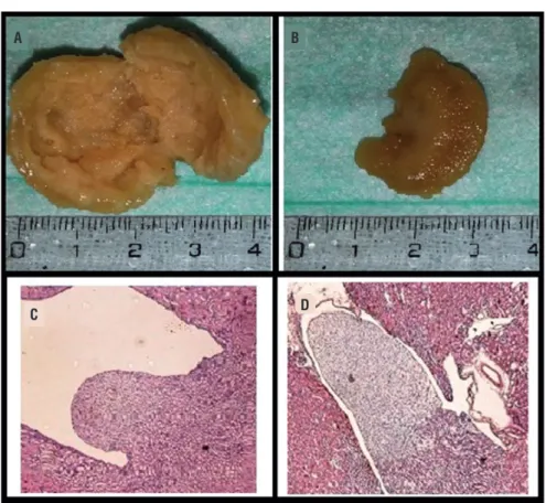
Figure 1 - a) Macroscopic view of kidney tissue of experimental group; b) Macroscopic view of kidney tissue of control group; c) Hematoxilin and eosin (H&E) stained histopathological images of kidney tissue of experimental group; renal pelvis dilatation and flattening; d) Hematoxilin and eosin stained histopathological images of kidney tissue of control group; there was no pathological findings on renal papilla, cortex and medulla.
A
C D
B
Tissue SOD Activity Assay
Tissue SOD activity was measured at 560nm by modifying the method determined by Sun et al. (13).
ADMA
ADMA levels in serum were measured by using a kit from BioVendor Research and Diag-nostic Products (Cat. No: REA 201/96) manufactu-red by DLD Diagnostica GMBH (Germany). Results were determined by the ELISA method and repor-ted as nanogram per millilitre (ng/mL).
Histopathological Investigation
After tissue samples were cleaned with saline they were immediately fixed in 10% for-maldehyde solution at room temperature for 72 hours and prepared for routine light microscope
examination. The tissue samples were dehydrated with alcohol and cleaned with xylene before being immersed in paraffin.
Five-micrometer slices of the tissues were made. The tissues were stained with hematoxyli-ne-eosine (H&E) and periodic acid-Schiff (PAS)
and the sections were examined and photogra-phed with a light microscope.
Statistical Evaluation
RESULTS
While there was no pathological changes in the kidney sections from the control group and sham group, in the experimental group induced PUUO, on histopathological examination of the H&E dyed kidney fibrosis, inflammation,
flatte-ning of the renal papillae and dilatation of the re-nal pelvis were observed with varying degrees On kidney sections stained with PAS sclerotic glome-rular changes, inflammation and fibrotic changes were observed (Figure-1).
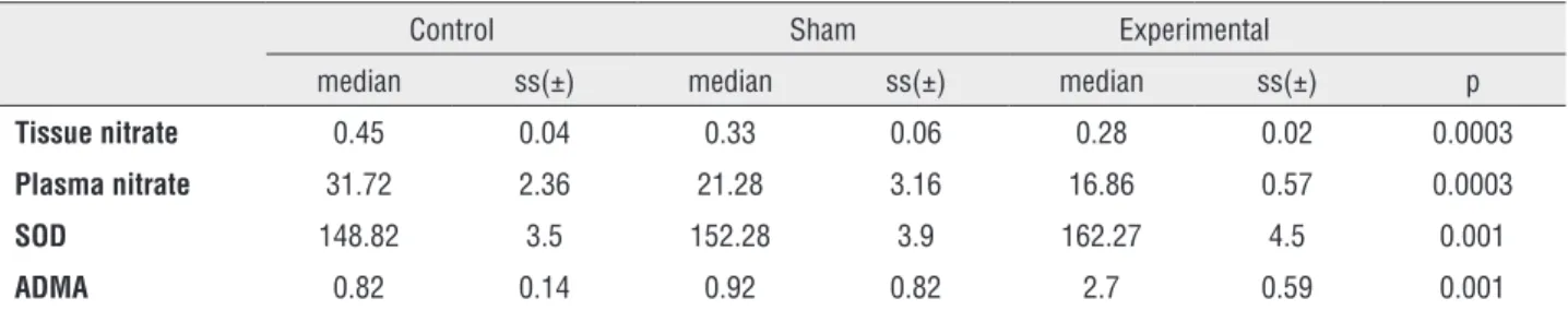
Linked to increased oxidative stress the tissue SOD values, the enzyme with primary an-tioxidant properties, are summarized in Table-1.
In the experimental group with induced PUUO, tissue levels of SOD were higher than in the sham and control groups (p=0.001) (Figure-2). Compa-ring the groups, there were differences between the experimental and control groups (p=0.001) and between the experimental and sham groups (p=0.003) (Table-1).
There was a clear increase in ADMA va-lues measured in blood in the experimental group compared to the control and sham groups (p=0.001) (Figure-3). In the sham group there were higher levels of ADMA than in the control group (Table-1).
Nitrite/nitrate levels, end products of NO, were identified in both renal tissue and in plas-ma. The nitrite/nitrate values in tissue were lower in the PUUO induced experimental group com-pared to the sham and control groups (p=0.0003) (Figure-4) (Table-1).
Assay results of plasma nitrite/nitrate le-vels are shown in Figure-5. These results show
Table-1 - Biochemical results of the three groups.
Control Sham Experimental
median ss(±) median ss(±) median ss(±) p
Tissue nitrate 0.45 0.04 0.33 0.06 0.28 0.02 0.0003
Plasma nitrate 31.72 2.36 21.28 3.16 16.86 0.57 0.0003
SOD 148.82 3.5 152.28 3.9 162.27 4.5 0.001
ADMA 0.82 0.14 0.92 0.82 2.7 0.59 0.001
Figure 2 - SOD values measured in tissue samples of the groups (µmoL/g tissue).
Figure 3 - ADMA values measured in blood samples of the groups (ng/mL).
(p=0.004)
(p=0.004)
Sham 165
160
155
150
145
140
Experimental Control
3
2,5
2
1,5
1
0,5
0
Sham Experimental Control
DISCUSSION
Different etiologic factors such as urinary tract stones, ureteropelvic junction or ureterovesical junction stenosis, tumors and iatrogenic factors lead to upper uriner tract obstruction that can cause or-gan failure in urology practice. Treatment planning is depend on duration, degree and level of
obstruc-tion. Upper urinary tract patologies have a broad tre-atment spectrum. The primary aim of tretre-atment is to protect or recover the functional reserve of kidneys by relieving the obstruction. In case of prolonged obstruction, functional reserve of kidney can decre-ase so much that nephrectomy is required to prevent morbidities of dysfunctional kidney.
Fibrosis, collagen and extracellular matrix components accumulation are the most leading in-terstitial changes in upper urinary tract obsruction. Besides cellular composition changes of interstitium, many biologically active molecules changes occur with interstitial fibrosis. It is considered that oblite-ration of tubules and interstitial capillaries due to in-terstitial fibrosis are the major determinants of renal function failure in kidney disease. Claesson et al. (14) induced left chronic partial ureteral obstruction in newborn rats. After two weeks they began to observe histopathological changes from ureteral obstruction and they reported that primarily papilla deformation occurred. In our study the rats with induced PUUO were sacrificed after 12 weeks. In the hydronephrotic kidney, histopathological changes observed included renal pelvis dilatation, flattened papillae, sclerotic glomerulus and fibrosis.
In upper urinary tract obstructions, increased ureteral pressure is the first stage of damage that le-ads to renal blood flow reduction resulting in tissue ischemia, cellular atrophy and eventually necrosis. TNF-α is a potent proinflammatory cytokine that is capable of stimulating renal tubular cell apoptosis and infiltration of inflammatory cells during ische-mic renal injury. After the development of obstruc-tive nephropathy, it is reported that there is a role of factors such as prostaglandins (PG), angiotensin (ANG) II, growth factors and NO in the accumulation of free oxygen radicals and leukocyte infiltration (15). ROS may release vasoconstrictor bioactive lip-ids such as prostaglandin, thromboxane and plate-let activating factors and inactivate NO inducing a reduction in glomerular blood flow and glomerular infiltration rate (16, 17). Ricardo et al. (18) after in-ducing a unilateral ureteral obstruction model in rats reported that ROS and overproduction of tubular ir-regular antioxidant enzymes cause increased intra-renal oxidative stress, leading to fibrogenesis, over expression of fibrogenic cytokines and loss of tubu-lar shape. Kinter et al. (19) in a UUO model found
Figure 4 - Nitrite/nitrate levels measured in tissue samples of the groups (µmoL/g tissue).
Figure 5 - Nitrite/nitrate levels measured in blood samples of the groups (µmol/L).
(KW-x2 = 16.06 ve P=0.0003)
(KW-x2 = 16.06 ve P=0.0003)
Tissue
Plasma Sham
Sham
Experimental
Experimental Control
Control 35
30
25
20
15
10
5
0 0,5
0,45
0,4
0,35
0,3
0,25
0,2
0,15
0,1
0,05
high levels of antioxidant enzymes (catalase, SOD, glutathione peroxidase) which can protect against the harmful effects of ROS. After UUO, ROS produc-tion was greater than the protective capacity of the antioxidant enzymes increasing renal damage with tubular atrophy and interstitial fibrosis observed. In our study a marker of oxidative stress (SOD level in tissue) was higher in the experimental group com-pared to the sham and control groups (p=0.001 and p=0.003). It was shown our study that upper urina-ry tract obstruction led to oxidative stress with dis-turbance of NO synthesis, creating fibrosis which could lead to renal damage.
Asymmetric dimethylarginine (ADMA) is an amino acid, like arginine, found in plasma, uri-ne and tissue. In 1992, Vallance et al. (4) identified ADMA in human plasma and urine endothelial tis-sue as endogenous inhibitors of endothelial nitric oxide synthase (eNOS). It was the first time that the importance of ADMA as endogenous inhibitör of NOS in patient with end stage renal failure was ci-ted by Vallance et al. (1). In these patients, increased plasma ADMA levels were reduced by dialysis with improvement of endothelial function. Later studies showed many times the relationship between in-creased ADMA levels and endothelial vasodilator dysfunction. In the endothelium NO is produced from L-arginine via the endothelial isoform (eNOS) of nitric oxide synthase. In humans NO synthesis may be disrupted by asymmetric dimethylarginine (ADMA), removing the endogenically-formed com-pound L-arginine from substrate linking points in-hibiting NOS activity. An infusion of ADMA dis-rupts vasodilation in the endothelium. Increased ADMA levels in plasma are not only linked to en-dothelial dysfunction, but also related to increased oxidative stress (20). Boger et al. (21) found that ox-idative stress damaged the cystein aminoacid in the active region of the DDAH enzyme responsible for ADMA catabolism reducing enzyme activity and thus reducing the ADMA disintegration. As a result of increasing oxidative stress in many degenerative diseases, ADMA levels were found to be high. In ureter obstruction, free oxygen radicals increase, as shown by many studies mentioned above (18, 19, 22). In our study, the findings are in accordance with these studies. Plasma ADMA levels in the ex-perimental group were higher than in the sham and
control groups (p=0.001 and p=0.001, respectively). ADMA inhibits NOS activity causing a reduction in NO levels, and as a result, disruption of endothelial function. Lin et al. (10) showed increased ADMA concentration in endothelial dysfunction was re-lated to increased ROS production in plasma. In ad-dition Takiuchi et al. (23) showed that endothelial dysfunction in coronary and peripheral vein dis-eases was linked to increased ADMA levels in plas-ma. The findings of our study are consistent with all these studies. When compared the experimental group to control and sham groups, we found incre-ased both ADMA and SOD levels (p<0.05).
CONCLUSIONS
Oxidative stress and disruption of NO syn-thesis play an important role in renal function and histopathological changes after obstructive uropa-thy. ADMA is an endogenous inhibitor of NOS. In-creased ADMA levels show the inhibitor effect on NOS, causing a fall in NO levels in the kidney. We believe that methods to reduce ADMA may be a new strategy to prevent renal complications that develop after obstructive nephropathy.
CONFLICT OF INTEREST
None declared.
REFERENCES
1. Chevalier RL, Forbes MS, Thornhill BA. Ureteral obstruction as a model of renal interstitial fibrosis and obstructive nephropathy. Kidney Int. 2009;75:1145-52.
2. Wongmekiat O, Leelarungrayub D, Thamprasert K. Alpha-lipoic acid attenuates renal injury in rats with obstructive nephropathy. Biomed Res Int. 2013;2013:138719.
3. Coplen DE, Synder HM. Ureteral Obstruction and Malformations. In: Ashcraft, Murphy, Sharp, Sigalet, Synder. Pediatric Surgery. 12th ed. Philadelphia: W.B. Saunders. 2000; pp. 690-706. 4. Chevalier RL, Thornhill BA, Gomez RA. EDRF modulates renal
hemodynamics during unilateral ureteral obstruction in the rat. Kidney Int. 1992;42:400-6.
5. Klahr S. Obstructive nephropathy. Intern Med. 2000;39:355-61. 6. Lanzone JA, Gulmi FA, Chou SY, Mooppan UM, Kim H.
Renal hemodynamics in acute unilateral ureteral obstruction: contribution of endothelium-derived relaxing factor. J Urol. 1995;153:2055-9.
7. Ekinci S, Ciftci AO, Atilla P, Muftuoglu S, Senocak ME, Buyukpamukcu N. Ureteropelvic junction obstruction causes histologic alterations in contralateral kidney. J Pediatr Surg. 2003;38:1650-5.
8. Araujo M, Welch WJ. Oxidative stress and nitric oxide in kidney function. Curr Opin Nephrol Hypertens. 2006;15:72-7. 9. Vallance P, Leiper J. Cardiovascular biology of the asymmetric
dimethylarginine:dimethylarginine dimethylaminohydrolase pathway. Arterioscler Thromb Vasc Biol. 2004;24:1023-30. 10. Lin KY, Ito A, Asagami T, Tsao PS, Adimoolam S, Kimoto M, et
al. Impaired nitric oxide synthase pathway in diabetes mellitus: role of asymmetric dimethylarginine and dimethylarginine dimethylaminohydrolase. Circulation. 2002;106:987-92. 11. Fleck C, Schweitzer F, Karge E, Busch M, Stein G. Serum
concentrations of asymmetric (ADMA) and symmetric (SDMA)
dimethylarginine in patients with chronic kidney diseases. Clin Chim Acta. 2003;336:1-12.
12. Ulm AH, Miller F. An operation to produce experimental reversible hydronephrosis in dogs. J Urol. 1962;88:337-41. 13. Sun Y, Oberley LW, Li Y. A simple method for clinical assay of
superoxide dismutase. Clin Chem. 1988;34:497-500.
14. Chertin B, Rolle U, Farkas A, Puri P. The role of nitric oxide in reflux nephropathy. Pediatr Surg Int. 2002;18:630-4.
15. Chevalier RL. Molecular and cellular pathophysiology of obstructive nephropathy. Pediatr Nephrol. 1999;13:612-9. 16. Baud L, Ardaillou R. Involvement of reactive oxygen species in
kidney damage. Br Med Bull. 1993;49:621-9.
17. Rabl H, Khoschsorur G, Colombo T, Petritsch P, Rauchenwald M, Költringer P, et al. A multivitamin infusion prevents lipid peroxidation and improves transplantation performance. Kidney Int. 1993;43:912-7.
18. Ricardo SD, Diamond JR. The role of macrophages and reactive oxygen species in experimental hydronephrosis. Semin Nephrol. 1998;18:612-21.
19. Kinter M, Wolstenholme JT, Thornhill BA, Newton EA, McCormick ML, Chevalier RL. Unilateral ureteral obstruction impairs renal antioxidant enzyme activation during sodium depletion. Kidney Int. 1999;55:1327-34.
20. Böger RH, Schwedhelm E, Maas R, Quispe-Bravo S, Skamira C. ADMA and oxidative stress may relate to the progression of renal disease: rationale and design of the VIVALDI study. Vasc Med. 2005;10:S97-102.
21. Böger RH, Maas R, Schulze F, Schwedhelm E. Elevated levels of asymmetric dimethylarginine (ADMA) as a marker of cardiovascular disease and mortality. Clin Chem Lab Med. 2005;43:1124-9.
22. Kawada N, Moriyama T, Ando A, Fukunaga M, Miyata T, Kurokawa K, et al. Increased oxidative stress in mouse kidneys with unilateral ureteral obstruction. Kidney Int. 1999;56:1004-13.
23. Takiuchi S, Fujii H, Kamide K, Horio T, Nakatani S, Hiuge A, et al. Plasma asymmetric dimethylarginine and coronary and peripheral endothelial dysfunction in hypertensive patients. Am J Hypertens. 2004;17:802-8.
24. Klahr S. The role of nitric oxide in hypertension and renal disease progression. Nephrol Dial Transplant. 2001;16:60-2. 25. Cherla G, Jaimes EA. Role of L-arginine in the pathogenesis and
treatment of renal disease. J Nutr. 2004;134:2801S-6S.