www.bjorl.org
Brazilian
Journal
of
OTORHINOLARYNGOLOGY
ORIGINAL
ARTICLE
Combined
ocular
and
cervical
vestibular
evoked
myogenic
potential
in
individuals
with
vestibular
hyporeflexia
and
in
patients
with
Ménière’s
disease
夽
,
夽夽
Tatiana
Rocha
Silva
a,∗,
Luciana
Macedo
de
Resende
b,
Marco
Aurélio
Rocha
Santos
aaUniversidadeFederaldeMinasGerais(UFMG),FaculdadedeMedicina,ProgramadePós-graduac¸ãoemCiências
Fonoaudiológicas,BeloHorizonte,MG,Brazil
bUniversidadeFederaldeMinasGerais(UFMG),FaculdadedeMedicina,DepartamentodeFonoaudiologia,BeloHorizonte,MG,
Brazil
Received15December2015;accepted9April2016 Availableonline31May2016
KEYWORDS
Vestibularnerve; Motorevoked potential;
Labyrinthvestibule; Vestibulardisorders; Sacculeandutricle
Abstract
Introduction:Thevestibular evoked myogenicpotentialisapotentialofmeanlatencythat measuresthemuscleresponsetoauditorystimulation.Thispotentialcanbegeneratedfrom thecontractionofthesternocleidomastoidmuscleandalsofromthecontractionofextraocular musclesinresponsetohigh-intensitysounds.Thisstudypresentsacombinedorsimultaneous technique of cervical and ocular vestibular evoked myogenic potential in individuals with changesinthevestibularsystem,foruseinotoneurologicdiagnosis.
Objective:Tocharacterizetherecordsandanalyzetheresultsofcombinedcervicalandocular VEMPinindividualswithvestibularhyporeflexiaandinthosewithMénière’sdisease.
Methods:Thestudyincluded120subjects:30subjectswithvestibularhyporeflexia,30with Ménière’sdisease,and60individualswithnormalhearing.Datacollectionwasperformedby simultaneouslyrecordingthecervicalandocularvestibularevokedmyogenicpotential.
Results:Thereweredifferencesbetweenthestudygroups(individualswithvestibular hypore-flexiaandindividualswithMénière’sdisease)andthecontrolgroupformostofwaveparameters incombinedcervicalandocularvestibularevokedmyogenicpotential.Forcervicalvestibular evokedmyogenicpotential,itwasobservedthattheprolongationoflatencyoftheP13andN23 waveswasthemostfrequentfindinginthegroupwithvestibularhyporeflexiaandinthegroup withMénière’sdisease.Forocularvestibularevokedmyogenicpotential,prolongedlatencyof N10andP15waveswasthemostfrequentfindinginthestudygroups.
夽 Pleasecitethisarticleas:SilvaTR,deResendeLM,SantosMA.Combinedocularandcervicalvestibularevokedmyogenicpotentialin
individualswithvestibularhyporeflexiaandinpatientswithMénière’sdisease.BrazJOtorhinolaryngol.2017;83:330---40.
夽夽StudyconductedattheAudiologyClinicoftheAnexoSãoGeraldooftheHospitaldasClínicasoftheUniversidadeFederaldeMinas
Gerais(UFMG),BeloHorizonte,MG,Brazil.
∗Correspondingauthor.
E-mail:[email protected](T.R.Silva).
PeerReviewundertheresponsibilityofAssociac¸ãoBrasileiradeOtorrinolaringologiaeCirurgiaCérvico-Facial.
http://dx.doi.org/10.1016/j.bjorl.2016.04.017
Conclusion: Combinedcervicalandocularvestibularevokedmyogenicpotentialpresented rel-evantresultsforindividualswithvestibularhyporeflexiaandforthosewithMénière’sdisease. Thereweredifferencesbetweenthestudygroupsandthecontrolgroupformostofthewave parametersincombinedcervicalandocularvestibularevokedmyogenicpotential.
© 2016 Associac¸˜ao Brasileira de Otorrinolaringologia e Cirurgia C´ervico-Facial. Published by Elsevier Editora Ltda. This is an open access article under the CC BY license (http:// creativecommons.org/licenses/by/4.0/).
PALAVRAS-CHAVE
Nervovestibular; Potencialevocado motor;
Vestíbulodo labirinto;
Doenc¸asvestibulares; Sáculoeutrículo
Potencialevocadomiogênicovestibularocularecervicalsimultâneoemindivíduos comhiporreflexiavestibulareemindivíduoscomdoenc¸adeMénière
Resumo
Introduc¸ão: Opotencialevocadomiogênicovestibularéumpotencialdemédialatênciaque avaliaarespostamusculardecorrentedeestimulac¸ãoauditiva.Estepotencialpodesergerado a partir da contrac¸ão do músculo esternocleidomastoideoe também apartir da contrac¸ão demúsculosextraocularesemrespostaasonsdeelevadaintensidade.Esteestudoapresenta umatécnicacombinadaousimultâneadepotencialevocadomiogênicovestibular cervicale ocularemindivíduoscomalterac¸õesnosistemavestibularparaqueestapossaserutilizadano diagnósticootoneurológico.
Objetivo: Caracterizaroregistroeanalisarosresultadosdopotencialevocadomiogênico ves-tibularcervicaleocularcombinadoemindivíduoscomhiporreflexiavestibulareemindivíduos comdoenc¸adeMénière.
Método: Participaramdoestudo120indivíduos,30comhiporreflexiavestibular,30comdoenc¸a deMénièree60comaudic¸ãodentrodospadrõesdenormalidade.Acoletadedadosfoifeitapor meiodopotencialevocadomiogênicovestibularcervicaleocularregistradossimultaneamente.
Resultados: Houvediferenc¸aentreogrupodeestudo(indivíduoscomhiporreflexiavestibular eindivíduoscomdoenc¸adeMénière)eogrupocontroleparaamaioriadosparâmetrosdas ondasnopotencialevocadomiogênicovestibularcervicaleocularcombinado.Paraopotencial evocadomiogênicovestibularcervicalobservou-sequeoprolongamentodalatênciadasondas P13eN23foiaalterac¸ãomaisencontradanogrupodeindivíduoscomhiporreflexiavestibulare nogrupodeindivíduoscomdoenc¸adeMénière.Paraopotencialevocadomiogênicovestibular ocularoprolongamentodalatênciadasondasN10eP15foiaalterac¸ãomaisencontradano grupodeestudo.
Conclusão:Opotencial evocado miogênicovestibular cervicale ocular combinado apresen-touresultadosrelevantesparaosindivíduoscomhiporreflexiavestibulareparaosindivíduos comdoenc¸adeMénière.Houvediferenc¸aentreogrupodeestudoeogrupocontroleparaa maioriadosparâmetrosdasondasnopotencialevocadomiogênicovestibularcervicaleocular combinado.
© 2016 Associac¸˜ao Brasileira de Otorrinolaringologia e Cirurgia C´ervico-Facial. Publicado por Elsevier Editora Ltda. Este ´e um artigo Open Access sob uma licenc¸a CC BY (http:// creativecommons.org/licenses/by/4.0/).
Introduction
Thevestibularevokedmyogenicpotential(VEMP)isformed
by myogenic responses activated by sound stimulation
throughhigh-intensitysounds.Theliteraturedescribestwo
typesofVEMP:cervicalandocular.1---3
CervicalVEMPactivatesthesaccularmacula,theinferior
vestibularnerve,andthedescendingvestibulospinal
path-ways,recordedbysurfaceelectromyographyofthecervical
muscles in the presence of muscle contraction.1,2 Ocular
VEMPactivatestheutricularmacula,thesuperiorvestibular
nerve,andtheascendingvestibularpathways,recordedby
surfaceelectromyographyontheextraocularmusclesinthe
presenceofmusclecontraction.4,5
Althoughrelatively old--- itwasdiscoveredinthe
mid-1960s--- VEMPisstilllittleknown,anditcomprisesa vast
universeofpossibleresearchandapplications.1---3
The significanceofelectrical responses,theneural
cir-cuitinvolved,andthebehavioroftheseresponsesinnormal
individualshavealreadybeenwelldemonstrated.6However,
althoughthefindingsofthisexamindifferentneurological
andotoneurological disordershave been described, much
remainstobeclarifiedandstudied.3,4,7
Instudiesconductedinpatientswithunilateralvestibular
dysfunction,ahighvariabilityofresponsesforcervicaland
ocular VEMP has been described.8---15 For individuals with
superior vestibular neuritis, a lack of response to ocular
VEMPandnormalresponsestocervicalVEMPwereobserved.
lackofresponsetocervical VEMPandnormalresponsesto
ocularVEMPwereobserved.10---12
For individuals with superior semicircular canal
dehis-cence syndrome,increased amplitudes were observed for
bothcervicalandocularVEMP.Accordingtotheliterature,
thereisasignificantcorrelationbetweenthesizeof
dehis-cenceandtheamplitudesofocularVEMPs.13---15
For individuals with vestibular schwannoma, a
pro-longedlatencyforcervicalVEMPwasobservedthatcanbe
attributedtotumorcompressionofthevestibulospinaltract
andthe compression of themyelin sheathon theinferior
vestibularnerve.RegardingocularVEMP,reducedorabsent
responseswereobservedformostindividualswithvestibular
schwannoma.16
ResearchershavebeenusingVEMPintheassessmentof
patientswithMénière’s disease.17---21 Giventhatthe
mech-anism through which endolymphatic hydrops develops is
controversialandthattheetiopathogenesisofthedisease
is still in the field of scientific speculation, new clinical
instrumentsareneededtoaidtheidentificationofsaccular
hydrops.18---20
InastudythatusedcervicalandocularVEMP toassess
patients with Ménière’s disease, the authors observed a
higher number of absent responses in ocular VEMP when
comparedwithcervical VEMP. Forthoseauthors, the
jus-tificationfor suchan occurrence wasprobablyduetothe
facttheutricleismoreassociatedwithhearingfunctionat
lowfrequenciesthanthesaccule.17
They also noted that VEMP response varied according
tothe stage of Ménière’s disease, whether acute or
dur-ingtheinterval betweenattacks.In theacutephase,the
amplitude of ocular VEMP (contralateral to the affected
ear) showed increased responses, while the amplitude
of cervical VEMP (ipsilateral to the affected ear) was
attenuated.17
Inastudythatevaluatedtheotolithfunctioninpatients
withMénière’sdiseaseduringtheacutephaseandduringthe
intervalbetweenattacks,itwasobservedthattheincrease
inN10waveamplitudewashigherintheacutephase.
How-ever,theincreaseinN10waveamplitudewasmuchhigher
in the contralateral side of the affected ear than in the
ipsilateralsideoftheaffectedear.18
Inanotherstudy,theauthorsfound ahighincidenceof
altered cervical VEMP responses in asymptomatic ears of
patients withMénière’s disease. Forthose authors, VEMP
canbeanaidinthestagingandfollow-upofMénière’s
dis-ease.Theyalsoobservedaverysmallnumberofdelaysin
P13andN23wavelatencies.19
Incontrast,another studyfound alteredcervical VEMP
responsesinasymptomaticearsofpatientswithMénière’s
disease,but found nosignificant differences betweenthe
latencies of P13 and N23 waves of affected and
asymp-tomaticears. In that same study,the authors observed a
lackof responseintheasymptomatic earin 20%ofcases.
Forthoseauthors,thisfactshowsthevalueofevoked
myo-genicpotentialsinthediagnosisofoccultsacculushydrops
withoutclinicalmanifestations.20
The present study was justified by the possibility of
simultaneouslyassessingtheipsilateraldescendingand
con-tralateralascendingvestibularpathwaysinindividualswith
otoneurologicalchanges,contributingtotheaccuracyofthe
combinedcervicalandocularVEMPtechnique,aswellasto
itscurrentuseinotoneurologicalassessment.
Thisstudyaimedtocharacterizetherecordandanalyze
theresultsofcombinedcervicalandocularVEMPin
individ-ualswithvestibularhyporeflexiaandinthosewithMénière’s
disease.
Methods
TheproceduresofthisstudywereapprovedbytheResearch
EthicsCommitteefromUniversidadeFederaldeMinasGerais
(UFMG),underCAAEProtocolN◦ 32505314.0.0000.5149,in
accordancewithResolution466/12 oftheNational Health
Council(ConselhoNacionaldeSaude[CONEP]).
Thiswasadescriptivestudywithqualitativeand
quan-titative analysis.One hundred andtwenty subjects, aged
18---59years,wereinvitedtoparticipate.
The sample comprised a study group of 30 individuals
withvestibularhyporeflexiaand30individualswith
unilat-eralMénière’sdisease,andanage-andsex-matchedcontrol
group of 60 individuals without a diagnosis of peripheral
disorders of the inner ear. The control group was
subdi-videdintotwogroupsof30individualseach.Controlgroup
1(CG1)waspaired withthegroupofindividualswith
ves-tibular hyporeflexiaand controlgroup 2(CG2)waspaired
withthegroupofpatientswithMénière’sdisease.
The participants were selectedat the UFMG School of
Medicine and at the Audiology Clinic at the Sao Geraldo
Annex fromthe University Hospital at UFMG and in
Diag-nosticCenterOtorhinolaryngologicalthroughanon-random
convenience sampling technique. The participants were
notifiedpersonallyoftheresearchobjectives,theabsence
ofdamagetotheirhealth,theassuranceofsecrecyoftheir
identities or any other characteristics that could identify
them,andabouttheresearchmethods.Afterthenecessary
clarifications,allparticipantssignedaninformedconsent.
Datacollection was conducted at the Audiology Clinic
at the SãoGeraldo Anexxfrom the University Hospitalat
UFMGandinDiagnosticCenterOtorhinolaryngological.The
individuals inthe studygroup underwent
otorhinolaryngo-logicevaluation,andallsubjects(studygroupandcontrol
group)underwentabasicaudiologicalevaluation.This
eval-uationconsistedof:medicalhistory,meatoscopy,puretone
audiometry,speechaudiometry,tympanometry,and
acous-ticreflexesassessment.
Forthemedicalhistory,the participantprovided
infor-mation such as personal data, audiological history, and
aspects related to health. A Heine® Mini 2000 otoscope
wasusedformeatoscopy.Puretoneaudiometryandspeech
audiometrywereperformed in asoundproof boothwitha
one-channel audiometer, Interacoustics® AD 229b model,
usingTDH-39earphones andaB-71bone vibrator.
Tympa-nometry and acoustic reflex assessment were performed
usinganInteracoustics® AZ7middleearanalyzer.
Forthe study group, inclusion criteriacomprised
indi-viduals with vestibular hyporeflexia defined by vestibular
assessment (electronystagmography or vector
electronys-tagmography) and another group was composed by
individuals diagnosed with Ménière’s disease according to
the Bárány Society criteria. The Bárány Society defines
disease:twoormorespontaneousepisodesofvertigo
last-ingfrom20minto12h;sensorineuralhearingloss(affecting
mainly the middle frequencies) in the affected ear onat
least one occasion before, during, or after one of the
episodesofvertigo;presenceofintermittentauditory
symp-toms such as hearing loss, ear fullness, and tinnitus on
the affected side; and signs and symptoms that are not
explainedbyanothervestibulardiagnosis.22
Forthecontrolgroup,inclusioncriteriacomprised
indi-vidualswithnohearingcomplaints,nohistoryofvestibular
and/or otologic disease,and audiologic evaluation within
normalstandards.Anaudiologicevaluationwasconsidered
tobewithinnormallimitswhenthepuretoneair-conduction
thresholds were 25dB HL or less in the frequencies of
250---8000Hz; pure tone bone-conduction thresholds were
15dB HL or less in the frequencies of 500---4000Hz, and
the differences between the thresholds for air and bone
conduction were less than or equal to 10dB; additional
requirements included type A tympanometric curve, and
presence of acoustic reflexes at 500, 1000, 2000, and
4000Hz. For the evaluation of pure tone thresholds, the
criteriaestablishedbySilmanandSilvermanwereused23;for
thetympanometriccurve,thecriteriaestablishedbyJerger
wereused.24
The exclusion criteria comprised participants who had
neurologicaldisorders,cancer, otitis,tympanicmembrane
perforation, those with history of craniocerebraltrauma,
previousotologicsurgery,andindividualswhowereunable
toperform cervical rotationand eye movements.
Consid-ering that Ménière’s disease may present with vestibular
hyporeflexia at thevestibular evaluation,individuals with
suspicion and/or diagnosis of Ménière’s disease were
excludedfromthevestibularhyporeflexiagroup.
Afterthebasicaudiologicalevaluation,participantswere
referredforelectrophysiologicalevaluationthroughthe
ves-tibularevokedmyogenicpotential(VEMP).
TheVEMPwasperformedinacomfortableandquiet
envi-ronment,withLabat® equipment,usingtwochannels.The
stimuliwerepresentedthroughER3Ainsertionphones,with
disposable foam eartips.Tone burst stimulus at an
inten-sity of 120dB nHL were used. In this study, a bandpass
filterof 10---1500Hz wasused.Toobtain each record,100
stimuliwerepresented at afrequencyof 500Hz ata rate
of four stimuli per second. The scan window was 50ms.
Eachsubjectunderwentatleasttwostimulationsperside,
toverify thereplication of the potential. The impedance
values werechecked before each record; theyhad to be
below5k.
ToperformtheVEMP,theparticipant’sskinwascleaned
with dehydratedalcohol followed by abrasive paste;
sur-face electrodes received a small amount of electrolyte
paste and were fixed with adhesive tape. For recording,
theactiveelectrode(negativeelectrode)inchannel1was
placedapproximately1cmbelowthelowereyelid,andthe
referenceelectrode(positiveelectrode)wasplacedata
dis-tanceofapproximately1cmfromtheactiveelectrode.The
activeelectrodeonchannel2wasplacedontheopposite
sidetothechannel1,at theanteriorborderofthe
stern-ocleidomastoidmuscleinitsupperthird,andthereference
electrodewasplacedinthesternalnotchregion.Theground
electrodewasplacedontheforehead(Fpz).Therefore,the
positioningof theelectrodes allowedforthesimultaneous
recordingofocularandcervicalVEMP;channel1wasused
forrecordingocularVEMPandchannel2,forcervicalVEMP.
Uponexamination,theparticipantwasinstructedtosit
on the chair and keep the head rotated to the opposite
sideofthestimulatedear,causingcontractionofthe
ster-nocleidomastoidmuscle.Atthesametime,theparticipant
wasinstructedtolookatastationarytargetlocatedonthe
wallofthetestroomandthenimmediatelytoafixedpoint
located above the target, which formed a vertical
view-ingangle ofapproximately30◦ abovethehorizontalplane
formedbytheparticipant’shead.Afterwards,the
contralat-eralcervicalandocularVEMPswererecordedusingthesame
technique.
Afterdatacollection,dataweretabulated and
submit-tedtostatisticalanalysis.Statisticalanalysiswasperformed
usingSPSSversion20.0.Initially,adescriptiveanalysiswas
performed, which included measures of central tendency
(mean and median), dispersion (standard deviation), and
position (maximum and minimum). The normality of the
samples was assessed using the Kolmogorov---Smirnov and
Shapiro---Wilktests.Inadditiontothedescriptivestatistics,
inferentialstatisticswasperformedthroughStudent’st-test
andtheMann---Whitneytestforcomparisonoftwo
indepen-dent samples, and through Student’s t-test and Wilcoxon
testforcomparisonofpairedsamples.Thechi-squaredtest
wasusedtocomparethefrequenciesobtainedby
calculat-ingtheasymmetryindexandtocomparehearinglosswith
theresultsofcombinedcervicalandocularVEMP.Thelevel
ofsignificanceof5%(p≤0.05)wasadopted.Results
signif-icantatthe10%level(p≤0.10)wereconsideredashaving
atrendtowardstatisticalsignificance.
Results
The mean age of the study population was 49.4 years
(SD=7.03)forthegroupofindividualswithpost-caloric
nys-tagmushyporeflexia;for theCG1, themeanagewas49.1
years(SD=7.12).Forthegroup ofpatientswithMénière’s
disease,the meanage was46.2 years(SD=8.66); for the
CG2,themeanagewas46.1years(SD=8.53).
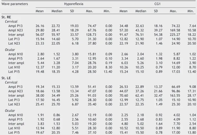
Descriptiveanalysisforthegroupofindividualswith
ves-tibular hyporeflexia and the CG1 are shown in Table 1.
Forcervical VEMP, it wasobservedthat themean latency
values for both P13 and N23 waves were higher in the
group of patients with vestibular hyporeflexia. For
ocu-lar VEMP, the meanlatency valuesof N10 andP15 waves
were higher in the group of individuals with vestibular
hyporeflexia.
Descriptive analysis for the group of patients with
Ménière’sdiseaseandfortheCG2isshowninTable2.For
cervicalVEMP, itwasobservedthatthemeanlatency
val-uesforbothP13andN23waveswerehigherinthegroupof
patientswithMénière’sdiseaseonlywiththerightear
stim-ulation.Forocular VEMP, the meanlatency values ofN10
andP15 waves were higherin the group of patients with
Ménière’s disease for both the right ear and the left ear
stimulation.
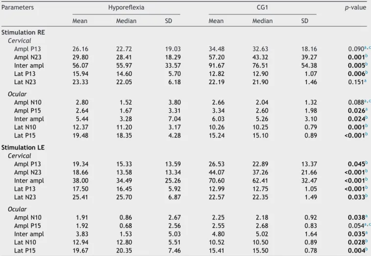
Intheinferentialstatisticalanalysisforthegroupof
sub-jectswithvestibularhyporeflexiaandtheCG1,adifference
wasobservedbetweenthegroupsformostwaveparameters
Table1 Measuresofcentraltendency,dispersion,andpositionforlatency(ms)andamplitude(V)forcombinedcervicaland
ocularVEMPforindividualswithvestibularhyporeflexiaandindividualsinthecontrolgroup(CG1).
Waveparameters Hyporeflexia CG1
Mean Median SD Max. Min. Mean Median SD Max. Min.
St.RE
Cervical
AmplP13 26.16 22.72 19.03 74.47 0.00 34.48 32.63 18.16 74.22 7.64 AmplN23 29.80 28.41 18.29 67.76 0.00 57.20 43.32 39.27 169.58 10.58 Interampl 56.07 55.97 33.57 128.73 0.00 91.67 76.51 54.38 225.27 18.22 LatP13 15.94 14.60 5.70 31.30 0.00 12.82 12.90 1.07 14.90 10.70 LatN23 23.33 22.05 6.18 37.80 0.00 22.19 21.90 1.46 24.90 20.50
Ocular
AmplN10 2.80 1.52 3.80 15.81 0.09 2.66 2.04 1.32 5.87 1.02 AmplP15 2.64 1.67 3.31 12.95 0.10 3.34 2.60 1.98 8.82 1.22 Interampl 5.44 3.28 7.04 28.76 0.19 6.03 5.26 3.10 14.69 2.90 LatN10 12.37 11.20 3.17 20.20 8.30 10.26 10.25 0.79 12.00 8.70 LatP15 19.48 18.35 4.28 28.50 13.40 15.24 15.10 0.89 17.03 13.40
St.LE
Cervical
AmplP13 19.34 15.33 13.59 51.41 0.00 26.53 22.89 13.37 66.69 9.08 AmplN23 18.66 13.58 13.34 47.07 0.00 44.07 37.26 21.66 96.86 17.31 Interampl 38.00 34.49 25.26 91.03 0.00 70.60 62.41 32.47 163.55 28.17 LatP13 17.50 16.45 5.92 28.30 0.00 12.99 12.75 1.05 15.10 10.90 LatN23 25.41 25.70 6.87 35.40 0.00 22.57 22.35 1.49 25.30 20.10
Ocular
AmplN10 1.91 0.86 2.67 12.19 0.00 2.25 2.18 0.92 4.02 1.04 AmplP15 1.92 0.68 2.56 10.60 0.00 2.55 2.68 0.83 4.09 1.12 Interampl 3.83 1.53 5.03 19.20 0.00 4.80 5.02 1.64 7.70 2.54 LatN10 12.94 12.80 5.51 28.30 0.00 10.52 10.50 0.89 11.90 8.80 LatP15 19.67 20.35 7.46 37.10 0.00 15.41 15.50 0.78 17.00 13.80
SD,standarddeviation;Max.,maximum,Min.,minimum;St,stimulation;RE,rightear;LE,leftear;Ampl,amplitude;Lat,latency.
andtheCG2,differencesbetweenthegroupsformostwave parameterswerealsoobserved(Table4).
Whencomparingtherightandleftearsinboththegroup
withvestibularhyporeflexiaandinthegroupwithMénière’s
disease, nodifferences between the ears in cervical and
ocularVEMPwereobserved.
Regardingtheasymmetryindex,nodifferencebetween
thestudyandcontrolgroupswereobservedatcervicalVEMP.
Theasymmetryindexrangedfrom0%to88%.
Fig.1showsthatthecombinedcervicalandocularVEMP
wasabnormalinasymptomaticearsoftheindividualsinthe
group with vestibular hyporeflexia and in the group with
Ménière’sdisease.
The main alteration observed at cervical VEMP in the
group with vestibular hyporeflexia and in the group with
Ménière’s disease was the prolonged P13 and N23 wave
latency (Fig. 2). The main alteration observed at ocular
VEMPin thegroupwithvestibularhyporeflexia andinthe
group withMénière’s disease was alsothe prolonged P13
andN23wavelatency(Fig.3).
It is noteworthy that 13 (43.30%) individuals from the
vestibularhyporeflexiagrouphadalterationsinboththe
cer-vicalandocularVEMP.InthegroupwithMénière’sdisease,
six(20%)patientshadchangesin bothcervical andocular
VEMP.
3 3
5
19
10
3
5
12
0 2 4 6 8 10 12 14 16 18 20
No alteration
Asymptomatic + Symptomatic Asymptomatic Symptomatic
Hyporeflexia Ménière’s disease
Figure1 Frequencydistributionoftheoutcomeofcombined cervicalandocularVEMP,pertheaffectedside,forindividuals withvestibularhyporeflexiaandforindividualswithMénière’s disease(n=30).
Regarding theside of the hearingloss, of the 30
indi-viduals with vestibular hyporeflexia, seven (23.35%) had
unilateral hearingloss, seven(23.35%) hadbilateral
Table2 Measuresofcentraltendency,dispersion,andpositionforlatency(ms)andamplitude(V)forcombinedcervicaland
ocularVEMPforindividualswithMénière’sdiseaseandindividualsinthecontrolgroup(CG2).
Waveparameters Ménière’sdisease CG2
Mean Median SD Max. Min. Mean Med. SD Max. Min.
St.RE
Cervical
AmplP13 25.96 24.54 13.18 52.02 4.31 43.68 45.02 19.06 77.98 7.64 AmplN23 34.59 31.42 20.25 85.63 6.90 62.28 60.49 27.16 111.38 10.58 Interampl 60.55 54.11 31.96 128.65 11.39 105.98 106.98 44.51 170.63 18.22 LatP13 15.08 13.90 3.12 23.50 11.60 13.40 13.45 0.96 14.90 11.60 LatN23 23.53 22.80 3.15 31.70 20.50 22.39 22.45 1.48 24.90 20.00
Ocular
AmplN10 4.40 1.87 7.63 28.81 0.14 2.33 2.05 0.80 5.25 1.04 AmplP15 5.95 1.89 12.32 45.18 0.15 3.21 3.05 1.18 8.48 1.13 Interampl 10.35 3.88 19.93 73.70 0.29 5.57 5.26 1.90 13.73 2.17 LatN10 12.18 10.95 3.49 22.60 9.10 10.40 10.40 0.84 12.10 9.20 LatP15 18.13 16.45 3.94 28.50 13.80 15.27 15.05 0.96 17.20 13.40
St.LE
Cervical
AmplP13 23.73 21.98 13.77 54.56 0.00 37.64 32.47 15.63 76.04 14.97 AmplN23 29.42 29.60 16.19 68.61 0.00 51.47 51.34 16.88 95.88 23.74 Interampl 53.15 50.75 28.91 123.17 0.00 89.11 83.84 29.77 158.22 39.60 LatP13 14.05 13.40 4.82 22.70 0.00 13.18 13.30 1.16 14.90 11.20 LatN23 21.40 20.95 6.47 29.30 0.00 22.65 22.60 1.36 24.80 20.50
Ocular
AmplN10 1.58 1.55 0.88 3.37 0.00 2.64 2.50 0.96 5.34 1.07 AmplP15 1.72 1.79 1.11 4.76 0.00 3.29 3.02 1.31 8.29 1.12 Interampl 3.30 3.61 1.91 7.95 0.00 5.93 5.53 2.21 13.63 2.53 LatN10 11.95 11.05 4.42 28.30 0.00 10.49 10.30 0.94 12.20 9.10 LatP15 17.42 16.80 5.62 37.10 0.00 15.38 15.25 0.83 17.10 13.80
SD,standarddeviation;Max.,maximum,Min.,minimum;St,stimulation;RE,rightear;LE,leftear;Ampl,amplitude;Lat,latency.
7
2
23
2
26
6
0
12
2
40
0 5 10 15 20 25 30 35 40 45
Increased latency P13
Increased latency N23
Increased latency P13 and N23
No response Normal
Hyporeflexia Ménière’s disease
Figure 2 Frequency distribution of the results of cervical VEMP,perear,for individualswithvestibularhyporeflexiaand forindividualswithMénière’sdisease(n=60).
the30patientswithMénière’sdisease,11(36.70%)had uni-lateralhearingloss,13(43.30%)hadbilateralhearingloss, andsix (20%) hadbilateral normal hearing.It is notewor-thythat hearinglosswassensorineuralin bothindividuals
0
10
26
2
22
1
7
15
1
36
0 5 10 15 20 25 30 35 40
Increased latency N10
Increased latency P15
Increased latency N10 and P15
No response Normal
Hyporeflexia Ménière’s disease
Figure3 FrequencydistributionoftheresultsofocularVEMP, perear,forindividualswithvestibularhyporeflexiaandfor indi-vidualswithMénière’sdisease(n=60).
withvestibularhyporeflexiaandindividualswithMénière’s disease.
Table3 Comparisonofindividualswithvestibularhyporeflexiaandthecontrolgroup(CG1)forlatency(ms)andamplitude (V)forcombinedcervicalandocularVEMP.
Parameters Hyporeflexia CG1 p-value
Mean Median SD Mean Median SD
StimulationRE
Cervical
AmplP13 26.16 22.72 19.03 34.48 32.63 18.16 0.090a,c
AmplN23 29.80 28.41 18.29 57.20 43.32 39.27 0.001b
Interampl 56.07 55.97 33.57 91.67 76.51 54.38 0.005b
LatP13 15.94 14.60 5.70 12.82 12.90 1.07 0.006b
LatN23 23.33 22.05 6.18 22.19 21.90 1.46 0.151a
Ocular
AmplN10 2.80 1.52 3.80 2.66 2.04 1.32 0.088a,c
AmplP15 2.64 1.67 3.31 3.34 2.60 1.98 0.026a
Interampl 5.44 3.28 7.04 6.03 5.26 3.10 0.024b
LatN10 12.37 11.20 3.17 10.26 10.25 0.79 0.001b
LatP15 19.48 18.35 4.28 15.24 15.10 0.89 <0.001b
StimulationLE
Cervical
AmplP13 19.34 15.33 13.59 26.53 22.89 13.37 0.045b
AmplN23 18.66 13.58 13.34 44.07 37.26 21.66 <0.001b
Interampl 38.00 34.49 25.26 70.60 62.41 32.47 <0.001b
LatP13 17.50 16.45 5.92 12.99 12.75 1.05 <0.001b
LatN23 25.41 25.70 6.87 22.57 22.35 1.49 0.033b
Ocular
AmplN10 1.91 0.86 2.67 2.25 2.18 0.92 0.038a
AmplP15 1.92 0.68 2.56 2.55 2.68 0.83 0.054a,c
Interampl 3.83 1.53 5.03 4.80 5.02 1.64 0.035a
LatN10 12.94 12.80 5.51 10.52 10.50 0.89 0.028b
LatP15 19.67 20.35 7.46 15.41 15.50 0.78 0.004b
SD,standarddeviation;RE,rightear;LE,leftear;Ampl,amplitude;Lat,latency. Boldvaluessignify(p≤0.05).
aWilcoxon’stest(p≤0.05). b t-test(p≤0.05).
c Valueswithtrendtowardstatisticalsignificance(p≤0.10)
differencein the results of combined cervical and ocular
VEMPbetweentheearswithnormalhearingandthosewith
hearingloss(Fig.4).
In turn, the comparison of the combined cervical and
ocular VEMP results regarding hearing in individuals with
Ménière’sdiseasedemonstratedthattherewasatendency
towardadifferenceintheresultsofcombinedcervicaland
ocularVEMPbetweentheearswithnormalhearingandthose
withhearingloss(Fig.5).
In the comparative analysis of the results of
com-binedcervical andocularVEMPandthe degreeofhearing
loss, Table 5 shows that, for individuals with vestibular
hyporeflexia, there was a higher frequency of changes
in the degree of mild hearing loss. For individuals with
Ménière’s disease,therewasa tendency for ahigher
fre-quencyof changesinthedegree ofmoderatehearingloss
(Table5).
WhencomparingtheresultsofcervicalandocularVEMP,
nodifferenceswereobservedfor thefrequencyofnormal
andalteredresponsesinthegroupwithvestibular
hypore-flexiaandinthegroupwithMénière’sdisease.
12
26
1
21
0 5 10 15 20 25 30
Altered VEMP Normal VEMP
P=.021
Normal hearing Hearing loss
Table4 Comparison ofindividuals withMénière’s disease and thecontrolgroup for latency (ms)and amplitude(V) for
combinedcervicalandocularVEMP.
Parameters Ménière’sdisease CG2 p-value
Mean Median SD Mean Median SD
StimulationRE
Cervical
AmplP13 25.96 24.54 13.18 43.68 45.02 19.06 <0.001b
AmplN23 34.59 31.42 20.25 62.28 60.49 27.16 <0.001b
Interampl 60.55 54.11 31.96 105.98 106.98 44.51 <0.001b
LatP13 15.08 13.90 3.12 13.40 13.45 0.96 0.010b
LatN23 23.53 22.80 3.15 22.39 22.45 1.48 0.083b,c
Ocular
AmplN10 4.40 1.87 7.63 2.55 1.94 1.29 0.713a
AmplP15 5.95 1.89 12.32 3.81 3.45 2.04 0.018a
Interampl 10.35 3.88 19.93 6.36 5.39 3.23 0.063a,c
LatN10 12.18 10.95 3.49 10.05 10.10 0.56 0.038a
LatP15 18.13 16.45 3.94 14.61 14.60 0.59 0.001b
StimulationLE
Cervical
AmplP13 23.73 21.98 13.77 37.64 32.47 15.63 <0.001b
AmplN23 29.42 29.60 16.19 51.47 51.34 16.88 <0.001b
Interampl 53.15 50.75 28.91 89.11 83.84 29.77 <0.001b
LatP13 14.05 13.40 4.82 13.18 13.30 1.16 0.043a
LatN23 21.40 20.95 6.47 22.65 22.60 1.36 0.316b
Ocular
AmplN10 1.58 1.55 0.88 2.64 2.50 0.96 <0.001b
AmplP15 1.72 1.79 1.11 3.29 3.02 1.31 <0.001b
Interampl 3.30 3.61 1.91 5.93 5.53 2.21 <0.001b
LatN10 11.95 11.05 4.42 10.49 10.30 0.94 0.047a
LatP15 17.42 16.80 5.62 15.38 15.25 0.83 0.002a
SD,standarddeviation;RE,rightear;LE,leftear;Ampl,amplitude;Lat,latency. Boldvaluessignify(p≤0.05).
a Wilcoxon’stest(p≤0.05). b t-test(p≤0.05).
c Valueswithtrendtowardstatisticalsignificance(p≤0.10).
P=.082#
14
9 14
23
0 5 10 15 20 25
Altered VEMP Normal VEMP
Normal hearing Hearing loss
Figure5 Comparativeanalysisoftheresultsofcombined cer-vicalandocularVEMPandhearing,perear,forindividualswith Ménière’sdisease(n=60).Chi-squaredtest(p≤0.05)orFisher’s exacttest(p≤0.05).#Valueswithtrendtowardstatistical sig-nificance(p≤0.10).
Itisnoteworthythatinthegroupwithvestibular
hypore-flexia,34(57%)earsshowedabnormalitiesincervicalVEMP
and38(63%)earspresentedchange inocularVEMP.Inthe
group of patients with Ménière’s disease, 20 (33%) ears
showedabnormalities in cervical VEMP and 24 (40%) ears
hadalterationsinocularVEMP.
Discussion
TheimportanceofVEMPisrelatedtothefunctional
assess-mentofthepathways involved inconductingthestimulus
from the inner ear to the reflex muscle response. The
advantageofthistestisthatalterationsyetundetected,or
thosethatarenotvisiblebyimagingtests,canbedetected
earlybyVEMP.3The analysisof theresponsesof the
com-binedcervicalandocularVEMPshowedsatisfactoryresults
forcomplementingthediagnosticassessmentofindividuals
withvestibular hyporeflexiaand thosewithMénière’s
dis-ease.
Sinceallsubjects inthe controlgroups showednormal
Table5 Comparisonofthedegreeofhearingloss,perear,investibularhyporeflexiaandMénière’sdiseaseinrelationtothe outcomeofcombinedcervicalandocularVEMP(n=60).
VEMPresults
Normal Altered p-value Oddsratio 95%CI
n(%) n(%)
Hyporeflexia
Hearingloss
Absent 12(20.00) 26(43.30) --- ---
---Mild 0(0.00) 12(20.00) 0.047 0.684 0.55---0.85
Moderate 0(0.00) 6(10.00) 0.167 ---
---Moderatelysevere 1(1.70) 1(1.70) 1.000 ---
---Severe 0(0.00) 2(3.30) 1.000 ---
---Ménière’sdisease
Hearingloss
Absent 14(23.30) 9(15.00) --- ---
---Mild 7(11.70) 11(18.30) 0.162 ---
---Moderate 4(6.70) 11(18.30) 0,052a 4.278 1.04---17.66
Severe 1(1.70) 2(3.30) 0.556 ---
---Deep 1(1.70) 0(0.00) 1.000 ---
---CI,confidenceinterval. Boldvaluessignify(p≤0.05).
Chi-squaredtest(p≤0.05)orFisher’sexacttest(p≤0.05). aValueswithatendencytostatisticalsignificance(p≤0.10).
theintegrityofthesaccularandutricularmacula,inferior
andsuperiorvestibularnerve,vestibularnuclei, vestibular
pathways,andeffectormuscle.Therefore,thisassumption
canexplainthedifferencefound,forthemajorityofwave
parameters,betweenthegroupwithvestibularhyporeflexia
andCG1,aswellasthedifferenceobserved(alsoformost
waveparameters)betweenthegroupwithMénière’sdisease
andCG2.
In the comparison between the right and left ears in
boththegroupwithvestibularhyporeflexiaandinthegroup
withMénière’sdisease,therewasnodifferencebetweenthe
ears.However,theasymptomaticearsofindividualsinthe
studygroupshowedalterationsinthecombinedcervicaland
ocularVEMPresponse.
InastudyofpatientswithMénière’sdisease,theauthors
observedprolongedP13wavelatencyinasymptomaticears
of 15% of subjects. Forthose authors, the high
endolym-phaticpressurethathindersthetransmissionofsoundwould
cause the prolonged P13 wave latency in asymptomatic
ears,providedthatthehearinginthenon-affectedsidewas
impaired.20
In thepresent study,it wasobserved thatseven (23%)
individualsfromthegroupwithMénière’sdiseasewhohad
prolongedP13wave latency in theasymptomatic earhad
hearing loss in the unaffected side. For some authors,
prolonged P13 wave latency suggests retro-labyrinthine
injury.21
For individuals with vestibularhyporeflexia, the
cervi-calandocular VEMPresponses varyaccordingtothetype
ofotoneurologicaldiseaseaffectingthevestibularsystem.
Theliteraturereportsalterationsinasymptomaticearsfor
individualswithvestibularneuritisandforthosewith
supe-riorsemicircularcanaldehiscencesyndrome.8---14Alterations
inasymptomaticearswerenotobservedinindividualswith
vestibularschwannoma.16
VEMP provides information that may be useful in the
assessment of saccule and utricle involvement in several
otoneurological diseases, both in the affected and in the
asymptomaticear.Otoneurologicaldiseasesinvolvethe
var-iouslabyrinthinesegmentsindifferentways,whichexplains
the heterogeneity of response in diseases withunilateral
involvement.8,9,19---21
Thealterationsobservedincombinedcervicalandocular
VEMPintheasymptomaticearsofthestudygroupmayalso
be explainedbythe fact thatVEMP assesses notonly the
neuralstructures,butmainlythesensorystructuresofthe
saccule andutricle,which aresensitiveand responsiveto
acousticstimulus, despitenot contributingtothe hearing
capacity.8,19
Regarding the asymmetryindex, it was observed that,
for both the group with vestibular hyporeflexia and the
group with Ménière’s disease, the value ranged from 0%
to88%.Theliteraturedescribesnormalvaluesasthoseup
to 47%.15 The variability of the responses is due to
indi-vidual differencesinthedegree ofcontraction, tone,and
massofthestudiedmuscle,despitethestandardizationof
the individualposture during the performance ofcervical
VEMP.1---3
The increase inthe asymmetry of amplitudeof
poten-tials index suggests hypersensitivity of saccular macula.
In Ménière’s disease, this increase indicates sacculus
hydrops.19---21Inthepresentstudy,two(7%)individualsinthe
groupwithMénière’sdiseaseshowedan increasein
asym-metryindex.Conversely,intwo(7%)individualsofthesame
group,anabsenceofipsilateralor contralateralresponses
maculaareflexia,andtherefore amoreadvancedstageof
thediseaseinthisorgan.19---21
Inthegroupofpatientswithvestibularhyporeflexia,it
wasobservedthatseven(23%)patientsshowedanincrease
inasymmetryindexandtwo(7%)subjectshadnoipsilateral
orcontralateralresponsesontheaffectedside.
The integrity of the sacculo-collic reflex is confirmed
by the presence of biphasic P13-N23 wave at cervical
VEMP.17,19,20 Thiswasobservedintheaffectedearofmost
individualswithvestibularhyporeflexiaandinmost
individ-ualswithMénière’sdisease.However,thebiphasicP13-N23
wavepresentedwithincreasedlatencyvalues(whetheronly
fortheP13wave,fortheN23wave,orforboth)in17(57%)
involvedearsinthegroupwithvestibularhyporeflexiaandin
ten(33%)involvedearsinthegroupwithMénière’sdisease.
ThelackofcervicalVEMPresponseisattributedto
insuf-ficient muscle contraction during the assessment, hidden
peripheralvestibulardisorder,orhyposensitivityofthe
sac-culeduetosaccularmaculaagingintheelderly.1---3,9Inthe
presentstudy,cervicalVEMPwasabsentinonepatient(3%)
inthegroupwithvestibularhyporeflexiaandinone(3%)in
thegroupwithMénière’sdisease.
The absence of cervical VEMP response in cases of
Ménière’sdiseasesuggestssaccular hydrops.Dependingon
the degree of severity of hydrops, some individuals may
presentanirreversibledegenerationofthesensory
epithe-liumofthesaccularmacula.17,19---21Thelackofresponsein
thenon-affectedearwasobservedin3%ofindividuals,both
inthegroupwithvestibularhyporeflexiaandinthe group
with Ménière’s disease. These findings may highlight the
value of cervicalVEMP in thediagnosis of occult sacculus
hydropswithoutclinicalmanifestations.
The integrityoftheutricularreflexis confirmedbythe
presence of a biphasic N10-P15 waveat ocular VEMP and
dependsonthestimulusmode(airorboneconduction)and
on the action of the inferior oblique muscles involved in
eye movement.4,5 The biphasic N10-P15 wave occurredin
theaffectedearofmostindividualswithvestibular
hypore-flexiaandMénière’sdisease.However,thebiphasicN10-P15
wave had increased latency values (whether only for the
N10wave,fortheP15wave,orforboth)in18(60%)affected
earsinthegroupwithvestibularhyporeflexiaandin11(37%)
affectedearsinthegroupwithMénière’sdisease.
OcularVEMPrepresentsthepathofthevestibular-ocular
reflex. When a missing or asymmetrical reflex is found,
injuriesatanypointalongtherouteneedtobeconsidered.
Delayedreflexes aretypicallyobservedin centralnervous
systemdiseases.Theliteraturedescribesavarietyofresults
forocularVEMP.Theresponsesvaryaccordingtothedisease
affectingthevestibularsystem(centralorperipheral),the
stageofthedisease,thestimulusused,andtheintensityand
durationofthestimulus.4---6Therefore,itbecomesdifficult
todrawcomparisons,sincestudieswithsimilar
methodolo-gieswerenotretrieved.
Althoughtherewerenodifferencesinthefrequencyof
normal and altered responses in the cervical and ocular
VEMP, more alterations were observed in the latter when
comparedwiththeformerbothinthegroupwithvestibular
hyporeflexiaandthegroupwithMénière’sdisease.Thisfact
isconsistentwiththeliteratureandsuggeststhatthe
utric-ularfunctionmaybemorerelatedtotheauditoryfunction
atlowfrequenciesthantothesaccularfunction.1,2,4,5,17
VEMPreliessolelyontheintegrityofthevestibular
sys-tem, which allows it to be measured in individuals with
hearingloss.1---3Inthepresentstudy,therewasadifference
betweentheresultsofcombinedcervicalandocularVEMP
inrelationtohearing,bothfortheindividualsinthegroup
withvestibular hyporeflexiaand thegroup withMénière’s
disease.
VEMPisnotinfluencedbythehearinglevelofthesubject
evaluated. However, the increase in the degree of
hear-ingloss may suggest a greater involvement of the organs
oftheinnerear,includingthesacculeandutricle.1,2,4Inthe
presentstudy,therewasahigherfrequencyofalterationsin
thecombinedcervicalandocularVEMPinthedegreeofmild
hearinglossofindividualswithvestibularhyporeflexia.For
individuals withMénière’s disease, alterations were more
frequentlyobservedwithamoderatedegreeofhearingloss.
Therefore,theseresultsshouldbeconsideredwithcaution,
sincethedataisnotconsistentwiththeliterature.1,2,4
Theliteraturedescribesthat,intheairconduction
stim-ulation, alterations in the middle ear cause changes in
therecord regarding the increased latency of this
poten-tial. However, the middle ear condition would have no
significant effecton the VEMP record in bone conduction
stimulus.6
Itisnoteworthythatairconductionstimuluswasusedin
thisstudy.Nonetheless,thesampleforthecontrolandstudy
groupsdidnotincludeindividualswithmiddleeardisorders.
Therefore,theresultswerenotinfluencedbyamiddleear
disorder.
ThecombinedcervicalandocularVEMPshoweddifferent
resultsforthestudiedgroups.Thisdiversityresultsfrom
dif-ferentpathophysiologicdiseaseprocesses.Thedifferences
canprovideinformationaboutwhichreceptorsand/or
path-ways present dysfunction. However, further studies with
similarmethodologyandinvolving morediverse
otoneuro-logicaldiseasesshouldbeperformed.
The morbidityrates ofvariousotoneurologicaldiseases
associatedwith late diagnosis justify the development of
increasinglyaccuratemethodsfortheirdiagnosis.Giventhat
vestibularschwannoma affects approximately twopeople
per100,000,thatvestibularneuritisisresponsiblefor15%of
causesofvertigo,andthatMénière’sdiseaseaffects
approx-imately43peopleper100,000,combinedcervicalandocular
VEMPemergesasamethodinthediagnosisandmonitoring
ofotoneurologicaldiseases.11,16,21
It is importantto notethat, aswith anyother evoked
potential, there is no specific correlation between
alter-ationsanddisease,becausemanyoftheabnormalitiesfound
aresimilarfor severaldiseases.Conversely,VEMPhas
sev-eraladvantagestobeconsidered:itiseasytoperformand
interpret,itisnon-invasive,anditisnotuncomfortablefor
thepatient.Thus,thismethoddeservestobeincluded in
routineclinicalotoneurologicalassessment.
Conclusion
Combined cervical and ocular VEMP presented relevant
results for individuals with vestibular hyporeflexia and
for those withMénière’s disease. There were differences
betweenthestudygroupsandthecontrolgroupsformostof
Conflicts
of
interest
Theauthorsdeclarenoconflictsofinterest.
References
1.RosengrenSM,WelgampolaMS,ColebatchJG.Vestibularevoked myogenic potentials: past, present and future. Clin Neuro-physiol.2010;121:636---51.
2.Mudduwa R, Kara N, Whelan D, Banerjee A. Vestibular evoked myogenic potentials: review. J Laryngol Otol. 2010;124:1043---50.
3.Martínez JR, López JR, Fernández NP, Guzmán RBD. ¿Cómo analizar un potencial evocado miogénico vestibular? apli-cación de un método no lineal. Acta Otorrinolaringol Esp. 2011;62:126---31.
4.KantnerC,GürkovR.Characteristicsandclinicalapplications of ocular vestibular evoked myogenic potentials. Hear Res. 2012;294:55---63.
5.CurthoysIS.Theoriginoftheocularvestibularevokedmyogenic potential.ClinNeurophysiol.2010;121:977---85.
6.ParkHJ,LeeIS,ShinJE,LeeYJ,ParkMS.Frequency-tuning char-acteristicsofcervicaland ocularvestibularevokedmyogenic potentialsinducedbyair-conductedtonebursts.Clin Neuro-physiol.2010;121:85---9.
7.IwasakiS, FujimotoC,KinoshitaM,Kamogashira T,EgamiN, YamasobaT.Clinicalcharacteristicsofpatientswithabnormal ocular/cervicalvestibular evokedmyogenicpotentialsinthe presenceofnormalcaloricresponses.AnnOtolRhinolLaryngol. 2015;124:458---65.
8.ChouCH,WangaSJ,YoungYH.Feasibilityofthesimultaneous ocular and cervical vestibular-evoked myogenic potentials in unilateral vestibular hypofunction. Clin Neurophysiol. 2009;120:1699---705.
9.Chiarovano E, Zamith F, Vidal PP, Waele C. Ocular and cervical VEMPs: a study of 74 patients suffering from peripheral vestibular disorders. ClinNeurophysiol. 2011;122: 1650---9.
10.OhSY,KimJS,YangTH,ShinBS,JeongSK.Cervicaland ocu-larvestibular-evokedmyogenicpotentialsinvestibularneuritis: comparison between air-and bone conductedstimulation. J Neurol.2013;260:2102---9.
11.ShinBS,OhSY,KimJS,KimTW,SeoMW,LeeH,etal. Cervi-calandocularvestibular-evokedmyogenicpotentialsinacute vestibularneuritis.ClinNeurophysiol.2012;123:369---75.
12.Halmagyia GM, Webera KP, Curthoyscn IS. Vestibular func-tion afteracute vestibular neuritis. RestorNeurol Neurosci. 2010;28:37---46.
13.Janky KL, Zuniga MG, Schubert MC, Carey JP. The effectof increasedintracranialpressureonvestibularevokedmyogenic potentialsinsuperiorcanaldehiscencesyndrome.Clin Neuro-physiol.2015;126:780---6.
14.Zuniga MG, Janky KL, Nguyen KD, Welgampola MS, Carey JP. Ocular versus cervical VEMPs in the diagnosis of supe-rior semicircularcanal dehiscence syndrome.Otol Neurotol. 2012;34:121---6.
15.Welgampola MS, Myrie OA, Minor LB, Carey JP. Vestibular-evokedmyogenic potentialthresholds normalize onplugging superiorcanaldehiscence.Neurology.2008;70:464---72.
16.ChiarovanoE,DarlingtonC,VidalPP,LamasG,WaeleC.Therole ofcervicalandocularvestibularevokedmyogenicpotentialsin theassessmentofpatientswithvestibularschwannomas.PLOS ONE.2014;9:105---26.
17.GürkovR,FlatzW,LouzaJ,StruppM,KrauseE.Invivo visu-alizationofendolymphatichydropsinpatientswithMeniere’s disease: correlation with audiovestibular function. Eur Arch Otorhinolaryngol.2011;268:1743---8.
18.ManzariL,TedescoAR,BurgessAM,CurthoysIS.Ocularand cer-vicalvestibular-evokedmyogenicpotentialstoboneconducted vibrationinMénière’sdiseaseduringquiescencevsduringacute attacks.ClinNeurophysiol.2010;121:1092---101.
19.Lin MY, Timmer FCA, Oriel BS, Zhou G, Guinan JJ, Kujawa SG, et al. Vestibular evoked myogenic potentials (VEMP) can detect asymptomatic saccular hydrops. Laryngoscope. 2006;116:987---92.
20.RibeiroS,AlmeidaRR,CaovillaHH,Gananc¸aMM.Dospotenciais evocadosmiogênicosvestibularesnasorelhascomprometidae assintomáticanaDoenc¸adeMénièreunilateral.BrazJ Otorhi-nolaryngol.2005;71:60---6.
21.Salviz M,Yuce T, Karatas A, Balikci HH, OzkulMH. Diagnos-ticvalueoffrequency-associatedvestibular-evokedmyogenic potential responses in Ménière’s disease. Audiol Neurootol. 2015;20:229---36.
22.Lopez-EscamezJA,CareyJ,ChungW,GoebelJA,MagnussonM, MandalàM,etal.Diagnosticcriteriafor Menière’sdisease.J VestibRes.2015;1:1---7.
23.SilmanS,SilvermanCA.Basicaudiologictesting.In:SilmanS, SilvermanCA,editors.Auditorydiagnosis:principlesand appli-cations.SanDiego:SingularPublishingGroup;1997.p.44---52.