997 Arq Neuropsiquiatr 2011;69(5):997
Images in neurology
Supratentorial primitive neuroectodermal tumor (PNET)
An uncommon location
Fabiano Reis
1, Guilherme Henrique Alves Vieira
4, Fabio Rogerio
3, Marilisa Mantovani Guerreiro
5,
Luciano de Souza Queiroz
2, Verônica de Araújo Zanardi
1A 10-year-old girl with headache. Diagnostic
im-aging demonstrated a right frontotemporal solid-cystic
lesion, in the cortex and white matter, with a component
with decrease in diffusion, attributed to high cellularity
and nuclear-to-cytoplasmic ratio, which may be seen in
PNET or lymphoma
1,2. A high signal component on T1
and on T2 was observed (extracellular methaemoglobin),
often seen in PNET.
Histological analysis led to the diagnosis of PNET .
Supratentorial PNETs are very rare
2. More than 50%
occur in the first 5 years of life
2. Our case also merits
at-tention because of the late age at presentation.
REFERENCES
1. Guo AC, Cummings TJ, Dash RC, Provenzale JM. Lymphomas and high-grade astrocytoma: comparison of water difusibility and histologic char-acteristics. Radiology 2002;224:177-183.
2. Klisch J, Husstedt H, Hennings S, Velthoven V, Pagenstecher A, Schum-acher M. Supratentorial primitive neuroectodermal tumours: difusion-weighted MRI. Neuroradiology 2000;42:393-398.
TUMOR NEUROECTODÉRMICO PRIMITIVO SUPRATENTORIAL: UMA LOCALIZAÇÃO INCOMUM
1MD, PhD, Professor of the Department of Radiology of the Clinics Hospital of the State University of Campinas, Faculty of Medical Sciences (HC-FMC/UNICAMP), Campinas
SP, Brazil; 2MD, PhD, Professor of the Department of Pathology, HC-FMC/UNICAMP; 3MD, PhD, Assistant pathologist Department of Pathology, HC-FMC/UNICAMP; 4Medical Student, Department of Radiology, HC-FMC/UNICAMP; 5MD, PhD, Full Professor of the Department of Neurology, HC-FMC/UNICAMP;
Correspondence: Fabiano Reis - UNICAMP / Departamento de Radiologia - Rua Tessália Vieira de Camargo 126 - 13083-887 Campinas SP - Brasil. E-mail: [email protected]
Received 3 June 2011. Accepted 21 June 2011.
Fig 1. [A] Axial T2 weighted image (WI) demonstrates a large right frontotemporal solid-cystic mass. On DWI [B] and ADC map [C] there is decreased diffusion in the lesion (arrows) (bright on DWI and low intensity in the ADC map). Axial T1 precontrast [D] showed a het-erogeneous mass, with a focal high signal (arrow) component (methaemoglobin) and [E] postcontrast T1 shows a hethet-erogeneous en-hancing mass. [F] Coronal T2 WI demonstrates heterogeneously low signal (arrow) and there is also right uncal herniation (small arrow).
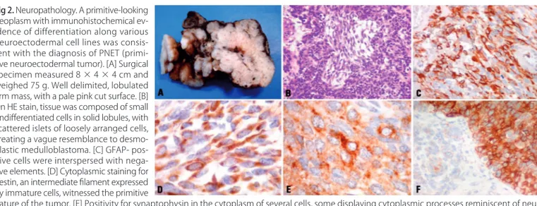
Fig 2. Neuropathology. A primitive-looking neoplasm with immunohistochemical ev-idence of differentiation along various neuroectodermal cell lines was consis-tent with the diagnosis of PNET (primi-tive neuroectodermal tumor). [A] Surgical specimen measured 8 × 4 × 4 cm and weighed 75 g. Well delimited, lobulated irm mass, with a pale pink cut surface. [B] On HE stain, tissue was composed of small undiferentiated cells in solid lobules, with scattered islets of loosely arranged cells, creating a vague resemblance to desmo-plastic medulloblastoma. [C] GFAP- pos-itive cells were interspersed with nega-tive elements. [D] Cytoplasmic staining for nestin, an intermediate ilament expressed by immature cells, witnessed the primitive