746
IMAGES IN NEUROLOGY
Ganglioglioma with anaplastic transformation
Ganglioglioma com transformação anaplásica
Fabiano Reis
1, Guilherme Henrique Alves Vieira
2, Ricardo Schwingel
2, Vinicius Trindade Gonçalves
2,
Luciano de Souza Queiroz
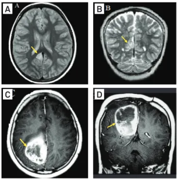
3A nine year-old male had refractory seizures for four
years. Diagnostic imaging (Fig 1A and B) demonstrated a
cor-tico-subcortical focal lesion at the isthmus of the right
cingu-late gyrus. Biopsy yielded diagnosis of ganglioglioma (Fig 2A).
Reoperation one year later showed similar features, but
in-cluded atypical and multinucleated cells (Fig 2B).
At the age of 13, the patient had symptom recurrence.
Diagnostic imaging (Fig 1C and D) demonstrated a
hetero-geneous enhanced lesion in the right parietal region, with
ne-crosis. Histology (Fig 2C to F) revealed a malignant glial
tu-mor with the appearance of glioblastoma multiforme. A few
tumor cells were positive for chromogranin. A diagnosis of
malignant transformation of ganglioglioma was made.
Gangliogliomas are rare tumors predominating in the
ear-ly decades of life, with strong association with long term
intrac-table epilepsy
1-3. hey are composed of variable proportions of
glial (mainly astrocytic) cells and mature or dysplastic neurons
2.
Malignant change is a rare, but well recognized, complication.
Transformation of the glial component from the low grade to a
higher grade is observed in most cases. Also, there is a case of
ma-lignant transformation secondary to degeneration of the
neuro-nal component into a neuroblastoma
4. Some reports in literature
5suggest that radiation may predispose to malignant degeneration.
In this case, the patient did not receive postoperative radiation.
1Professor of the Department of Radiology of the Clinics Hospital, Faculty of Medical Sciences, Universidade Estadual de Campinas (Unicamp), Campinas SP, Brazil. 2Medical student; Department of Radiology of the Clinics Hospital, Faculty of Medical Sciences, Unicamp, Campinas SP, Brazil.
3Professor of the Department of Pathology of the Clinics Hospital, Faculty of Medical Sciences, Unicamp, Campinas SP, Brazil.
Correspondence: Fabiano Reis; Departamento de Radiologia, Faculdade de Ciências Médicas, Unicamp; Rua Tessália Vieira de Camargo 126; 13083-887 Campinas SP - Brasil; E-mail: [email protected]
Conflict of interest: There is no conflict of interest to declare.
Received 29 February 2012; Received in final form 17 May 2012; Accepted 25 May 2012
Fig 1. (A) Axial proton density: a small well circumscribed lesion
in the right cingulus; (B) Coronal T2: a small lesion in the right
cingulus, with high signal intensity in the mass. (C) and (D) Axial
and coronal contrast-enhanced, 5 years after: a large mass
in the right parietal region, with heterogeneous enhancement
and component of necrosis.
C
A
D
B
Fig 2.
Neuropathology. (A) First biopsy. Moderately cellular low grade glial tumor with scanty atypical cells and thin capillaries. No
mitotic figures or necrosis. HE x 100. Inset. Neuron in deep area of the tumor. HE x 400. (B) Second biopsy, one year later. It keeps same
features as original specimen. At center, atypical neuron with eccentric nucleus. HE x 100. Inset. Aberrant cell with four nuclei, lineage
uncertain. HE x 400. (C–F) Third biopsy. (C) Highly cellular tumor with moderate atypia. HE x 100. Inset. Atypical mitotic figure. HE x 400.
(D) Abnormal vessel with thickened walls and occlusive thrombosis. HE x 100. Necrotic areas were present nearby. (E) Tumor cells are
strongly positive for glial fibrillary acidic protein indicating astrocytic lineage. X 100. Inset. Isolated tumor cell positive for chromogranin
(suggests neuronal differentiation). X 400. (F) About 10% of nuclei were marked by Ki-67 (mib1) antibody. X 100.
747
1. Miller DC, Lang FF, Epstein FJ. Central nervous system gangliogliomas. Part I: Pathology. J Neurosurg 1993;79:859-866.
2. Lantos PL, VandenBerg SR, Kleihues P. Tumors of the nervous system. In: Graham DI, Lantos PL (Eds). Greenfield’s neuropathology. 6th ed. London, England: Arnold 1997:583-794.
3. Luyken C, Blümcke I, Fimmers R, et al. The spectrum of long-term epilepsy-associated tumors: long-term seizure and tumor outcome and neurosurgical aspects. Epilepsia 2003; 44:822-830.
4. David KM, de Sanctis S, Lewis PD, Noury AM, Edwards JM. Neuroblastomatous recurrence of ganglioglioma. Case report. J Neurosurg 2000;93:698-700.
5. Rumana CS, Valadka AB. Radiation therapy and malignant degeneration of benign supratentorial gangliogliomas. Neurosurgery 1998;42:1038-1043.