ABSTRACT
Speech therapy for compensatory articulations
and velopharyngeal function: a case report
1 2 !"3, Josiane Denardi Alves
NEVES4, Jeniffer de Cássia Rillo DUTKA5, Maria Inês PEGORARO-KROOK6
1- Graduate student, Department of Speach-Patology and Audiology, Bauru School of Dentistry, University of São Paulo, Bauru, SP, Brazil.
2- PhD, Speech-Language Pathologist, Palatal Prosthesis Department, Hospital for Rehabilitation of Craniofacial Anomalies, University of São Paulo, Bauru, SP, Brazil.
3- DDS, Palatal Prosthesis Department, Hospital for Rehabilitation of Craniofacial Anomalies, University of São Paulo, Bauru, SP, Brazil.
4- Speech-Language Pathologist, Palatal Prosthesis Department, Hospital for Rehabilitation of Craniofacial Anomalies, University of São Paulo, Bauru, SP, Brazil. 5- PhD, Communication Sciences and Disorders, Speech-Language Pathologist, Post Graduate Program at Hospital for Rehabilitation of Craniofacial Anomalies, University of São Paulo, Bauru, SP, Brazil.
6- PhD, Human Communication Disorders, Full Professor, Department of Speach-Patology and Audiology, Bauru School of Dentistry, University of São Paulo, Bauru, SP, Brazil.
Corresponding address: Melina Evangelista Whitaker - Rua Dr. Antonio Prudente, 4-152 - Jardim Estoril II - Bauru - SP - 17016-010 - Phone: (14) 3235-8096 - e-mail: [email protected]
#$%&'()(*+*08#$&&+)(*++
T
! "# $ % procedure (pharyngoplasty). The program of intensive speech therapy involved 3 phases, &' &*+ + of compensatory articulations. Evaluation before the program indicated the use of ! & !$ ' and hypernasality, all compromising speech intelligibility. To address place of articulation, $ '$0 % "- $ "1 $ the objective of achieving velopharyngeal closure during speech consistently. After the $ "
Key Words: Cleft palate. Children. Speech bulb. Speech therapy.
INTRODUCTION
Among the areas of Dentistry, Odontopediatrics, Orthodontics and Maxillofacial prosthesis are closely "# and his partner, the prosthetic technician, are $ during treatment of oncologic and neurologic cases,
anomalies like cleft palate.
$7$ such as the velopharyngeal dysfunction (VPD), feeding disorders.
V P D i s c h a r a c t e r i z e d b y i n a d e q u a t e during production of oral speech sounds and during sucking and deglutition, also affecting middle ear function. Particularly in relation to speech, signs of VPD can include hypernasality, nasal air escape, ' production, and use of atypical place of production (Compensatory Articulation – CA)2,3,6.
91 (for example, an absence of musculus uvulae or a short or hypoplastic palate) or to an increase in nasopharyngeal space (for example, a deep ;!$ ' !$ 7 procedure (surgical or prosthetic) to establish 7 velopharyngeal functioning. VPD can also be the result of velopharyngeal incompetency. In this < 7 $ $ functioning does not occur. Incompetency can be the result of neuro-sensory-motor alterations (paralysis or paresis) or even the result of incorrect learning of velopharyngeal functioning for speech production. Finally, VPD can be caused by an "
Treatment of insufficiency may involve a secondary surgical procedure or a pharyngeal obturator. The obturator is made by the dentist !$ an intermediate portion (connecting the anterior portion to the bulb) and a pharyngeal bulb (the portion shaped to occupy the velopharyngeal gap)1. When functional, the bulb favors the correction of VPI related obligatory speech errors. Sometimes physical correction alone is not enough and speech therapy is needed to establishing adequate velopharyngeal functioning during speech production. The combination of behavioral and resulting from mislearning. It is also an excellent alternative to optimize a future surgical correction 7. That is, once velopharyngeal
$ the speech pathologist can use behavioral strategies functioning of the velopharynx improving excursion recomendation. This intervention requires inter
' dentistry and speech pathology. It may be the only due to the presence of incompetency resulting from learned velopharyngeal inadequacy, such as hypodynamic velopharynx.
OBJECTIVE
# process of intensive speech therapy for a 6 year ! and a history of cleft lip and palate.
MATERIAL AND METHODS
Participant
# presented classic signs of VPI after primary palatal surgery. The girl started treatment at a craniofacial center receiving a primary lip procedure at 6 months > 7 ? @' 7" 1 ' $ & G months old.
During the evaluation performed before the "> $ interventions. At 6 years of age, a nasoendoscopic ; incompetency, revealing a large gap associated limited velar elevation characterizing a condition
';14. With a bad
$ the combined behavioral and prosthetic treatment "
Assessment procedures
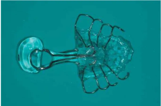
Figure 1- Pharyngeal obturator (speech bulb)
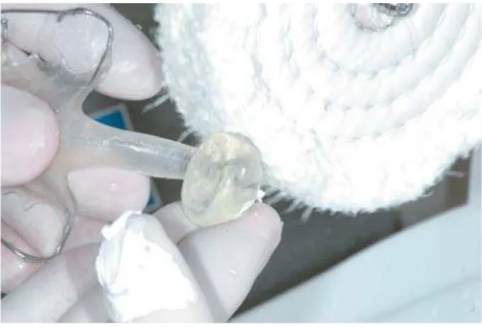
Figure 2- Intraoral speech bulb The auditory perceptual assessment of speech
for identification of presence or absence of " - $ pathologist identifying presence or absence of the use of compensatory articulation and phonological errors not expected for the child’s age. Using nasometric assessment, a speech pathologist calculated the nasalance score to quantify the relative amount of nasal acoustic energy during oral speech production. A maximum nasalance score &JK ; speech samples in Brazilian Portuguese language. &JK of hypernasality12.
# 7 !$ by Warren and Dubois13 (1964) using a PERCI-SARS computerized system. The minimum cross-sectional ;! determined during the production of the plosive /p/ X"Y-+ Z"?
mm2 7
velopharyngeal closure, areas of 5 to 9.9 mm2 adequate velopharyngeal closure, and areas of 10
to 19.9 mm2
borderline and inadequate velopharyngeal closure, and areas of 20 mm2 as representative of inadequate velopharyngeal $ by Warren14 (1997).
N a s o e n d o s c o p i c a n d v i d e o f l u o r o s c o p i c 0 the velopharyngeal structures during speech $ the velum to elevate and ability of the pharyngeal ;" During both exams, the presence, pattern and size "
Prosthetic treatment of VPI
Once favorable dental conditions to support a $ G $ each corresponding to one visit to the rehabilitation center. Phase one involved the development of the anterior portion of the device involving an acrylic "# ! oral sounds that favor adequate velopharyngeal "# material and later acrylized and polished, respecting the gap shape and size (Figures 1 and 2).
The three phases for development of the
prosthesis took approximately 6 months, involving 3 visits of 4 days each to the center. Each visit G $ & " # \ $ initially caused gagging and discomfort. Once patient used the obturator daily and consistently, "
Behavioral treatment of VPD
- conducted at the craniofacial center, consisting of G &'$ " > *+ minutes and initially targeted the elimination of compensatory articulations.
Figure 3- Pharyngeal obturator reduction with bur – phase of fray
Figure 4- Pharyngeal obturator reduction with sandy paper
Figure 5- Pharyngeal obturator reduction with grinding paste – phase of polishing
7 $ 7 "
` " @ ; adequately produced during conversational speech.
The program involved orientations and training of the family and the child to monitor and to contrast adequate (target) and inadequate (CA) productions. With this program, all sounds trained during therapy ! " # $ the center during school vacation for periods no &' \ ' intensive therapy.
- & '$ consistent use of oral place of production for all " #$ addressing velopharyngeal (VP) functioning. Perception of oral and nasal air pressure and % $ tactile feedback involving a plastic tube for auditory %X escape” and nasometry for visual enhancement of %"_ $ closure and VP opening. Once consistent adequate the use of the pharyngeal obturator, a program for "
Speech bulb reduction program
# dentist and the speech pathologist. The prosthesis asked to shave off about 1 mm of acrylic around the surface creating a second bulb slightly smaller "#
"_ 0 $ $ " w' %" _ $ ' { than the reduced bulb.
# ;
Figure 6- Speech bulb original X Speech bulb with reduction
Assessment BEFORE Intervention AFTER Intervention
Auditory perceptual assessment of resonance Hypernasality Normal
Auditory perceptual assessment of articulation
Presence of compensatory articulation – glottal stops
Use of correct oral place of articulation
Nasometry – oral sample (27% or less = normal)
37% Indicative of hypernasality
23% Indicative of adequate
resonance
Aerodynamic assessment velopharyngeal area /papa/
29 mm2
Indicative of VPI
0.08 mm2
Indicative of borderline closure
Nasoendoscopic assessment Large VP gap
Limited movement of pharyngeal walls and velum
Adequate VP closure at the speech bulb with good displacement of pharyngeal
walls
Large VP Gap
Limited velar elevation and limited wall displacement
Touch of the velum and pharyngeal wall to the bulb
Speech intelligibility Severely compromised Normal
Figure 7- Results of speech and velopharyngeal function assessment before and after intensive therapy and bulb reduction
RESULTS
- + completed during the 3 phases of the program. # performed before and after the intensive speech therapy and bulb reduction program. As presented and described in Figure 7 all speech alterations the program (Figure 7).
DISCUSSION
The role of the primary surgeries in cases of CLP 7 to support oral functions like feeding and speech. @ *K *+K
correct cleft palate (average of 20%) still present VPD after primary repair due to insufficiency, incompetency or both. Correction of VPD can involve a physical (surgery or prosthesis) and/or behavioral treatment4,5,9,15. In some cases the management 1 ! ! requiring the use of prosthetic management. The behavioral manipulation of the velopharyngeal mechanism.
and optimizing future surgical treatment. The device can be easily individualized according to the anatomical and functional needs of the patient. The functional adaptation of a pharyngeal obturator, $ 7 ' and the patient.
If, on one hand, there are many advantages for the use of a pharyngeal obturator, on the other $ need to be considered before recommending the procedure. The treatment itself is usually planned in 3 phases, each requiring one visit to the craniofacial center one, for the construction of each portion of the palatal prosthesis. The $$ " Acceptance and understanding by the family and patient of the type of treatment and the burden it entails are essential for its success. The device area), leading to discomfort and gagging at the
beginning8"1 $
0$ the patient must be compliant during all phases of the treatment, particularly during functional requires speech production. In the present case to the treatment prescribed. Furthermore the therapy addressing the use of adequate place of sound production involved daily practice at home, requiring the compliance and commitment of the child and her family. Orientation and training of ; "
The bulb reduction program, in particular, is described in the literature as an alternative to elicit $ $ 0 a future surgical correction of a velopharyngeal ' respiratory obstruction10,16. For some patients, like the one in this case report, the presence of learning errors regarding velopharyngeal functioning can lead to unsuccessful surgical results independent of the surgical procedure used, particularly due to velopharyngeal hypodynamism. In such cases treatment is an excellent alternative to establish adequate velopharyngeal function for speech.
Final Consideration
1
to improve speech intelligibility and to optimize "
REFERENCES
1- Bzoch KR. Communicative disorders related to cleft lip and palate. Austin: Pro-Ed; 2004.
2- Golding-Kushner KJ. Therapy techniques for cleft palate speech and related disorders. San Diego: Singular-Thompson Learning; 2001.
3- Kuehn DP, Moller KT. Speech and language issues in the cleft palate population: the state of the art. Cleft Palate Craniofac J. 2000;37:1-35
4- Mazaheri M. Indications and contraindications for prosthetic speech appliances in cleft palate. Plast Reconstr Surg Transplant Bull. 1962;30:663-9.
5- Park S, Saso Y, Ito O, Tokioka K, Takato T, Kato K, et al. The " Surg. 2000;105:12-7.
6- Peterson-Falzone SJ, Hardin-Jones MA, Karnell MP. Cleft palate speech. Saint Louis: Mosby; 2001.
7- Peterson-Falzone SJ, Hardin-Jones MA, Karnell MP. " Peterson-Falzone SJ, Hardin-Jones MA, Karnell MP. Cleft palate speech. Saint Louis: Mosby; 2001. p. 162-98.
8- Pinto JHN, Dalben GS, Pegoraro-Krook MI. Speech Intelligibility prosthesis. Cleft Palate Craniof J. 2007;44:635-41.
9- Pinto JH, Pegoraro-Krook MI. Evaluation of palatal prosthesis for the treatment of velopharyngeal dysfunction. J Appl Oral Sci. 2003;11:192-7.
10- Shelton RL, Lindquist AR, Arndt WB, Elbert M, Youngstrom KA. of the pharynx and posture of tongue. Cleft Palate J. 1971;8:10-7. {{ > $ ' `$ @ w>" "w` S Paulo. 1972;27: 5-6.
12- Trindade IEK, Genaro KF, Dalston RM. Nasalance scores of normal brazilian portuguese speakers. Braz J Dysmorphol Speech Hear Disord. 1997;1:23-34.
{G 9 19$ 1 -" - 7 " Cleft Palate J. 1964;16:52-71.
14- Warren DW, Rouget AH, Hinton VA. Aerodynamics. In: McNeil M, ed. Clinical management of sensorimotor speech disorders. '#`{??J"
15- Witt PD, Marsh JL, Marty-Grames L, Muntz HR, Gay WD. Management of the hypodynamic velopharynx. Cleft Palate Craniofac J. 1995;32:179-87.