ULTRASTRUCTURAL STUDY OF THE EFFECTS
OF CYCLOSPORINE IN THE BRAINSTEM OF
WISTAR RATS SUBMITTED TO THE ETHIDIUM
BROMIDE DEMYELINATING MODEL
Eduardo Fernandes Bondan
1, Maria Anete Lallo
2, Dominguita Lühers Graça
3Abstract – The ethidium bromide-demyelinating model (EB) was used to study remyelination in the brainstem under the use of cyclosporine (CsA). Wistar rats were submitted to intracisternal injection of 0.1% EB or 0.9% saline solution, and others were taken as histologic controls (group I). Within those injected with EB, some have not received immunosuppressive treatment (II); some were treated by intraperitonial route with CsA (III.E - 10 mg/kg/day). Rats from group III.C were injected with saline solution and treated with CsA. The animals were perfused from 15 to 31 days post-injection collecting brainstem sections for light and transmission electron microscopy studies. After EB injection it was noted the presence of macrophages and non-degraded myelin debris, demyelinated axons, oligodendrocyte or Schwann cell remyelinated axons, groups of infiltrating pial cells, hypertrophic astrocytes and few lymphocytes. Tissue repair of EB-induced lesions in group III.E was similar to that of group II, but with the presence of a higher density of oligodendrocytes near remyelinating areas. KEY WORDS: central nervous system, myelin sheath, oligodendroglia, Schwann cells, cyclosporine, ethidium bromide.
Estudo ultra-estrutural dos efeitos da ciclosporina no tronco encefálico de ratos Wistar submetidos ao modelo desmielinizante do brometo de etídio
Resumo – Empregou-se o modelo desmielinizante do brometo de etídio (BE) com o objetivo de estudar a remielinização no tronco encefálico frente ao uso de ciclosporina (CsA). Foram utilizados ratos Wistar, submetidos à injeção de BE a 0,1% ou de solução salina na cisterna pontina, assim como controles histológicos (grupo I). Dos animais injetados com BE, alguns não receberam tratamento imunossupressor (II); outros foram tratados por via intraperitoneal com CsA (III.E - 10 mg/kg/dia). O grupo III.C incluiu animais injetados com salina e tratados com CsA. Os animais foram perfundidos dos 15 aos 31 dias pós-injeção, com colheita de material do tronco encefálico para estudos de microscopia de luz e eletrônica de transmissão. Após injeção de BE, foram observados macrófagos e restos de mielina não-degradada, axônios desmielinizados ou remielinizados por oligodendrócitos e por células de Schwann, grupos de células piais infiltrantes, astrócitos hipertróficos e poucos linfócitos. O processo de reparo das lesões no grupo III.E apresentou-se similar ao do grupo II, porém com maior densidade de oligodendrócitos próximos às áreas de remielinização.
PALAVRAS-CHAVE: sistema nervoso central, bainha de mielina, células de Schwann, oligodendróglia, ciclosporina, brometo de etídio.
1Professor Titular da Universidade Paulista, São Paulo SP, Brazil (UNIP) e da Universidade Cruzeiro do Sul, São Paulo SP, Brazil (UNICSUL); 2Professora Titular da UNIP e da UNICSUL; 3Professora Titular, Departamento de Patologia da Universidade Federal de Santa Maria RS, Brasil (UFSM).
Received 20 February 2008, received in inal form 17 April 2008. Accepted 5 May 2008.
Dr. Eduardo Fernandes Bondan – Rua Caconde 125 / 51 - 01425-011 São Paulo SP - Brasil. E-mail: [email protected]
Several studies on the biology of demyelination and re-myelination in the central nervous system (CNS) have been made based on the use of the gliotoxic agent ethidium bromide (EB)1-11. In this experimental model, it was noted
the presence of lymphocytes during the process of myelin loss and removal after EB injection, as well as an intense macrophagic activity in the damaged areas2,3,6. The
pres-ence of lymphocytes remains controversial, maybe just
part of the inlammatory response induced by the agent. Despite the toxic nature of this model, it can not be ruled out the possibility of some participation of these lympho-cytes as a feature of antigenic recognition or as effectors in some immune-mediated responses to the detached my-elin sheaths3. Immunosuppressive agents have been
char-Thin slices of the brainstem (pons, mesencephalon and trap-ezoid body) were collected and post-ixed in 1% osmium tetrox-ide, dehydrated with graded acetones and embedded in Araldite 502 resin, following transitional stages in acetone. Thick sections were stained with 0.25% alkaline toluidine blue. Selected areas were trimmed and thin sections were stained with 2% uranyl ac-etate and lead citrate and examined using a Philips EM-201 trans-mission electron microscope.
RESULTS
Observations on group II
(EB injection and no CsA treatment)
The examination of semithin sections from rats of group II revealed the appearance of lesions of variable extent (from the mesencephalon into the trapezoid body), but affecting mostly the ventral surface of the pons, and allowed the establishment of 2 areas with very distinct morphological characteristics. The center of the lesion presented an extended extracellular space, with many foamy macrophages, demyelinated axons and some my-elin debris (Fig 1). At the periphery, phagocytic cells were cellular processes involved in CNS repair after local EB
injection under an attempt of pharmacological interfer-ence with cyclosporine (CsA) in the rat brainstem remy-elinating process.
METHOD
This experiment was approved by the Ethics Comission of the Universidade Paulista (UNIP). Thirty male Wistar rats, 4 to 6 months old, were used. They were divided into 4 groups - I (n=2), including animals taken as histologic controls; II (n=8), constituted of ani-mals injected with EB into the cisterna pontis; III.E (n=16), aniani-mals equally injected with EB, but treated with CsA; and III.C (n=4), in-cluding animals injected with saline solution and treated with CsA. The rats were anaesthetized with ketamine and xylazine (5:1, 0.1 mL/100g) and a burr hole was made on the right side of the skull, 8 mm rostral to the fronto-parietal suture. Injections were performed freehand using a Hamilton syringe, itted with a 35º angled polished gauge needle into the cisterna pontis, an en-larged subarachnoid space below the ventral surface of the pons. Ten microliters of 0.1 EB solution were injected into the cis-terna pontis of rats from groups II and III.E and the same volume of 0.9% saline solution was injected in rats from group III.C.
less conspicuous and thinly remyelinated axons could be seen, some clearly associated with Schwann cells.
Ultrastructural analysis of the central area revealed the appearance of macrophages containing myelin in dif-ferent stages of degradation, from loose lamellae to the accumulation of fat neutral droplets in the cytoplasm. In the vicinity of these macrophages, there were many de-myelinated axons, some with clear signs of degeneration, as well as concentrations of myelin-derived membranes in the extracellular space of some areas.
At peripheral sites, it was found groups of demyelinat-ed axons in association with astrocytic processes and ax-ons exhibiting thin myelin sheaths of oligodendrocyte or-igin. Lymphocytes were commonly observed in perivas-cular locations and all over the neuropil. Iniltrating pial cells were also found as monotypical nests or as diffuse
arrangements joined by desmossome-like juncions. Around blood vessels and in areas of expanded extra-cellular space, where the glia limitans was disrupted and astrocytic processes had disappeared, Schwann cells were seen related to one or more naked axons or at initial re-myelinating stages.
Axons related to Schwann cells presented faster re-myelination, with thicker myelin sheaths than those pro-duced by oligodendrocytes at the same period, although there was an evident preponderance of oligodendroglial remyelinated axons. Naked axons either clumped or sep-arated by astrocytic prolongations were also noted.
Observations on group III.E (EB injection and CsA treatment)
By 15-17 days after EB injection, light microscopy
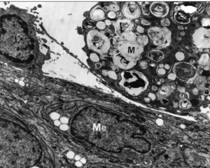
re-Fig 3. A lymphocyte (L) and a macrophage (M) contacting a group of iniltrating pial cells (Me). Lesion of 15 days - Group III.E. Electron micrograph - 7.450x.
vealed lesions similar to those in group II and also present-ing naked axons, macrophages and few myelin debris in the extracellular space of the central area. At the periphery, some dark cells of dificult identiication by light microsco-py could be noticed, as well as pial iniltration, mostly peri-vascular, demyelinated ibers and thinly remyelinated axons. Ultrastructural examination showed as the most prom-inent aspect of the lesion (in relation to animals from group II) the high density of round cells identiied as oli-godendrocytes, close to small remyelinated axons and/or astrocytic processes and illed with long cisternae of en-doplasmic reticulum and evident Golgi apparatus.
Other common peripheral features included areas of oligodendrocyte-remyelinated axons, the presence of hypertroic astrocytic prolongations and of perivascular Schwann cells, associated with one or more naked
axo-ns and even forming redundant myelin loops (Fig 2). In addition, lymphocytes were seen around blood vessels and in the neuropil contacting macrophages, myelin de-bris and agglomerates of pial cells (Fig 3). Demyelinated axons, associated or not with astrocytic prolongations, were also found.
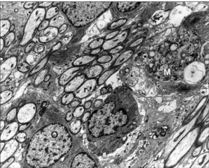
By 21-31 days post-injection, there was a massive pre-dominance of oligodendroglial remyelination at peripher-al sites in rats that received CsA. Oligodendrocytes contin-ued to show a prominent endoplasmic reticulum and they appeared in groups or nests or were separated by astro-cytic processes and axons with slim myelin sheaths (Fig 4). Thin and thick astrocyte prolongations were associ-ated to remyelinassoci-ated ibers, oligodendrocytes and blood vessels. However, some blood vessels remained unrelat-ed to astrocytic prolongations. In addition lymphocytes
Fig 4. Oligodendrocytes (O) at the periphery of the lesion close to groups of remyelinating axons (r). Note the pres-ence of a macrophage (M) containing myelin in different de-grees of breakdown. Lesion of 21 days - Group III.E. Electron micrograph - 4.875x.
were observed in fewer numbers than those found at 15-17 days and they appeared frequently contacting astro-cytes and lamellae of vesiculated myelin.
Many axons persisted demyelinated and some of them were already related to Schwann cells individually or in groups. Thicker myelin sheaths than those produced by oligodendrocytes were deposited by Schwann cells (Fig 5), mainly in perivascular areas, illed with collagen ibers and sometimes close to the nests of pial cells.
Observations on group III.C
(saline solution injection and CsA treatment)
From the 4 rats injected with saline solution, just 2 (those perfused at 15 and 21 days) presented a focal le-sion due to injection procedure. This lele-sion was circum-scribed to the pons and showed a light expansion of the extracellular space, containing some loose myelin lamel-lae and clearing of cellular debris by phagocytic cells, but with no evidence of either primary demyelination or loss of the neuroglia (Fig 6).
No mortality was recorded in the groups that received CsA treatment using the drug administration scheme pre-viously mentioned.
DISCUSSION
In general terms, the results obtained in the present in-vestigation conimed and complemented those described in former studies with the EB model in the brainstem5-11.
The most important feature observed in rats injected with EB and treated with CsA was the higher density of oligodendrocytes on the edges of the lesions, in relation to the immunocompetent animals from group II and those treated with cyclophosphamide5 or dexamethasone6 from
previous studies.
CsA has as its major known effect the capacity of re-ducing the synthesis and liberation of interleukin-2 (IL-2) from T CD4+ lymphocytes (Th - helper)12, with consequent
suppression in the generation of T cytotoxic lymphocytes (CD8+), although relatively sparing the suppressor ones and
even the cytotoxic lymphocytes already formed13. Other
components of the immune system, such as monocytes, macrophages, NK (natural killer) cells and B lymphocytes, can be equally affected, but by an indirect manner, due to the reduction of IL-2 and other cytokines, like GM-CSF, IL-3, IL-4 and IFN-g, among others14.
IL-2 may be present in the CNS in pathological states following disruption of the blood-brain barrier (BBB) and iniltration of activated T cells in this site, more precisely of those producing the referred cytokine such as Th1 cells and T cytotoxic cells15,16.
Injection of IL-2 in the rat brainstem induces sleep, which seems to be mediated by receptors in the locus coeruleus. In addition IL-2 mRNA was localized in some areas of the murine encephalon, being found in neuronal bodies and astrocytes17.
In the CNS there are no evidences that IL-2 may modu-late the function or gene expression of any other glial cell excepting oligodendrocytes and their progenitors15,16.
Human recombinant IL-2 appears to inluence prolif-eration and differentiation of rat oligodendrocytes in vi-tro, increasing as far as in 3 times their numbers in cultures containing IL-2 and stimulating their maturation, as shown by the augmented expression of myelin basic protein (MBP) and of MBP mRNA15,16. Additionally two lineages
of human glioblastoma with oligodendroglial phenotype proliferate in the presence of IL-2 and also presented re-ceptors for this cytokine15,16.
to cross the BBB decreases the number of these patrolling cells in normal CNS, as just a minority of T circulating cells are in state of activation19.
Lymphocytes are present in EB-induced lesions, many times contacting myelin debris in the extracellular space and activated macrophages containing phagocytosed my-elin, in a relationship suggestive of antigenic recognition3.
In the CsA-treated group, the number of lymphocytes did not differ from that found in non-immunosuppressed ani-mals, which may indicate that the CsA dosage used in this investigation was not suficient to cause adequate immu-nosuppression or maybe that lymphocytes present in these lesions belong to a CsA-unresponsive lymphocytic subpop-ulation. Besides, lymphocytes recruited to the CNS might escape from the immunosuppressive action of the drug, as, despite its high lipossolubility, Csa paradoxically seems to not pass through intact BBB in signiicant amounts20.
The possibility of insuicient CsA plasmatic levels to cause immune depression in the CNS can be refuted by the fact that similar administration schemes have been widely used to avoid rejection of mice cerebral grafts in rat CNS21 and of glial xenografts in myelin deicient rats22.
Mechanical damage to the BBB is inevitable when mi-croinjections are applied in the nervous tissue and, thus, it is believed that, in the present investigation, BBB disrup-tion occurred as a result of injecdisrup-tion trauma and of astro-cytic disappearance induced by EB gliotoxic action. Astro-cytes are recognized to play a key role in the induction and maintenance of BBB characteristics in the CNS23.
Blood vessels devoided of nearby astrocytic prolonga-tions were seen until the 31st day after EB injection,
sug-gesting the lack of a completely developed BBB, as it is known that astrocyte processes must be in proximity to CNS endothelial cells for the expression of BBB tight junc-tions23. Although tight junctions could be easily visualized
from the 15th to the 31st day post-injection, it is
worth-while mentioning that simple morphological observation of these junctions is not suficiently conclusive to allow functional approaches on the permeability of CNS blood
incidence and severity of clinical signs in animals already sensitized, with usual recurrence of symptoms after the end of treatment12. In Theiler`s murine encephalomyelitis,
the same drug, given during the infection period, was re-sponsible for the suppression of inlammation and demye-lination, although it did not decrease myelin loss if admin-istered after the establishment of inlammatory reaction26.
Even during inlammation, the total number of lym-phocytes in the CNS is comparatively small in relation to other body sites. So the amount of cells capable of reacting to a given antigen is proportionally reduced and lymphocytic response is restricted to a relatively small number of clones (oligoclonal response)27.
In the present study, there was no evidence that im-munosuppression with CsA was prejudicial to macrophag-ic activity, because, on the contrary to that observed with the use of cyclophosphamide5 or dexamethasone6, it was
seen much smaller amounts of myelin-derived membranes using CsA, in a pattern similar to group II.
Even though it was not seen a remarkable difference in the extension of oligodendrocyte remyelinated areas be-tween animals from groups II and III.E, those treated with CsA presented a greater proportion of oligodendroglial cells on the edges of the lesions. These oligodendrocytes showed a cytoplasmic density that had resemblance with the appearance of cells referred by Mori and Leblond28 as
medium and dark oligodendrocytes. These authors attrib-uted the notable morphological variation on oligoden-droglia of young rats to different stages of cellular differ-entiation. The light ones would represent those of bigger size and greater mitotic activity; as maturation occurred they were progressively transforming themselves into smaller and darker cells with lesser proliferative capacity28.
Nests of oligodendrocytes similar to those observed in our study were seen in attempts of remyelination in MS29 and EAE30. Such distribution is not considered a
In the EB model, it was described a cell population with membrane immunoreactivity to ganglioside D3 (GD3) from
6 to 12 days after the gliotoxic injection. These cells were considered as probable oligodendroglial progenitor cells that came out in response to the demyelinating process9.
A possible explanation for the high density of oligo-dendrocytes in rats submitted to CsA treatment could be the inhibition of IL-2 secretion induced by the drug. This requires that lymphocytes in the EB-induced lesions had the capacity of secreting the cytokine (such as the Th1 subpopulation) and were activated by antigenic stimula-tion. Since the referred cytokine inhibits in vitro prolifera-tion of oligodendroglial progenitor cells of rats15,16,18, it is
possible that depletion of IL-2 in the lesion site facilitates division and migration of such cells. Paradoxically, mature oligodendrocytes are capable of accelerating their capac-ity of myelination or remyelination in the presence of IL-215,16. This would be beneicial in demyelinating diseases,
although it is not observed in immune-mediated condi-tions characterized by abundant lymphocytic iniltrates, specially of Th1 cells, as it is seen in MS and EAE16. It is
important to note that studies connecting IL-2 and oli-godendroglial cells are commonly performed in vitro and the environment in vivo is much more complex and un-predictable due to the intricate interactions between the different cell types involved and their secreted factors.
CsA could affect directly or indirectly the inal balance between proliferative and antiproliferative factors related to oligodendroglial populations in the lesion microenvi-ronment, maybe facilitating the irst ones and by doing this resulting in greater numbers of oligodendrocytes.
Oligodendrocyte proliferation has been described in adult animals28, as well as from bipotential progenitor cells
(O-2Aadult cells). The latter have the capacity of presenting
division, differentiation and migration under appropriate stimulus therefore creating the necessary oligodendro-cytes for myelin repair following a demyelinating event8.
On the other hand Schwann cells kept a similar behaviour and distribution on groups II and III.E. However, comparing to group II, it was more common the appearance of redun-dant myelin loops with the CsA treatment (already seen at 15 days), suggesting that somehow CsA could have some posi-tive effect on the remyelinating activity of Schwann cells. Although the use of CsA in this study changed the dy-namics of the nervous tissue repair following EB injection, it is not possible to assume that these alterations were directly due to the suppression of lymphocytic activity.
REFERENCES
1. Blakemore WF. Ethidium bromide induced demyelination in the spi-nal cord of the cat. Neuropathol Appl Neurobiol 1982;8:365-375. 2. Gevehr CG, Graça DL, Pereira LAVD. Desmielinização e remielinização
após múltiplas injeções intramedulares de brometo de etídio em ratos Wistar. Arq Neuropsiquiatr 1997;55:189-202.
3. Graça DL. The presence of lymphocytes in a toxically induced demy-elinating process of the central nervous system. Micr Electr y Biol Cel 1988;12:17-22.
4. Graça DL, Blakemore, WF. Delayed remyelination in the rat spinal cord following ethidium bromide injection. Neuropathol Appl Neurobiol 1986;12:593-605.
5. Bondan EF, Lallo MA, Sinhorini IL, Pereira, LAVD, Graça DL. The ef-fect of cyclophosphamide on the rat brainstem remyelination follow-ing local ethidium bromide injection in Wistar rats. J Submicrosc Cytol Pathol 2000;32:603-612.
6. Bondan EF, Lallo MA, Baz, EI, Sinhorini IL, Graça DL. Estudo ultra-es-trutural do processo remielinizante pós-injeção de brometo de etídio no tronco encefálico de ratos imunossuprimidos com dexametasona 2003. Arq Neuropsiquatr 2003;62:131-138.
7. Bondan EF, Lallo MA, Trigueiro AH, Ribeiro CP, Sinhorini IL, Graça DL. Delayed Schwann cell and oligodendrocyte remyelination after ethid-ium bromide injection in the brainstem of Wistar rats submitted to strep-tozotocin diabetogenic treatment. Braz J Med Biol Res 2006;39:637-646.
8. Pereira, LAVD, Dertkigill MS, Graça DL, Cruz-Höfling MA. Dynam -ics of remyelination in the brain of adult rats after exposure to ethid-ium bromide. J Submicrosc Cytol Pathol 1998;30:341-348.
9. Reynolds R, Wilkin GP. Cellular reaction to an acute demyelinating /
remyelinating lesion of the rat brain stem: localization of GD3
ganglio-side immunoreactivity. J Neurosci Res 1993;36:405-422.
10. Yajima K, Suzuki K Demyelination and remyelination in the rat cen-tral nervous system following ethidium bromide injection. Lab Invest 1979a;41:385-392.
11. Yajima K, Suzuki K Ultrastructural changes in oligodendroglia and my-elin sheaths induced by ethidium bromide. Neuropathol Appl Neuro-biol 1979b;5:49-62.
12. Borel JF. Cyclosporine. In: Dale MM, Foreman JC. Textbook of immu-nopharmacology. 2. ed. Oxford: Blackwell, 1989: 300-311.
13. Kupiec-Weglinski JW, Filho MA, Strom TB, Tilney NL. Sparing of suppressor cells: a critical action of cyclosporine. Transplantation 1984;38:97-101.
14. Nussller AK, Thomson AW. Immunomodulatory agents in the labora-tory and clinic. Parasitology 1992;105:5-23.
15. Benveniste EN. Cytokines: Inluence on glial cell gene expression and
function. In Blalock JE. Neuroimmunoendocrinology. 2. ed. Basel: Karg-er, 1992:106-153.
16. Benveniste EN. Role of cytokines in multiple sclerosis, autoimmune en-cephalitis, and other neurological disorders. In Aggarwall BB, Puri RK. Human cytokines: their role in disease and therapy. Cambridge: Black-well, 1995:195-215.
17. Fabry Z, Raine CS, Hart MN. Nervous tissue as an immune compart-ment: the dialect of immune response in the CNS. Immunol Today 1994;15:218-224.
18. Saneto RP, Altman A, Knobler RL, Johnson HM, De Vellis J. Interleu-kin 2 mediates the inhibition of oligodendrocyte progenitor cell prolif-eration in vitro. Proc Nat Acad Sci 1986;83:9221-9225.
19. Wekerle H, Linington C, Lassmann H, Meyermann R. Cellular immune reactivity within the CNS. TINS 1986;9:271-276.
20. Fazakerley JF, Webb HE. Cyclosporin, blood-brain barrier, and multi-ple sclerosis. Lancet 1985;2:889-890.
21. Inoue H, Kohsaka S, Yoshida K, Ohtani M, Toya S, Tsukada Y. Cyclo-sporin A enhances the survivability of mouse cerebral grafted into the third ventricle of rat brain. Neurosci Letters 1985;54:85-90.
22. Rosenbluth J, Liu Z, Guo D, Schiff R. Myelin formation by mouse glia in
my-elin-deicient rats treated with cyclosporine. J Neurocytol 1993;22:967-977.
23. Risau W. Induction of blood-brain barrier endothelial cell differentia-tion. Ann NY Acad Sci 1991;633:405-419.
24. Felts PA, Smith KJ, Tilt E. Blood-brain barrier function in central de-myelinating lesions repaired by Schwann cell remyelination. Ann NY Acad Sci 1991;633:615-616.
25. Poser CM. Pathogenesis of multiple sclerosis. A critical reappraisal. Acta Neuropathologica 1986;71:1-10.
26. Rodriguez M, Quddus J. Effect of cyclosporin A, silica quartz dust and macrophage protease inhibitors on virus induced demyelination. J Neu-roimmunol 1986;13:159-174.
27. Leibowitz S, Hughes RAC. Immunology of the nervous system. Lon-don: Edward Arnold, 1983.
28. Mori S, Leblond CP. Electron microscopic identiication of three classes
of oligodendrocytes and a preliminary study of their proliferative activ-ity in the corpus callosum of young rats. J Comp Neurol 1970;139:1-30. 29. Raine CS, Scheinberg L, Waltz JM. Multiple sclerosis. Oligodendrocyte
survival and proliferation in an active established lesion. Lab Invest 1981;45:534-546.