S
USCEPTIBILITY OF THE
CEBUS APELLA MONKEY TO DIFFERENT
STRAINS OF r CWZLiFTER SINGLE
OR REPEATED INOCULATIONS
A. FaZasca,2 D. Grdna, 3 J. BUCCOZO,~ M. Gidi, 5 A. Merlo, 6 J. Zoppi, 7 aad E. Mareso
I
NTRODUC’I’ION
Since 1909, when the find- ings of Carlos Chagas (a physician of the Brazilian Health Service and an investi- gator at the Oswald0 Cruz Institute) be- came known (I), Chagas’ disease has been reported in every country of Central and South America.
In Argentina, two physicians (Maggio and Rosenbusch- la) reported in 1914 that although numerous TV- panosoma cruzz’ could be observed in the vector Tritoma infestam existing in 10 Argentine provinces, no associated dis- ease could be detected in humans. Later another Argentine (Mazza-2) contin- ued Chagas’ work and confirmed Ameri- can trypanosomiasis to be a serious en-
’ The investigation reported here received support from the UNDPlWorId B~&WHO Special Program for Re- search and Training in Tropical Diseases, and from El Salvador University in Buenos Aires, Argentina. This article wEI also be published in Spanish in the Bo/eth de la Ojkina Samtank Panamericana, 1987. 2 Director, Latin American Medical Research Institute of
El Salvador University, Medical School, El Salvador University, Buenos Aires, Argentina. Address: IL- AIMUS: Facultad de Medicina. Universidad de1 SaIva- dor, Tucuman 1859 (1050). Buenos Aires, Argentina. 3 Veterinarian, Latin American Medical Research Insti-
tute.
4 Radiologist, Latin American Medical Research Insti- tute.
demic pathology. He worked at the Misi6n de Estudios de Patologia Regional in Jujuy from 1929 to 1946, where he re- corded some 1,400 cases and performed about a hundred necropsies of acute and chronic cases.
Chagas’ disease is now widely recognized as one of Latin America’s most important public health problems. It is estimated that at present over 65 million people live in endemic areas where the risk of infection exists, and that at least 20 million people living in both rural and urban parts of Latin America are infected @a). The number of infected people in Argentina has been estimated at about three million (3).
) Cardiologist, Latin American Medical Research Insti- 1
tute.
6 Biochemist, Latin American Medical Research Insti- s
tute. %
’ Pathologist. Latin American Medical Research Insti- .,$
tute. 9,
8 Pathologist, Latin American Medical Research Insti- a x
tute.
2
3
The acute phase of the infec- tion occurs most often in childhood and is generally mild, passing unnoticed in about 85 % of the cases (4). It is esti- mated that only some 5 % of all the acute infections are actually diagnosed; of the resulting acute disease cases, past work has classified 75 % as mild, 18 % as mod- erately severe, and 6% as severe (3). As a consequence, the disease poses a far greater threat to adults, as may be in- ferred from the high morbidity and mor- tality rates found among chronically infected patients (5). Furthermore, che- motherapeutic treatments now available for combating the disease, especially in the chronic phase, are far from satisfactory.
P
AST ANIMAL
EXPERIMENTS
Most of the animals infected experimentally to date have been mice (G), rabbits (7)) or dogs (8), all of which are very susceptible to ir: crmzi. However, observations about the infections in these hosts are generally limited to the acute phase of the disease, because of the high mortality produced by the parasite. In general, what chronic infection has been observed in these animals has shown an erratic course involving slight disturbances.
g ch findings have indicated that autoim- In addition, other authors’ Y
- munity probably plays an important role 3
c
in the pathogenesis of the human cardiac and gastrointestinal lesions associated
.g with chronic Chagas’ disease. These
P)
P findings suggest that development of an :
118
immunoprophylactic method, if it were possible, would require further investi- gation , using an experimental animal model capable of developing clinical manifestations of the chronic human dis- ease (9).
Dorland (10) studied the viru- lence of T. CWZ’ strains isolated in Texas and California for monkeys by inoculat- ing the feces of naturally infected bugs through the animals’ ocular conjunctiva. All of the monkeys developed the infec- tion and some showed Romana’s sign. Histopathologic studies demonstrated a slight diffuse chronic myocarditis.
Torres and Tavares (11) stud- ied the course of the infection in Cebtis monkeys inoculated several times, using different routes and inocula. The ani- mals, which died between 95 and 243 days after infection, presented a slight diffuse chronic myocarditis with marked individual variations. This made it diffi- cult to evaluate the results, even though more pronounced lesions were observed in the animals that had been reinocu- lated over short periods of time.
Other researchers (12, 13) have infected rhesus monkeys with T. crmzz’and have followed the infections for long periods. Megaesophagus has been reported at least twice in these primates. However, this species is not native to the areas endemic for Chagas’ disease, and it is very expensive and difficult to obtain.
Program has been the development and evaluation of such animal models for re- producible experiments. This work is es- sential for the improvement of our knowledge about Chagas’ disease patho-
genesis and immunopathology, espe-
cially for discerning the mechanism responsible for chronic disease manifes- tations, evaluating the chemotherapeutic effects of drugs, and developing a safe and effective vaccine.
M
ATERIALS
AND METHODS
The work being reported here was directed primarily at making the New World nonhuman primate Cebzcs upeZZa serve as an experimental model for chronic chagasic pathology in man. Our aim was to come as close as possible to establishing an ideal animal model-an ideal model being defmed as one that would do the following:
1) support a long-lasting sub-
clinical parasitemia detectable by xenodiag- nosis and/or hemoculture as well as by con- ventional serology;
2) present cellular and/or hu-
moral immune reactions;
3) develop the cardiac and di- gestive forms of the disease with typical histo- pathologic lesions;
4) survive the acute phase of the infection;
5) display lesions in a relatively short period of time;
6) develop the disease in a man- ner pretty much independent of the age and sex of the particular infected animal in- volved;
7) utilize animals native to the endemic area and easy to obtain; and
8) be available at a reasonable cost.
Specifically, we attempted to induce experimental chagasic infections
in G&J ape& monkeys by means of single or repeated inoculations with dif- ferent strains of ?Y crmzi (using two dif- ferent inoculation routes and different numbers of parasites in the inocula) in order to determine the susceptibility of this species to infection with lY c~.G.
Forty-eight Cebzls upeZZu
monkeys (40 adult males, six mostly ju- venile males, and two juvenile females) that had normal hematologic and sero- logic enzyme parameters (14), were free of chagasic infection (as determined by serology and xenodiagnosis), and were also free of ECG pathology, were selected from an outdoor colony. (This colony, sit- uated in Escobar about 30 kilometers from Buenos Aires and supported by the uNDp/ World Bank/WHO tropical disease research program, maintains a breeding population of about 200 Cebus mon- keys.) The 48 animals were placed in an indoor colony in individual cages, with water udZibitzcm and food provided ac- cording to a standard pellet diet (25 % protein, 290 calories/ 100 g) prepared by Cargill (Buenos Aires, Argentina) and supplemented with fresh fruit.
Temperature, humidity, and light conditions were provided that were adequate for the needs of the experi- ment. Periodic and orderly control of the monkeys’ behavior, body weight, and in- testinal parasites found in feces was car- ried out in the cages.
10 years (the average estimated age being eight years), and their weights ranged from 2,110 to 3,320 g.
Parasite Strains and Inoculations
The animals were divided into four different groups-one control group and three groups inoculated with three different 1: cru.zi strains (Table 1). The control group consisted of 30 adults; another group, which received i’: crzlzi strain “CA1 ,” was composed of 10 adults; the third group, which received the “Co- lombian” strain until May 1984, in- cluded four juveniles (two males and two females); and the fourth group, which received the “Tulahuen” strain, was made up of four young males. All of these ‘I: C~ZLZZ’ strains had been main- tained in the laboratory by periodic pas- sage through Swiss mice.
The 10 adult monkeys receiv- ing the CA1 strain were inoculated with the parasite’s metacyclic forms by the conjunctival route. Three received one inoculation of 4 x lo4 parasites; four re- ceived one inoculation of 1 x 10” para- sites; and the last three received two in- oculations, the first of 4 x lo* parasites
and the second of 1 x 10’ parasites one year later.
The two other inoculated groups (receiving the Colombian and Tu- lahuen strains) were repeatedly inocu- lated with 3 x 10” blood forms of the parasite by the intraperitoneal route-a route intended to produce better absorp- tion of the parasite. The four young males receiving the Tulahuen strain were inoculated 10 or 11 times at intervals ranging from a few days to 30 weeks (see Figure 3), and the four juveniles inocu- lated with the Colombian strain were in- oculated 18 or 19 times at intervals rang- ing from three to 24 weeks (see Figure 2). The periodic reinoculations were per- formed in order to approximate condi- tions found by people living in endemic areas, where the periods of natural rein- fection vary.
TABLE 1. Basic characteristics of the Cebus ape//a monkeys inoculated with the cnl , Colombian, and Tulahuen r cruzi
strains and of the uninoculated controls. Data on the inoculations administered are shown below.
Number of animals Sex
Estimated age at first inoculation (years) Weight (g)
Date of first inoculation Number of inoculations
as of 6/84
Number of % cruzi administered (per inoculation)
Route of inoculation
7: cruzi strains inoculated
Controls CA1 Colombian Tulahuen (no inoculations)
IO 4 4 30
male 2 male, 2 female male male
6-10 3.3 I,2 4-5 5-9
2,110-3,320 940-l ,800 1,660-i ,950 2,250-2,570 6/80 to 7/81 9182 to lo/82 11/82 to 12182 -
1 to 2 18 to 19 10 to 11
Subsequent Monitoring and
Testing
Patent parasitemia, serologic changes, electrocardiographic and echo- cardiographic alterations, and cardiac or gastrointestinal disorders detectable by radiology were monitored as follows:
Parasitology. The parasitemia was mon- itored in each monkey, initially by direct observation of the T. cm.zi parasite via the fresh-drop or Strout test (IS). This was to be done three times a week after the first inoculation, before each reinoc- ulation, and at the end of the first and each subsequent month. When the di- rect testing yielded negative results, each negative animal was tested by xenodiag- nosis using 40 third-instar nymphs of Triatoma infestam. In this case, each ani- mal was exposed to the bugs for 30 min- utes every three months. A pool of feces from each group of bugs was then exam- ined for 1: cmczi at 30 and 60 days after the bugs’ exposure.
Serology. Sera were collected from all of the monkeys (including the controls) at regular intervals. Each animal was bled every seven days for the first month after inoculation or initiation of the experi- ment. Thereafter, each animal was bled twice a month.
The collected blood was al- lowed to clot at room temperature, se- rum was withdrawn, and each collected serum was divided into small samples and stored at - 70” C. All of the col- lected sera were tested for antibodies against T. cTzczz’ by indirect hemaggluti-
nation (Cellognost-Chagas, Behring-
werke) and ELBA (16). These tests were performed weekly during the first month and monthly thereafter.
Hematology. Each monkey’s hemato- crit, hemoglobin level, white blood cell
count, and red blood cell count were de- termined monthly with a Clay Adams Hematology Analyzer HA-5 Counter.
Serum enzymes and proteins. The ac- tivity in plasma of various enzymes (GOT,
GPT,LDH, alpha HBDH, andgamma GT)
was assessed by means of Boehringer ki- netic tests. This was done once a week during the acute and subacute phases of the infection, and twice a month thereaf- ter during the first year. The level of plasma proteins was assessed monthly.
Electrocardiography and echocardi- ography. ECGS were recorded every week for the three first months after inocula- tion and twice a month thereafter. For this purpose the animals were given 10 mg/kg of ketamine hydrochloride anes- thesia (Ketalar@, Parke Davis, Buenos
Aires, Argentina). They were then
placed in a dorsal decubitus position on an adequate stretcher; and, after five minutes of rest, an ECG was recorded with a Fukuda FJC-7100 monitor at a speed of 25 mm/s.
Needle electrodes were placed in the subcutaneous cellular tissue in or- der to reduce dermic impedance. Limb electrodes were placed on the anterior portion of the forearms and on the inner portion of both legs. The precordial leads used were Vl-V2-V4 and V6, and occa- sionally V3R-the latter in order to ob- tain a better evaluation of the right cavi- ties.
The possible existence of sub- clinical conduction disturbances was in- vestigated through intravenous injection of 1.0 mg/kg of Ajmalin aspartate
[(17 R) Ajmalan-17, 21-diol aspartate;
Craveri, Buenos Aires, Argentina], a RmwoZfia derivative that depresses in- traventricular conduction.
Developments related to car- diac pathology were also studied by echocardiography; animals with altera- tions detected by this method or by ECG
were grouped together; and animals to be sacrificed for anatomopathologic studies were selected at random from this group. A Berger Cl 17 echocardiograph
(M mode), made in Argentina with a SMHZ transducer 0.5 cm in diameter, was used for this purpose.
The monkeys were prepared for this examination in the same way they were prepared for ECGS. Each ani- mal was then placed in a dorsal and left lateral decubitus position during the ex- amination .
For purposes of comparison, we considered as normal a collection of electrocardiograph and echocardiograph patterns obtained from the control group of 30 monkeys in our indoor colony (Ta- bles 2 and 7).
Radiology. In order to evaluate the car- diac and gastrointestinal alterations pro- duced by the infection, we performed ra- diologic examinations of the control and infected groups one and three years after inoculation by means of: (1) uncon- trasted radiographs of the thorax (front view) and (2) contrast radiographs (video images) of the esophagus-stomach transit area, and also of the large intestine.
The first type of radiograph was obtained by placing each monkey in a vertical position at a focal distance of
1.80 m. An approximate indication of
heart size relative to chest size (the car- diothoracic index) was derived by apply- ing the equation
RD + LD CT1 =
TD
where CT1 is the cardiothoracic index; TD (thorax diameter) is a line drawn parallel to the transverse diameter of the heart that connects the most lateral extremities of the thorax; RD is the maximum trans- verse diameter of the right half of the heart; and LD is the maximum transverse diameter of the left half of the heart. A diagram of these three lines, drawn over an appropriate radiograph, is shown in Figure 1.
The second category of radio- graphs provided contrast-images of the upper and lower gastrointestinal tract (by
means of CGR 1,000 mAmp equipment
that was also employed for the thorax and esophagus-stomach X-rays), using serioscopy and image intensification with closed-circuit television. No antispas- modic drugs were used; the barium per- fusion was controlled by pressure.
Anatomopathology. Animals to be sac- rificed were selected at random from the inoculated monkeys that showed echo- cardiographic or ECG disturbances. Two controls were also sacrificed.
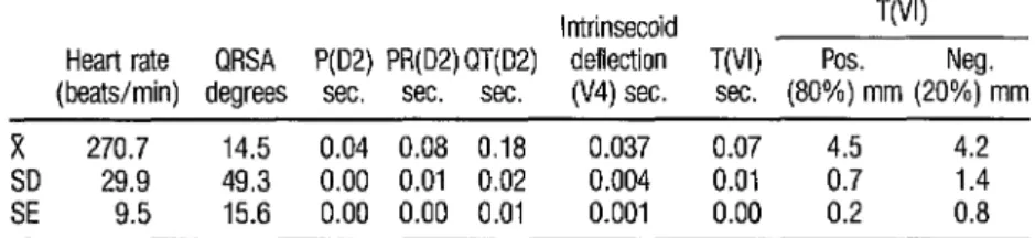
TABLE 2. ECG parameters found for the 30 control group monkeys, showing the average value (X) of each together wth the standard error (SE) and one standard deviation (SD).
lntrinsecoid VW Heart rate QRSA P(D2) PR(D2) QT(D2) deflection T(N) Pos. Neg. (beats/min) degrees sec. sec. sec. (V4) sec. sec. (80%) mm (20%) mm
x 270.7 14.5 0.04 0.08 0.18 0.037 0.07 4.5 4.2
29.9 49.3 0.00 0.01 0.02 0.004 0.01 0.7 1.4
FIGURE 1. An X-ray showing the measurements used to derive the cardiihoracic index. RD is the maximum trans- verse diameter of the right half of the heart; LD is the maxi- . mum transverse diameter of the left half of the heart; and
TD is the thoracic diameter.
A complete autopsy was per-
formed on each animal. All organs and
tissues were examined for gross lesions
and were then fixed in a conventional
formaldehyde buffer solution and Zam-
boni’s fixative (16a) for application of
hematoxylin-eosin and Masson’s trichro-
mic stain.
After fixation, three sections
were taken from the walls of each cham-
ber of the heart. Special attention was
devoted to the septum, which was sec-
tioned serially in the area located near
the septal portion of the tricuspid valve;
and a similar series of sections were also
made in the portion of the auri’cular wall
near the sinoatrial node.
In order to facilitate examina-
tion of the esophagus and colon, these
organs were rolled along their longitudi-
nal axes before being sectioned. Routine
single sections were also taken from each
monkey’s brain, lungs, kidneys, adre-
nals, spleen, lymph nodes, and psoas
muscles.
RE
SULTS
Control Group
None of the aforementioned
examinations revealed any alterations
outside established parameters in the
control group during the period of the
experiment.
Parasitology
Seven of the
10adult animals
inoculated with the
CA11: crzczi strain
showed positive parasitemia by xenodi-
agnosis-one doing so at week five, five
at week nine, and one at week 60. None
of the
10yielded positive fresh-drop or
Strout test results during the acute phase
(Table 3).
Similarly, none of the four ju-
venile monkeys inoculated with the Co-
lombian ZY crzlxt’ strain yielded positive
fresh-drop or Strout test results; but all
four demonstrated parasitemia by xeno-
diagnosis-one
at weeks
15and
18;one
at weeks
18,21, and 39; one at weeks 35
and 54, and one at week 39 (Table 4 and
Figure 2).
L
a. PAHO Bdetin 20(2), 1986
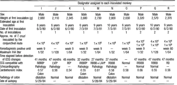
TABLE 3. Summary of test results obtained with the 10 adult Cebus ape//a monkeys inoculated with the CAM strain of 1 cruzi.
Designator assigned to each inoculated monkey
I J K L M N 0 P Q R
Sex Male Male Male Male Male Male Male Male Male Male Weight at first inoculation (g) 2,660 2,110 2,345 2,680 2,730 2,800 2,030 3,320 2,570 2,370 Estimated age at first
inoculation 8 years 6 years 9 years 6 years 9 years IO years 6 years 9 years 10 years 9 years Date of first inoculation 6/2/80 6/2/80 6/2/80 7/3/81 7/3/81 7/3/81 7/3/81 6/2/80 6/2/80 6/2/80
No. of inoculations 1 I I 1 1 1 I 2 2 2
Approx. no. of 1 cruzi fnoculated by the
conjunctival route 4x10⁴ 4x10⁴ 4x10⁴ 1x10” 1x10” 1x10” 1x10” 4x10⁴ 1x10⁶ 4x10⁴ 1x10” 4x10⁴ 1x10” Xenodiagnosis positive until week 9 - week 9 week 9 week 9 - week 5 week 9 - week 60 Maximum IHA titer i/32 i/128 i/64 i/32 l/64 i/32 i/64 i/32 l/i28 l/64 Time elapsed before detection
of ECG changes 41 months 47 months 48 months 32 months 27 months 27 months - 47 months 47 months 47 months ECG compatible with RBBBa LVOa RDa lRBBBa RBBB+ lAHa RBBB Normal IRBBB RBBB RD Echocardiography Pathology Pathology - Pathology Pathology Pathology - Normal Normal - Cardiothoracic index 0.57 0.56 0.54 0.55 0.61 0.58 0.58 0.66 0.55 0.56
Colon Colon Colon
Radiology of colon dilatation Normal Normal dilatation Normal dilatation Normal Normal Normal Normal Date of autopsy 5/29/84 - - - 5/28/84 5/29/84 - - - -
TABLE 4. Summary of test resulfs obtained with the four juvenile Ce&us ape//a monkeys inoculated with the Colombian strain of ?I cruzi.
Designator assigned to each inoculated monkey
E F G H
Sex Male
Weight at first inoculation (g) 1,750 Estimated age at first inoculation 3 years Date of first inoculation g/10/82 No. of inoculations 19 Appmx. no. of r cruzi inoculated
by the i.p. route 3x10⁶ Xenodiagnosis positive until week 39 Maximum IHA titer II128 Time elapsed before detection of ECG changes 20 months ECG compatible with RP Echocardiography Pathology Cardiothoracic index 0.51 Radiology of colon Normal Date of autopsy 6/23/84
Male Female Female 1,800 940 940 3 years 1 or 2 years 1 or 2 years g/10/82 10/4/82 IO/4182
19 18 18
3x10⁶ 3x16 3x10⁶ week 39 week 54 week 18
l/128 l/128 II128 - 13 months 18 months - LAHa LVoa Pathology Pathology Pathology
0.59 0.64 0.56 Normal Normal Normal
- 6/23/84 -
a RD=repolanzation disturbance; iAH=left anterior hemiblock: LVO=left ventricle overload.
that varied from eight to 30 weeks (Fig- ure 3). Later, as the figure shows, xenodi- agnosis yielded positive results through week 46 for one pair of monkeys and through week 49 for the other pair; and the Strout test yielded positive results again for five to seven weeks in three of the four monkeys following reinocula- tions that began on weeks 75 and 78 (E- ble 5).
Serology
IHA testing indicated that the 10 adult monkeys inoculated with the CAM strain developed fairly high levels of serum antibodies against the parasite within 20 days of the first inoculation. These titers ranged from 1: 32 to 1: 128 (see Table 3). In general, the levels of these antibodies remained high for about three months and then tended to decline. The 10 monkeys also had ele-
.a
. 0
OIQ 0 0 am P 0. CD 00
. ---
IfI I+, I--I (+I I+) ___, e , , , , 1+, , ~ r”““‘,‘/“/‘/‘~~““““““““/~’
0 7 4 6 8 10 12 14 16 18 M 22 24 26 2s 30 32 34 16 38 a0 42 44‘ m 48 50 52 54 56 58 60 71 76 18 80 82 84 86 88 $4 92
2
Weeks after font lnoculatlon
FIGURE 3. Resuh obtained from xenodiagnosis, IHA tests, and Strout tests performed on the four Cebus ape/la monkeys inoculated repeatedly with the Tulahuen strain of 7: cruzi. The boxes at upper left show each monkey’s sex and letter designator, together with the shade (black or white) used to display data for that animal. The small circles show the IHA titers obtained at varying numbers of weeks after the initial inoculation. The bars at bottom indicate weeks when the Strout test yielded positive resufts. The plus and minus signs in parentheses show whether xenodiagnostfc tests conducted in particu-
126 lar weeks yielded positive or negative results; and the small arrows show when reinoculations were administered.
vated IgG levels, as determined by ELISA testing: that remained high over the course of the infection, ranging from 1.97 to 2.77. This indicates that low se- rum titers persisted throughout the chronic phase of the infection.
IHA testing revealed a some- what different response in the four juve- niles receiving the Colombian strain. Specifically, these monkeys appeared to develop high levels of serum antibodies between the second and the eighth week after the first inoculation, and these lev- els tended to stay fairly high over the course of subsequent reinoculations for 54 to 60 weeks before tending to decline. As in the previous case, these IHA titers ranged from 1:32 to 1:128 (see Figure 2). Also, the ELISA test revealed rising IgG levels over the course of the infection (Figure 4).
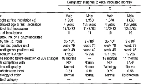
TABLE 5. Summary of test results obtained with the four Cebus ape/la monkeys inoculated with the Tula- huen strain of E cruzi.
Designator assigned to each inoculated monkey
A B C D
Sex
Weight at first inoculation (g) Estimated age at first inoculation Date of first inoculation No. of inoculations
Approx. no. of T cruzi inoculated by the i.p. route
Strout test positive until Xenodiagnosis positive until Maximum IHA titer
Time elapsed before detection of ECG changes ECG compatible with
Echocardiography Cardiothoracic index Radiology of colon Date of autopsy
Male 1,900 4 years 11/8/82
11
3x 10⁶. week 79 week 49 if256 18 months
RDa Normal
0.61 Normal
-
Male 1,950
4’12 years 11/8/82
11
3x16 week 79 week 49 II256
- Normal Normal 0.61 Normal
-
Male 1,670 4 years 1212182
10
3x10” week 76 week 46 i/1,024 18 months
RP Pathology
0.59 Normal
-
Male 1,660
4112 years 12/2/82
10
3x10" week 76 week 46 11256 11 months
LAHa Normal
0.56 Dolichocolon
-
a RD=repolarization disturbance, LAH=lett anterior hemiblock
I@ TULAHUEN
5-
COLOMBIAN
0 IO 20 30 40 50 60 70 Weeks
FIGURE 4. Positive IgG results obtained by ELISA testing of the eight Cebus ape//a monkeys inoculated repeatedly with the Colombian and Tulahuen strains of 7: cruzi (triangles and circles, respectively), as compared to values obtained by testing the 30 controls (shaded band). Negative results obtained from the test monkeys are not shown.
ure 3); IgG levels (see Figure 4) were also elevated over the course of the infection.
Hematology, Serum Enzymes, and
Serum Proteins
No significant hematologic or serum enzyme changes were detected in the inoculated monkeys. Likewise, the levels and electrophoretic fractions of plasma proteins determined immedi- ately before sacrifice did not differ signif- icantly in the inoculated monkeys as compared to the controls.
Radiology
No alterations were observed in the control group, regarding either the cardiothoracic index (CTI) obtained from the chest X-ray or the diameter and mo- tility of the esophagus and colon as re- vealed by contrast radiography of the gastrointestinal tract.
Only two of the inoculated animals yielded notably altered CTIs. One with a CT1 of 0.66, inoculated with the CA1 strain, had an intermittent right bundle branch block (see Photo A). The other, inoculated with the Colombian strain, had a CT1 of 0.64 and a left ante- rior hemiblock (Photo B). Regarding hu- man subjects, who are not strictly com- parable, Maresh-Washburn (cited by Mosca et al.-1 7) states that a maximum CT1 in the range of 0.53 to 0.58 can be m
3
found among infants weighing up to 11 kilograms.
s The test of esophagus transit
G time with a barium meal showed no evi- .$j dence of alterations, and none of the in-
u
* 23 oculated animals showed any noteworthy 2
increase in esophagal diameters.
Of five inoculated animals that had ECG alterations and were sacri-
128
ficed, two showed an enlarged colon di- ameter in the contrast study of that organ (Photos C and D).
One animal inoculated with the CA1 strain that had an intermittent right bundle branch block yielded a ra- diologic image compatible with megaco- lon; and another monkey, this one inocu- lated with the Tulahuen strain and having a left anterior hemiblock, ap- peared to have dolichocolon (Photos E and F). The contrast studies of the colon did not reveal any noteworthy alterations in any of the other animals. In each case where alterations were detected radiolog- ically, the findings were corroborated his- topathologically, and lesions of the mesenteric plexus responsible for those images were found.
Electrocardiography
The observed electrocardio- graphic patterns were found not to be al- tered by administration of the drug uti- lized as an anesthetic. However, ECG
alterations were detected in most
(83.3%) of the infected monkeys. None of the control animals showed similar disturbances during the course of the ex- periment.
The ECG alterations observed in each group of inoculated monkeys are listed in Table 6. These alterations in- cluded intermittent right bundle branch block (IRBBB), right bundle branch block (RBBB), left ventricle overload (LVO), re- polarization disturbance (RD), and left anterior hemiblock (LAH). Ajmaline did not produce any alterations in the mon- keys with normal traces.
TABLE 6. Summary of the EGG disturbances observed in the 15 Cebus ape&monkeys inoculated with the three 7I cruzi strains, at periods corresponding to the chronic phase of the disease.
T cruzi strain inoculated
CA1 Colombian Tulahuen
Animals Months Animals Months Animals Months affected elapsed affected elapsed affected elapsed since first since first since first ECG finding No. % inoculation No. % inoculation No. % inoculation
lRBBBa 2 20 32-47 0 0 20 0 0 18
RBBBa 4b 40 27-47 0 0 20 0 18
LVoa 1 IO 47 1 25 18 0
0” 18
RP 2 20 47-48 1 25 20 2 50 18
L4Ha lb 10 27 1 25 13 1 25 11
Total (all pathologic
findings) 9 90 27-48 3 75 13-20 3 75 11-18
a IRBBB=intermtient right bundle branch block; RBBB=right bundle branch block. LVO=left ventricle overload, RO=repolanzatron d6turbance. and LAH = left antenor hemlblock
b One of the animals inoculated with the cnl strain came to exhibrt both right bundle branch block and left anterior hemlblock.
strain, while the juveniles receiving the Colombian and Tulahuen strains most commonly exhibited repolarization dis- turbances or left anterior hemiblock. It should also be noted that the delay in the appearance of electrocardiographic pa- thology following the first inoculation was shorter (11 to 20 months) in the ani- mals inoculated repeatedly i.p. with larger numbers of parasites (in those re- ceiving the Colombian and Tulahuen strains), and was longer (27 to 47 months) in the adults receiving one con- junctival inoculation of the CA1 strain. m
%
Overall, the electrocardiographic distur- bances observed in these groups of mon-
2 keys resembled those found in human
2 Chagas’ disease (18-20).
$j u
b
Echocardiography
P
2 Nine of the fifteen inoculated
2
monkeys that were tested showed echo- cardiographic disturbances. The mean values for the echocardiographic parame- ters exhibited by these animals, and their 130 statistical significance relative to the
same echocardiographic data for the con- trol group, are shown in Table 7.
The frequencies of specific
echocardiographic disturbances-in-
creased right ventricle capacity @WC), in- creased left ventricle capacity (~JYc), de-
creased shortening fraction (dSF),
decreased intraventricular septum motility
(dWSM), and increased mitral valve point
E-septum distance (&SD)-are indicated in Table 8. In general, these are the distur- bances most frequently observed in hu- man Chagas’ disease (21).
Anatomopathology
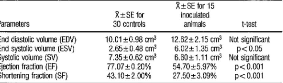
TABLE 7. Echocardiographic parameters found for the 30 control monkeys and the 15 inoculated monkeys, showing the average value (X) of each parameter, the standard error (SE), and the statisti- cal significance of the differences between the values found for each group.
Parameters
XkSE for 30 controls
X&SE for 15 inoculated
animals t-test
End diastolic volume (ED!!) 10.01+0.98 cm3 12.62-12.15 cm3 Not significant End systolic volume (ESV) 2.6520.48 cm3 6.02k1.35 cm3 p< 0.05 Systolic volume (SV) 7.3520.62 cm3 6.601k1.11 cm3 Not significant Ejection fraction (EF) 77.07+0.20% 54.70+5.97% p< 0.001 Shortening fraction (SF) 43.10+2.00% 27.50+3.09% p< 0.001
TABLE 8. Echocardiographic disturbances observed in monkeys inoculated wfth differ- ent 7: cruzi strains and examined during the chronic phase of the disease.
Disturbance
iRVCa iLVCa dSFa dlVSMa iE-SDa
Total
7: cruz~ strain inoculated
CA1 Colombian Tulahuen No. of No. of No. of animals % animals % animals % affected affected affected affected affected affected
3 43 1 25 0 0
4 57 0 0
2 29 0” 0 0 0”
5 71 3 75 1 25
1 14 3 75 0 0
5 71 3 75 1 25
a nWC=~ncreased right ventricle capacily: iLVC=wz.ased leff ventricle capacity; dSF=decreased shortening fraction. dlVSM =decreased Waventncular septum motllfty. and iE-SD=increased mltral valve pornt E-septum distance.
hibited various histopathologic altera- tions. These were as follows:
All the auricular sections showed subpericardial infiltrates that were especially noteworthy in the zone adjacent to the sinus node (Photo G). One of the monkeys was found to have a large area characterized by lymphocyte- plasmocyte infiltrates arranged in a com- pact manner in the auricular pericardium (Photo H).
All five animals had lympho- cyte-plasmocyte infiltrates in the inter- auricular septum. These could be ob- served among the muscular fibers and close to the autonomic neurons of the
atrioventricular node, where they were particularly marked, surrounding and even replacing the neural elements (Photo I). This was especially evident in the two animals that had been sacrificed soonest after their inoculation with T. G?-i74~i.
while fibrosis and the presence of atrophic myocardial fibers in the fibrotic tissue became more marked (Photos J and K).
Pictures similar to those found in the ventricular walls were ob- served in the muscular interventricular septum. Again, the animals that had been inoculated a shorter time showed only isolated lymphocyte-plasmocyte in- filtration, while those that had been in- oculated for over 27 months showed con- spicuous fibrosis.
In all five animals the sections of the sinoauricular node showed a pre- served architecture with some lympho- cyte-plasmocyte elements among the conductive fibers. These elements were more prominent in the connective tissue adjacent to the node, especially in the subpericardial zone. Alterations ob- served in the atrioventricular node and the bundle of His were similar to those found in the sinus node.
In two of the three animals in- oculated with the CAM strain, serial sec- tions of the colon showed an area 3 cm from the anal margin and 1 cm in length where the elements corresponding to the mesenteric plexus of Auerbach were ab- sent or markedly diminished (Photo L). In both cases contrast X-ray of the colon showed images compatible with mega- viscera.
D
ISCUSSION
As previously noted, experi- mental 2: crmzi infection has often been studied in the mouse (16) to evaluate drug and vaccine therapy; but the severe infection that it produces is usually lethal within a few weeks. Furthermore, 2Y
crmzi infection of mice does not resemble human Chagas’ disease, and therefore the results obtained by using mice to as- sess the benefits of drugs or vaccines are usually inconclusive (22).
The high susceptibility of dogs to I: CTZZ~ infection was established by Marsden et al. (8), who infected beagle puppies with the virulent Peru strain of the parasite. Nine of the 10 in- fected puppies died within 35 days.
Rabbits show strong natural resistance to 2: crzGzi infection, and para- sitemia is often difficult to detect by di- rect microscopic examination of the pe- ripheral blood. Teixeira (7) studied chronic infection in these animals; he found a diffuse myocarditis without par- asites, as well as megacolon in two of the
test animals. Thromboembolism and
congestive cardiac insufficiency seemed to be the cause of spontaneous death.
Other authors have experi- mentally inoculated primates of differ- ent genera (CaZWmk, Macaca, Cebm) with different 2: c~zczz’ strains. In most cases a slight myocarditis was observed. In one of the experiments, however, the animals died between 95 and 243 days after inoculation, with results that varied greatly and were difficult to interpret (1 I). These findings, taken together, led workers to theorize that primates had a natural resistance to this disease.
In the study reported here, Cebzls ape&a monkeys showed serologic conversion by IHA and ELISA following inoculation with ir: crz&, irrespective of the T. C~ZJZZ strain used. In all, 83% of the inoculated monkeys yielded positive parasitologic results by the Strout test or xenodiagnosis.
It is of considerable interest that survival of the infected Cebzcs apel’l’a monkeys was related neither to the level nor to the length of patent parasitemia. Therefore, if the virulence of the parasite
Chagas’ disease pathogenicity, such viru- lence could not be related in our work to the levels of parasitemia attained, proba- bly as a consequence of the host’s im- mune response.
For example, no parasitemia was detected in three of the 10 monkeys inoculated with the CA1 strain. However, all three of these monkeys developed car- diac disturbances during the chronic phase of the disease, and one of them also exhibited histologically confirmed megaviscera. (Human patients usually survive the acute phase of infection when the parasitemia is high-3, J-and die many years later when no parasitemia can be demonstrated by conventional methods.)
Regarding the electrocardio- graphic disturbances observed in 83 % of the inoculated animals, it should be noted that the two groups receiving re- peated intraperitoneal injections with 3 x lo6 parasites of the Colombian or Tulahuen strains showed altered electro- cardiographic parameters sooner (11 to 20 months after the first inoculation) than did the 10 monkeys inoculated once or twice by the conjunctival route with fewer parasites of the CAI strain; the lat- ter monkeys showed alterations 27 to 48 months after the first inoculation.
These findings suggest that variations in the numbers of parasites in- oculated, the inoculation route, the fre- b
2
quency of the inoculations, and the viru- lence of the !i’Y crzlzi strain used could s play an important role in the natural evo- z lution of the disease, as well as in its
-$ pathogenesis and immunopathology. It
P,
h 23 should also be noted that the terns obtained with this model resemble ECG pat-
2 those produced by Chagas’ disease in
2
man (J&20), and that the anesthesia used in our work did not appear to pro- duce any alterations in these patterns.
Regarding echocardiography, 134 a noninvasive method employed in order
to assess its experimental usefulness, it was found that nine of the 15 inoculated monkeys (60 % ) exhibited echocardio- graphic disturbances. This conclusion was reached after comparing these mon- keys’ echocardiographic patterns with those found in a control group of 30 monkeys exhibiting normal ECGS. In general, echocardiography was found to require relatively continuous and long evaluation, but the correlation found be- tween the observed disturbances in pri- mates and those in cases of human cha- gasic pathology (21) indicates that echocardiography could prove a very use- ful tool for procuring better knowledge of the pathogenesis of Chagas’-related cardiac lesions.
With respect to the contrast radiology performed, this showed nei- ther organic dilatation nor motor distur- bances of the esophagus; but it did show pictures of the colon compatible with megaviscera in three cases and with doli- chocolon in one case. In three of the four animals, two inoculated with the CA1 2: C~ZLZZ’ strain and one with the Tulahuen strain, the pictures of colon dilatation and dolichocolon were corroborated by observation of pathologic lesions. Also, in the two monkeys inoculated with the CA1 strain, diminution or absence of the neural elements of the mesenteric plexus. was observed, and in the monkey with dolichocolon, perineural infiltrates of the esophagus and colon were seen.
interventricular septum. The pericar- dium showed mononuclear infiltrates. It appears that the degree of evolution of the disease could be evaluated by assess- ing the intensity of the infiltrating and fibrotic lesions, because the former be- came progressively less marked while the latter progressively increased.
Two of these monkeys, both inoculated with the CAM strain, also showed histopathologic lesions of the mesenteric plexus approximately 3 cm from the anal margin-lesions wherein the number of neurons and structures corresponding to the Auerbach plexus were markedly diminished.
For purposes of comparison, it is worth noting that among 1,700 autop- sied human patients dying from Chagas’ disease, the incidence of cardiac lesions was found to be 90%, while the inci- dences of megacolon, megaesophagus, bronchiectasis, and other megaforma- tions were found to be 20%) 18%) 7%, and 5 % , respectively (23).
Considering the results re- ported here, it appears reasonable to con- clude that inoculation of Cebus apeZZa with three T. crmzi strains did not pro- duce demonstrable clinical alterations ,during the acute phase of the infection, but that it did lead to both cardiac and colon disturbances compatible with the chronic stage of human Chagas’ disease, and that the histopathologic lesions found afterwards were similar to those observed in human chagasic pathology.
It should be emphasized that the susceptibility of the primates tested appeared independent of age and sex. However, the parasite strain inoculated, the nature of the inoculum, the number of inoculations, and the route of inocula- tion could have conditioned the natural evolution of the disease and could have influenced the times at which the elec- trocardiographic and histopathologic le- sions developed.
It also appears that our test animals developed the pathology in a relatively short time, considering that humans generally do not manifest symp- toms of the chronic stage until several years after natural infection. In addition, it is worth recalling that Cebzls ape&a is native to zones endemic for Chagas’ dis- ease, that it has been adapted to indoor colony conditions, and that in comparf- son with other primates it reproduces well in captivity and can be maintained at fairly low cost.
Overall, therefore, Cebas
ageZZa appears to offer the most suitable available animal model for chronic Cha- gas’ disease, one that survives the acute infection and develops the chronic car- diac and gastrointestinal form of the dis- ease, apparently independently of age and sex.
A
CKNOWLEDGMENTS
The authors wish to thank Drs. S. M. Gonzalez Cappa, Rita W. de Cunio, and Ziltron Andrade for supply- ing the CAl, Tulahuen, and Colombian 1: crz& strains; Dr. Miriam Postan, Fel- low of the World Health Organization at the Argentine National Institute of Health, for performing the ELISA; Miss Elena Gomez, Mr. Angel Di Martino, and Washington Sesma of Argentina’s National Council of Scientific and Tech- nical Research (CONICET) for technical as- sistance; and all the personnel of our in- stitute who made this investigation possible.
S
UMMARY
A principal obstacle to re- search on the prevention and treatment of human Chagas’ disease has been lack of a good experimental animal model ca- pable of developing symptoms of the disease’s chronic stage. In seeking to de- velop such a model, the authors inocu- lated three groups of Cebzls apeZZa mon- keys with the CAl, Colombian, and Tulahuen strains of 1: CTZLZZ by the con- junctival and intraperitoneal routes. They then reinoculated some of these monkeys from time to time in order to more closely approximate normal pat- terns of human infection with the para- site, and conducted a battery of tests de- signed to detect the parasite and trace the course of the disease.
Xenodiagnosis or other para- sitology tests succeeded in detecting the parasite in 15 of the 18 inoculated mon- keys at intervals ranging from one to 18 weeks after the last previous inoculation. Also, IHA tests indicated high levels of antibodies against the parasite lasting for about three months following infection, and ELISA tests detected elevated IgG lev- els in some of the infected monkeys over the course of the infection.
Radiology detected unusually large heart diameters in only two of the
18 inoculated monkeys, but electrocardi- ography detected ECG alterations in 15 and echocardiography detected distur- bances in nine. These changes took con- siderable time to emerge, the ECG altera- tions being detected at times ranging from 11 to 48 months after the initial in- oculation. Subsequently, histopathologic examinations of heart tissue from five of the 15 monkeys with ECG alterations re- vealed lymphocyte and plasmocyte infil-
trates and fibrosis, the former appearing to diminish and the latter to increase over time.
Contrast X-rays of the inocu- lated monkeys also revealed four appar- ent cases of enlarged viscera (three of megacolon and one of dolichocolon). In two of these cases, histopathologic exam- ination of the. colon showed a marked re- duction in elements pertaining to the Auerbach plexus in a part of the colon some three centimeters from the anal margin.
Overall, the results indicate that inoculation of Cebzls apella mon- keys with three 27 cz~zz’ strains did not produce demonstrable clinical alterations during the acute phase of the infection, but did lead to both cardiac and colon disturbances similar to those seen in chronic human cases of Chagas’ disease. Histopathologic lesions found at autopsy were also similar to those observed in cases of chagasic pathology in man. These findings, together with the mon- key’s availability and the relatively low cost of maintaining it in captivity as com- pared to other monkeys, suggest that Ce- bzcs apella currently constitutes the most suitable available animal model for chronic Chagas’ disease.
1
la
2
Rx
FERENCES
Chagas, C. Nova trypanosomiaze humana: Estudos sobre a morfologia e o ciclo evolutivo do Schizotripanum cruzi n.
aiente etioloeico de nova et-m ade morbida kY’> n. sp,v do homen. %iem Inst Oswald0 Crzlz 1:159- 218, 1909.
Maggio, C., and F. Rosenbusch. Studien uber die Chagas krankheit in Argentinien und die Trypanosomen der “vinchucas” (Wanten, Triatoma in&tans Klug. Z&j Bakt 77:40-46, 1915.
2a 3 4 5 6 7 8 9 10 11 12
Pan American Health Organization. Report
of a Study Group on Chagas ’ Disease. PAHO
Scientific Publication 195. Washington, D.C., 1970.
Lu ones, H. S. Enfermedad de Chagas en la i2
r ancra. AnaLes de Sanidad 13(l), 1979.
Teixeira, A. R. L., G. Teixeira, V. Macedo, and A. Prata. Acauired cell-mediated im- munodepression in’ acute Chagas’ disease. J CLin Znvest 62:1132-1141, 1978.
Prata, A. Natural History of Chagasic Car- diomvonathv. In: Pan American Health Or- ganiiat fan. ‘New Approaches in Amerikan Tvpanosomiasis Research. PAHO Scientific Publication 318. Washington, D.C., 1978, pp. 191-193.
Brener, Z., E. Chiari, and N. J. Alvarenga. Observations on T. cruzi strains maintained over an 8 year period in experimentally inocu- lated mice. Rev Inst Med Trap Sdo Pado 16:39-46, 1974.
Teixeira, A. R. L., E Figuereido, J. Retende Filho, and V. Macedo. Chagas’ disease: A clinical, parasitological, immunological and pathological study in rabbits. Am J Fop Med Hyg 32:258, 1983.
Marsden, P., and J. W. C. Hagstrim. Experi- mental Tvpgnosoma cruzi infection in beagle pu
i pies: The effect of variations in the dose an source of infecting trypanosomes and the route of inoculations on the course of the in- fection. Trans R SOG Trap Med Hyg 62:816, 1968.
Laguens, R. I?, P Cabeza Meerkert, M. A. Basombrio, G. L. Chambo, P. A. Cossio, R. M. Arana, and R. Gelpi. Infecci6n cronica de1 raton con Tvpanosoma c17czi: Modelo experi- mental de enfermedad de Chagas. Medicina (Buenos Aires) 40:33, 1980.
Dorland, D. Infection in monkeys with ?i
strains of rypanosoma cruzi isolated in the United States. Pub& Health Rep 58:1006-
1010, 1943.
Torres, C. M., and B. M. Tavares. Miocardite no macaco Cebzcs apos inoculacoes repetidas con Schizot7ypanum cruzi. Mem Inst Os- Waldo Crzz 56:85-152, 1958.
Marsden. P., S. K. K. Seach, C. C. Draper, L. E. Pettitt, M. A. Miles, and A. Voller. Experi- mental T. cmzi infections in rhesus monkeys: II. The early chronic phase. l?am R SOG Trap Mea’Hyg 70:247-251, 1976.
13 14 15 16 16a 17 18 19 20 21 22 23
Miles, M. A., P D. Marsden, L. E. Pettitt, C. C. Drawer. S. Watson. S. K. K. Seach. M. S. R. H&t, and J. M. Fowler. Experimental T cruzi infection in rhesus monkeys: III. Elec- trocardiographic and histopathological find- ings. Trans R Sot Trap Mea’ Hyg 73(5):528- 532, 1979.
Falasca, C. A., D. Grana, E. Gomez, B. Merlo, and J. Mino. NormalLaboratory Pa- rameters of the New Work Non-human Pri-
mate Cebus apella. In press.
Strout, R. G. J. A method for concentrating hemoflagellates. J Parasiitol48( 1): 100, 1962.
Voller, A., C. C. Draper, D. E Bidwell, and A. Bartlett. Microplate enzyme linked im- munosorbent assay for Chagas’ disease. Lun- cet 1~426-427, 1975.
Zamboni, L., et al. Abstract. J Cell Biol 35:148A, 1976.
Mosca, L. G., L. E. Mosca. and 0. Brasseur. Tecnica radiologica: Teoria y practica (second ed.). Ldpez Editores, Buenos Aires, 1978, Chapter 19, Coratdn, p. 341.
Rosenbaum, M. B., and A. Alvarez. The ECG in chronic chagasic myocarditis. A M Heart 4:50, 1955.
Rosenbaum, M. B., J. 0. Lattari, and M. V. Elizari. Los hemib!oqueos. Editorial Paidos, Buenos Aires, 1970.
Capris, T. A., and A. J. Fernandez Moores. Alteraciones electrocardiograficas en la car- diopatia chag%sica cr6nica. Rev Arg Cardol’ 37:200, 1967.
Puigbo, J.? E. Hirschaut, I. Boccalandrok, R. Valccillo. H. Giordano, and C. SuPrez. Diag- nosis of Chagas’ cardiomyopathy: Noninva- sive techniques. Postgraduate Mea’J 5 3 : 5 37. 1977.
Laguens, R., P. Cabeza Meeker, G. Chambo, and R. Gelpi. Chronic Chagas’ disease in the mouse: IV. Effect of trypanocidal drugs. Me- dicina (Buenos Aires) 43: 126, 1983.
Koberle, E Pathogenesis of Chagas’ Disease. CIBA Foundation Symposium 20, 1974.