www.jcol.org.br
Journal of
Coloproctology
Technical Note
Human fi brinogen and thrombin patch for extraluminal
protection of intestinal anastomosis
Paulo Gustavo Kotze
a,*, Ivan Folchini de Barcelos
a, Renato Vismara Ropelato
a,
Claudio Saddy Rodrigues Coy
ba Colorectal Surgery Unit, Cajuru University Hospital, Catholic University of Paraná, Curitiba, PR, Brazil b Colorectal Surgery Unit, Campinas State University (UNICAMP), Campinas, SP, Brazil
a r t i c l e i n f o
Article history:
Received 15 July 2013 Accepted 2 August 2013
Keywords:
Anastomosis, surgical Colorectal surgery
Dehiscence, surgical wound
a b s t r a c t
In spite of recent advances regarding equipment and surgical techniques in colorectal sur-gery, rates of anastomotic dehiscence (AD) have remained stable throughout the years. The development of products to protect anastomosis aiming the reduction of AD rates has shown to be promising. Human i brinogen and thrombin patch (HFTP - Tachosil®) have been used in experimental studies in animals and small case series in humans, with prom-ising results. In this study, the authors describe the technique of HFTP use in details, aiming the protection of colorectal anastomosis, and retrospectively demonstrate the preliminary results in a pilot case series. HFTP was used in 4 patients submitted to conventional sur-gery. The procedures performed were: left colon resection, segmental colectomy (both for colorectal cancer), enteral anastomosis for i stula closure and right ileocolectomy. Anasto-motic healing and absence of complications were observed in 3 patients, and the patient submitted to right ileocolectomy developed AD and died after reoperation. The use of HFTP is safe and can be indicated in selected cases. However, AD can occur even after the use of this strategy. Randomized controlled trials with larger samples of patients are needed in order to properly dei ne the real benei ts of this strategy in dehiscence prevention
© 2013 Elsevier Editora Ltda. All rights reserved.
* Corresponding author.
E-mail: [email protected] (P.G. Kotze)
2237-9363/$ - see front matter. © 2013 Elsevier Editora Ltda. All rights reserved. http://dx.doi.org/10.1016/j.jcol.2013.08.004
Membrana de fi brinogênio e trombina humanos para proteção extra-luminal de anastomoses intestinais
Palavras-chave:
Anastomose cirúrgica Cirurgia colorretal
Deiscência da ferida operatória
r e s u m o
em 4 pacientes para proteção das anastomoses, todos operados por via de acesso conven-cional. Os procedimentos realizados foram: retossigmoidectomia abdominal, colectomia segmentar, enteroanastomose para fechamento de fístula enteral e ileocolectomia direita. Observou-se cicatrização das anastomoses em 3 casos, e DA com necessidade de reope-ração e óbito no paciente submetido a ileocolectomia direita. A utilização da MFTH para proteção de anastomoses colorretais é segura, e pode ser utilizada em casos selecionados. DA pode ocorrer mesmo com a utilização da membrana. Aguarda-se estudos randomizados e controlados com maior amostragem de pacientes para se avaliar o real papel dessa estra-tégia na prevenção da deiscência.
© 2013 Elsevier Editora Ltda. Todos os direitos reservados.
Introduction
Although in recent years surgeons have enjoyed greater suc-cess in performing intestinal anastomosis when compared to centuries ago, the results have never been perfect.1
Anas-tomotic dehiscence (AD) is the most feared complication in intestinal surgical procedures and can cause serious conse-quences to patients, from reoperations to need for stoma cre-ation, pelvic sepsis and deaths from various causes.2
In colorectal operations, depending on the location of the anastomosis, the incidence of dehiscence may vary from 1 to 24%.2 When symptomatic, they are associated with mortality
in 6 to 22% of cases.3
The integrity of the anastomosis results from a complex interaction between the surgeon, the patient and the under-lying disease.1 Although the accurate prediction of risk is
im-possible, certain factors related to the patient, such as age, male gender, smoking, hypoalbuminemia, corticosteroid use, ASA (American Society of Anesthesiology) statuses III and IV are related to AD. Elements related to the surgical proce-dure itself (tension at the anastomosis site, blood supply, pro-longed surgery) and the intestinal condition (inl ammatory disease, distal neoplasia, metastases, peritonitis) also play a key role for successful healing of anastomoses.1,4,5
Stapled anastomoses have gained more room in the last 30 years, with the introduction of new disposable staplers. Even with good acceptance in the current scenario, recent studies have failed to demonstrate the superiority of this technique when compared with hand-sewn anastomosis in colorectal surgeries.6-9 Although some studies have reported
a higher rate of stenosis,7 low anastomoses performed by
stapling seem to technically facilitate the procedure and re-duce surgical time, especially in patients with a narrow pel-vis. The decision regarding the technique should be based on the surgeon’s expertise, clinical circumstances and the availability of staplers.7
Several materials have been proposed to obtain internal or external anastomosis reinforcement, aiming to reduce the risk of dehiscence and/or minimize its consequences. Intra-luminal sealants,5,10 biodegradable material compounds with
barrier function, aim to prevent the contact of the fecal con-tents with the anastomosis, thereby avoiding the extravasa-tion of fecal matter in the case of AD.5,11 A prospective,
multi-center, randomized controlled trial is in progress to evaluate the effectiveness of this device.11
Extraluminal sealants function as an external coating of the anastomosis and have been proposed aiming to i ll the gaps between the staples/stitches, reduce bleeding and the rate of AD (extravasation).10 This method can be particularly
valuable for anastomosis of high risk, as in the case of some degree of AD, the defect would be sealed by the coated ma-terial, thereby preventing the clinical outcome.12 Included in
this group are multiple devices, such as i brin glue, expand-able polytetral uoroethylene (ePTFE), oxidized regenerated cellulose (Curacel®), platelet-rich plasma, the omentum,
hy-aluronic acid/carboxymethyl cellulose and collagen matrix-bound coagulation factors, also called human i brinogen and thrombin patch (HFTP - TachoSil®).10
Most studies performed with these materials are experi-mental models in animals and have not shown convincing results.12,13 Evidence of efi cacy and safety through
prospec-tive, controlled and randomized trials are scarce.10,12,13
Fi-brin sealants are the most often studied of these materials, showing positive results in humans, although without sta-tistical signii cance.14
The HFTP (TachoSil®) is an equine collagen sponge coated
with human thrombin and i brinogen, primarily developed for secondary hemostasis. Thrombin converts i brinogen into i brin, thereby creating a i brin clot. This clot maintains the collagen sponge i rmly adhered to the tissue, which provides an extra layer of sealing.12 Its usefulness, efi cacy
and safety have been demonstrated in some studies, for different types of procedures, from liver and heart surgery and transplants to abdominal and urological procedures.15
When used to protect high-risk intestinal anastomoses, it seems to have a benei cial effect in the prevention of AD, although most of these results were obtained from experi-mental studies.16,17
Clinical data on the use of HFTP - Tachosil ® in these anastomoses is limited. In one of the few studies carried out in humans, De Stefano et al.,18 in a nonrandomized trial,
described the use of this device in 24 patients undergoing conventional colorectal operations. The use of the product was considered effective as a sealant of anastomoses in these patients, assessed by the shorter hospital stay com-pared to the control group (7.2 × 9.3 days).
In spite of advances in surgical technology and all anas-tomosis creation and protection mechanisms, the occur-rence of AD after procedures in the colon and especially the rectum9 continues to show unacceptably high
tech-niques, products and studies that demonstrate the real benei t of the available devices, as the currently endorsed mechanisms have not been able to reduce this important complication.
The aim of this article was to describe in details the tech-nique of HFTP - TachoSil ® use as an extraluminal protec-tive device in intestinal anastomoses, showing preliminary results in four patients submitted to surgical procedures, with varying diagnoses and clinical conditions.
Technical description
To use HFTP as an extraluminal protection device in intestinal anastomoses, the surgical steps to perform the latter should occur without any changes in the usual technical standards in their respective topographies. The anastomoses can be per-formed manually or mechanically, according to the surgeon’s experience and preference and the local conditions of the segments to be anastomosed. This protection strategy can be used in both anastomoses performed through conventional or laparoscopic surgery.
Thereafter, the device is removed from its sterile packag-ing and sectioned into pieces of determined sizes accord-ing to the type of anastomosis to be protected (Fig. 1A). The original size of the product is 9.5 × 4.8 cm, with a rectangular aspect. The active surface of the product, which triggers the coagulation that results in the sealing of the anastomotic site, has a yellowish color due to its ribol avin coverage used only as a marker.
On the back table, the membrane fragments are washed in a container with approximately 20 mL of isotonic saline solu-tion at 0.9% for several seconds (Fig. 1B). From this point on, with the active yellowish surface in contact with the anasto-mosed segment, the membrane segments are adapted around
the anastomosis and gently compressed with gauze wrapped in a surgical glove for 3-5 minutes (Fig. 1C). There can be over-lapping of the product segments without any detrimental ef-fects. After this time, the soft compression is removed and a i rm clot adhered to the applied segment can be observed, and then the anastomosis surrounded by the product is re-viewed. One should emphasize the need for coverage of about 1 to 2 cm on each side of the anastomosis for proper adhesion of the membrane to the intestine and sealing of the sutured segment (Fig. 1D).
All types of anastomoses of the small and large intes-tine (entero-enteric, entero-colic, colo-colic, ileo-rectal and colorectal) can be protected with HFTP.
Preliminary results
According to the previously described technical principles, four patients that underwent intestinal anastomoses were submitted to the use of HFTP for extraluminal reinforce-ment and comprised the preliminary sample of this techni-cal description study. Two female patients were operated for colorectal neoplasms (left colon) and underwent convention-al colectomy. One mconvention-ale patient was operated for an anasto-motic i stula secondary to right ileocolectomy due to previous complicated appendectomy, being submitted to a new ileo-colic resection, also conventionally. Finally, a female patient with enteral i stula and previous peritoneostomy underwent enteroanastomosis for i stula closure. The details of each pa-tient are given in Table 1 and in Figs. 2, 3 and 4.
As it can be observed, two patients underwent hand-sewn anastomosis and two were submitted to stapled ses. Patient 1 underwent high colorectal stapled anastomo-sis at the level of the sacral promontory and HFTP was ap-plied after the use of the stapler, without removing the latter. This technical detail allows better i tting when applying the device. The stapler was removed only after the complete ad-hesion of the membrane. Patient 2 underwent a colo-colic end-to-end hand-sewn anastomosis, and the membrane was applied without difi culty (Fig. 2). Patient 3 underwent an entero-enteral anastomosis in an area of enteral i stula sec-ondary to previous complex abdominal surgery. After primary suture with 3.0 polypropylene yarn, HFTP was applied with a closed bandage over the i stula (Fig. 3). These i rst 3 patients recovered uneventfully postoperatively without any general complications or complications at the anastomotic site.
Patient 4 received the HFTP after an ileo-transverse anas-tomosis performed by the side-to-side technique with a 100-mm linear stapler. Although the technical conditions of the anastomosis were the best possible (Fig. 4), the patient devel-oped AD and abdominal abscess, underwent reoperation with externalization of the two anastomotic mouths on the right l ank (Mikulicz stoma) and died due to acute bacterial endo-carditis and heart failure.
Discussion
In the year 2013, the anastomoses performed on the small intestine, colon and especially on the rectum still show
nii cant rates of dehiscence. In recent years, developments in the i eld of colorectal surgery were mainly concentrated in staplers and disposable equipment mainly for laparoscopic techniques. Despite signii cant advances in this area, AD is found in a signii cant proportion of patients. Its consequences include reoperations, longer hospital stay and consequent in-crease in treatment costs, as well as the possibility of
mortal-ity.1 Therefore, any technology or device aimed at reducing AD
will have an impact on the outcome after these procedures. Colorectal anastomoses by compression (Valtrac ™ ring, magnetic rings, AKA-2™, endoluminal compression ring − endoCAR™, anastomotic compression clip − CAR) have also been studied and although they appear to be safe, they do not show superiority to hand-sewn or stapled anastomosis when AD rates are compared.9
Products designed to seal anastomotic sites are classii ed as intraluminal and extraluminal. HFTP (TachoSil ®) is among the latter, used for secondary hemostasis mainly in liver sur-gery. In recent years, some publications have demonstrated the safety and feasibility of using this device in digestive anastomoses.
Pantelis et al.16 evaluated the use of HFTP in rats for
high-risk anastomoses (with only 4 separated absorbable suture points), compared to traditional anastomoses (with 8 sepa-rate absorbable suture points). The experiment showed that the use of the product in high-risk anastomoses resulted in lower mortality and dehiscence rates when compared to anastomotic procedures without the sealing mechanism.
Parker et al.10 described the feasibility of applying HFTP in
24 patients undergoing anterior resection of the rectum. The product was well tolerated in procedures performed by
lapa-Table 1 – Baseline characteristics of the four patients submitted to procedures with intestinal anastomoses and extra-luminal use of HFTP - TachoSil®.
Patient Gender Age (years) Procedure Type of anastomosis
Diagnosis Postoperative complications
#1 F 59 Anterior rectal resection Stapled Neoplasia No
#2 F 67 Left segmental colectomy Hand-sewn Neoplasia No
#3 F 43 Enteroanastomosis Hand-sewn Enteral i stula with
peritoneotomy
No
#4 M 41 Right ileocolectomy Stapled Enteral i stula Yes (anastomosis
dehiscence)
F, female; M, male.
Fig. 4 – Final aspect of the functional termino-terminal ileo-transverse anastomosis in patient 4. In spite of the perfect technical condition and adequate protection with HFTP membrane, there was evolution to anastomotic dehiscence and death.
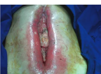
Fig. 3 – Final aspect of patient 3 after enteroanastomosis for enteric fi stula closure in peritoneotomy area with HFTP protection.
roscopic and conventional techniques, with no signii cant adverse effects. According to the authors, the use of HFTP seems to be facilitated by the use of circular staplers, except in male patients with a narrow pelvis. In our initial series of cases, the membrane was used only in conventional pro-cedures, despite the possibility of using it with the laparo-scopic approach.
The use of products for anastomosis protection may also be indicated in attempts to close enteral i stulae. There are scarce data in the literature regarding this indication, but the rational use of this strategy in high-risk patients with bad nutritional status represents the possibility of future studies. In our preliminary series of 4 patients, we used the HFTP in a patient with enteral i stula and peritoneotomy with good results.
Evidently, the use of products and devices such as HFTP does not replace adequate surgical technique and the basic principles for AD prevention, such as adequate blood supply in the anastomosed segment, no tension and good local con-ditions. Furthermore, the use of these alternative techniques is no guarantee of a healed anastomosis, as seen in patient 4 of this preliminary series. Even with the use of HFTP, dehis-cence can occur, with consequent mortality.
The cost of these devices must also be considered. The routine use of products such as HFTP is not recommended, as anastomoses on the small and large intestine have been performed over the past 50 years with high success rates.
However, it is believed that patients with elevated risk of AD, such as those with dei cient nutritional status, prior ra-diotherapy in the case of the anastomoses on the rectum and inl ammatory bowel disease with previous use of corticoids, for instance, can benei t from this type of strategy here de-scribed. Randomized and controlled studies, with signii cant sample of patients, are required for more extensive conclu-sions regarding the actual role of anastomosis protection de-vices such as the HFTP.
Conl icts of interest
The authors declare no conl icts of interest.
R E F E R E N C E S
1. Davis B, Rivadeneira DE. Complications of Colorectal Anastomoses. Surg Clin N Am 93 (2013) 61–87.
2. Shogan BD, Carlisle EM, Alverdy JC, Umanskiy K. Do We Really Know Why Colorectal Anastomoses Leak? J Gastrointest Surg. 2013 May 21 [Epub ahead of print]
3. Cauli eld H, Hyman NH. Anastomotic Leak After Low Anterior Resection. JAMA Surg. 2013;148(2):177-182
4. Slieker JC, Daams F, Mulder IM, Jeekel J, Lange JF. Systematic review of the technique of colorectal anastomosis. JAMA Surg. 2013 Feb;148(2):190-201.
5. Daams F, Luyer M, Lange JF. Colorectal anastomotic leakage: Aspects of prevention, detection and treatment World J Gastroenterol 2013 April 21; 19(15): 2293-2297.
6. Neutzling CB, Lustosa SA, Proenca IM, et al. Stapled versus hand-sewn methods for colorectal anastomosis surgery. Cochrane Database Syst Rev 2012;(2):CD003144.
7. Lustosa SA, Matos D, Atallah AN, Castro AA. Stapled versus handsewn methods for colorectal anastomosis surgery. Cochrane Database Syst Rev 2001; (3): CD003144
8. MacRae HM, McLeod RS. Handsewn vs. stapled anastomoses in colon and rectal surgery: a meta-analysis. Dis Colon Rectum 1998; 41: 180-189.
9. Ho YH, Ashour MAT. Techniques for colorectal anastomosis. World J Gastroenterol 2010 April 7; 16(13): 1610-1621. 10. Parker MC, Pohlen U, Borel Rinkes IHM, Delvin T. The
application of TachoSil® for sealing colorectal anastomosis: a feasibility study. Colorectal Disease 2013:15(2):252-257. 11. Bakker IS, Morks AN, Hoedemaker HOC, Burgerhof JGM,
Leuvenink HG, Ploeg RJ, Havenga K. The C-seal trial: colorectal anastomosis protected by a biodegradable drain i xed to the anastomosis by a circular stapler, a multi-center randomized controlled trial. BMC Surg. 2012; 12: 23.
12. Pommergaard HC, Achiam MP, Rosenberg J. External coating of colonic anastomoses: a systematic review. Int J Colorectal Dis 2012; 27:1247–1258.
13. Vakalopoulos KA, Daams F, Wu Z, Timmermans L, Jeekel JJ, Kleinrensink GJ, Ham AVD, Lange JF. Tissue adhesives in gastrointestinal anastomosis: a systematic review. Journal IF Surg Res 2013; 180:290-300.
14. Huh JW, Kim HR, Kim YJ. Anastomotic leakage after laparoscopic resection of rectal cancer: the impact of i brin glue. Am J Surg 2010; 199(4):435–441.
15. Rickenbacher A, Breitenstein S, Lesurtel M, Frilling A. Efi cacy of TachoSil a i brin-based haemostat in different i elds of surgery - a systematic review. Expert Opin Biol Ther 2009; 9: 897–907.
16. Pantelis D, Beissel A, Kahl P, Wehner S, Vilz TO, Kalff JC. The effect of sealing with a i xed combination of collagen matrixbound coagulation factors on the healing of colonic anastomoses in experimental high-risk mice models. Langenbecks Arch Surg 2010; 395(8):1039–1048.
17. Nordentoft T, Rømer J, Sørensen M. Sealing of gastrointestinal anastomoses with a i brin glue-coated collagen patch: a safety study. J Invest Surg 2007; 20: 363–9.