Obesity and gut microbiota - what do we know so far?
Vicente Lopes da Silva-JuniorI, Fernanda de Azevedo Marques LopesI, Rodolpho Mattos AlbanoII, Maria das GraçasCoelho de SouzaIII, Carolina Monteiro de Lemos BarbosaIII, Priscila Alves MaranhãoIV, Eliete BouskelaIII, Luiz Guilherme
Kraemer-AguiarV
DOI: 10.5935/MedicalExpress.2017.04.01
I Universidade do Estado do Rio de Janeiro, Graduate Program in Clinical and Experimental Physiopathology (FISCLINEX), Rio de Janeiro, RJ, Brazil. II Universidade do Estado do Rio de Janeiro, Instituto de Biologia Roberto Alcantara Gomes, Departmento de Bioquímica, Rio de Janeiro, RJ, Brazil. III Universidade do Estado do Rio de Janeiro, Instituto de Biologia, Departmento de Ciências Fisiológicas, Rio de Janeiro, RJ, Brazil.
IV Universidade do Porto, Faculdade de Medicina, Departamento de Ciências da Informação e da Decisão em Saúde, Porto, Portugal. V Universidade do Estado do Rio de Janeiro, Faculdade de Ciências Médicas, Departamento de Medicina Interna, Rio de Janeiro, RJ, Brazil.
In the history of medicine, only recently has obesity been recognized as a disease. We know now that it is a pandemic condition, partly explained by the so-called Western lifestyle and related to multiple other comorbidities in various systems. This lyfestyle includes eating large portions, rich in saturated fats and reined sugar, all coupled with sedentary habits. In recent years, the gut microbiota has been indited as a new culprit in pathophysiological aspects involved in obesity. From studies with animals free of bacteria in the digestive tract, known as “germ-free animals”, the relevance of intestinal microbiota in the regulation of body fat became evident and its importance has also been extended to the pathophysiology of diseases such as diabetes mellitus and coronary heart disease. Characterization of Toll-like receptors led to the discovery of mechanisms that link the immune system with some metabolic pathways and opened new avenues of a previously unknown world to biological sciences. Increased knowledge about interactions between gut microbiota and the host can certainly reveal, in a not too distant future, new therapeutic perspectives for obesity and its related diseases.
KEYWORDS: obesity; gut; microbiota
Silva-Junior VL, Lopes FAM, Albano RM, Souza MGC, Barbosa CML, Maranhão PA, Bouskela E, Kraemer-Aguiar LG. Obesity and gut microbiota - what do we know so far? MedicalExpress (São Paulo, online). 2017;4(4):M170401.
Received for Publication on June 24, 2017; First review on July 13, 2017; Accepted for publication on July 22, 2017; Online on August 4, 2017
E-mail: lgkraemeraguiar@gmail.com
■
INTRODUCTIONObesity is a condition known for millennia in humans, but only recently it started to be considered as a disease.1 It is estimated that genetic factors may account
for 25% to 40% of body mass index (BMI) variance, by determining factors such as differences in basal rate of metabolism and response to overfeeding. It is believed that changes in eating habits and sedentary lifestyle acting on susceptibility genes are the main determinants in the growth of obesity worldwide.2,3
The theory of “economic genotype” was introduced by Neel et al. in 1962,4 according to which
individuals with greater ability to accumulate energy during periods of food scarcity would be more likely to survive in such conditions. This concept reinforces the theory of Darwin’s natural selection, according to which
the fittest individuals are selected by nature to maintain their offsprings.
According to recommendations of the World Health Organization5 for health maintenance, at least
150 minutes of moderate intensity physical activity or 75 minutes of high intensity activity per week must be performed in populations aged 18 to 64 years; however, anthropological studies have shown1 that isolated African
tribes in regions of Kenya and Ethiopia, who keep living habits to very similar those of their ancestors maintain their BMI ranging from 17.8 to 19,1 kg/m2 due to an
■
DISCUSSIONGut microbiota
Until birth, the gastrointestinal tract of a normal fetus is sterile. During labor and shortly after delivery, bacteria from the mother and surrounding environment colonize the gut of the newborn. The type of delivery seems to influence decisively in this colonization: the gastrointestinal tract of normally delivered babies appears to be colonized predominantly by the mother’s gut microorganisms that seem to influence its microbiota up to about one month after birth; in contrast, the gastrointestinal tract from babies delivered by cesarean section appears to be preferentially colonized by bacteria of the surrounding environment, the air, other children and medical staff, and may have its composition changed until the sixth postnatal month. By the second year, the intestinal flora is similar to that found in adults.9
The gastrointestinal tract is initially colonized by aerobic bacteria, including facultative aerobic bacteria. Their expansion consumes oxygen and creates a favorable environment for the growth of strictly anaerobic organisms, most of them belonging to the genera Bifidobacterium,
Bacteroides, Clostridium and Ruminococcus.10
The pioneer microorganisms may act to induce or modulate the expression of genes in epithelial cells of the host, which may create a favorable environment while preventing the growth of bacteria introduced later in this ecosystem. Thus, the initial colonization is of fundamental importance in establishing the permanent flora of adults.11,12
Colonization in the gastrointestinal tract diverges according to its segments. Among the microorganisms, bacteria are the majority with over 90% of the species belonging the phyla Firmicutes and Bacteroidetes.8 The
colonization of gastrointestinal tract has an impact in energy use of non-digestible food such as cellulose and in the synthesis of short chain fatty acids, such as propionic acid that serves as substrate for gluconeogenesis and acetate, a substrate for de novo lipogenesis in hepatocytes and adipocytes. Furthermore, the gastrointestinal tract microbiota acts as a barrier against colonization of pathogens and stimulates the development of the immune system.13,14 Therefore, the intimate contact between
commensal bacteria and the intestinal epithelium seems to play a key role in regulating host-commensal bacteria against pathogens (Figure 1).8
Obesity and gut microbiota
The intestinal microbiota of obese subjects is characterized by a higher Firmicutes/Bacteroidetes ratio when compared to lean individuals. Weight loss programs and chromosomal regions associated with human obesity
phenotypes.6
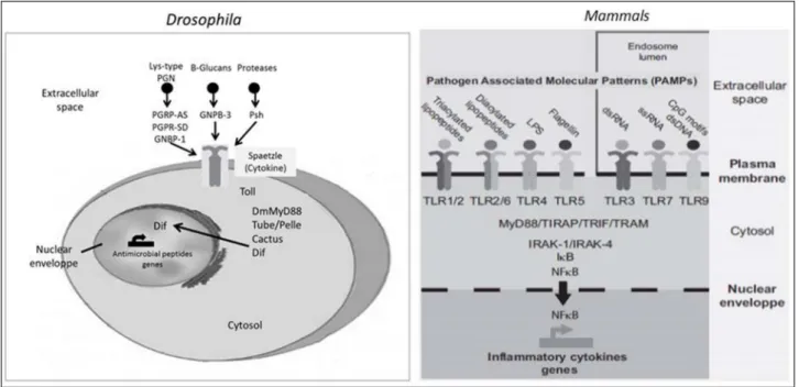
From the observation of Drosophila, in which the same tissue is responsible for providing energy and defense against invading microorganisms, emerged the hypothesis of a “memory” ancestor that connects the immune system and the development of obesity.1 Actually, we already
know that the adipose tissue is capable of producing many substances previously associated solely with the immune system. A family of membrane proteins called Toll-like receptors (TLR) may be the link between the immune system and body metabolism. These receptors are capable of recognizing pathogens, of initiating the immune response and to respond to both lipopolysaccharide (LPS) present in Gram-negative bacterial cell walls and to some types of fatty acids.7
Accumulation of excessive adipose tissue is due to hyperplasia and hypertrophy and the latter is more susceptible to lipolysis, which broadens the pool of circulating fatty acids to bind to the subtype 4 of TLR (TLR-4); this triggers an immune response that results in a low-grade inflammatory state even in the absence of any infection. This low-grade or subclinical inflammation is often asymptomatic and closely related to insulin resistance. One of the clearest evidences of the importance of TLR-4 in obesity and diabetes has been demonstrated by experiments with TLR-4 knockout mice, which have been shown to be protected from developing diabetes or obesity-related morbidities when exposed to a high-fat diet.1,8
The human gastrointestinal microbiota has been physiologically recognized as a true organ that produces local and systemic mediators, which may contribute or cause damage to host metabolism. Among the main signaling molecules, LPS is present in the outer membrane
of Gram-negative bacteria; in addition to being involved in
pyrogenic mechanisms in sepsis, LPS was recently related to low level systemic inflammation present in obesity and metabolic diseases.
This review article aims to discuss the main links between obesity and gut microbiota and how they can change the perspectives of treatment of this disease that has a huge impact on Western civilization.
■
MATERIALS, METHODS AND RESULTSFigure 1 - Schematic representation of Toll/TLR pathways in Drosophila and mammals. Toll and TLRs activate an evolutionarily conserved signaling pathway. In Drosophila, Toll is activated by Spaetzle cytokine, through a proteolytic cascade. In mammals, TLRs are activated on direct binding of microbial molecular patterns. PGN indicates peptidoglycan; dsRNA, double-stranded RNA; ssRNA, single-stranded RNA; Dif, Dorsal-related immunity factor (Adapted from Ley et al.15).
are associated with changes in the proportion of both phyla related to how much weight was lost and not with lower calorie intake.15
Animals with sterile intestines (e.g., germ-free mice) are protected from diet-induced obesity and its associated comorbidities, whereas when colonized by the intestinal microbiota of other mice, they show significant weight gain 2 weeks after colonization. Interestingly, the weight gain of these colonized animals varies from 40% when the microbiota came from lean mice, to 60% when it came from genetically obese mice (ob/ob mice). Germ-free animals have reduced ability to extract dietary energy associated with reduced energy supply in the liver and skeletal muscle.16,17 These findings link the germ-free condition
to caloric restriction regimen in which there is resistance to develop obesity. Furthermore, caloric restriction is associated with longevity due to improvements in general health.17,18 However, this resistance to become obese
induced by feeding seems to be dependent on specific interactions between diet and microflora.18
The greater ability to obtain energy from nutrients observed in the ob/ob mouse appears to be related to genes that encode enzymes that process indigestible polysaccharides, causing an increase in the production of fermentation products, mainly short chain fatty acids. Some authors observed that the amount of calories in the feces of these animals was smaller than what is found in lean animals19 while others found no differences in
the amount of fecal energy of germ-free mice compared to controls.17 These obervations suggest that other
mechanisms underlying food energy extraction capacity
could be responsible for weight gain related to intestinal microbiota composition. It has also been reported that intestinal colonization of germ-free animals resulted in significant changes in hormone levels with increased plasma levels of insulin, leptin and glucose, in capillary density and, especially, in the expression of genes that regulate lipogenesis and “energy sensing”.16 An example of
such changes is that germ-free animals, when colonized, had reduced expression of the fasting-induced adipocyte factor (FIAF) in the gut epithelium, which is capable of inhibiting the activity of lipoprotein lipase, thus reducing the release of fatty acids from triglycerides.16,20 Furthermore, higher
expression of FIAF stimulates 1α coactivator peroxisome proliferator-activated receptor, which causes up-regulation of genes encoding regulators of mitochondrial fatty acid oxidation.17 In contrast, lower levels of FIAF lead to higher
lipoprotein lipase activity, causing an increase in storage of triglycerides in the adipocytes,
Another feature that makes germ-free animals more resistant to western diet-induced obesity appears to be related to higher expression of phosphorylated AMP-activated protein kinase (AMPK) in skeletal muscle and liver,17 since AMPK upregulates fatty acid oxidation and
glucose uptake in muscle tissue and inhibits the synthesis of fatty acids and hepatic gluconeogenesis. In adipose tissue, AMPK inhibits fatty acid synthesis and lipolysis; it also inhibits insulin secretion mediated by fatty acids, playing an important role in body weight maintenance and reduction of lipotoxicity.21 In summary, the protection from
of fasting-induced adipocyte factor and increased AMPK activity work independently, they result in reduced storage of triglycerides.
Taken together, the results of the study conducted with germ-free animals directs attention to the active role of the microbiota in oxidation and storage of nutrients and not only to an increased absorption of nutrients by the gut. Contrary to what was believed, germ-free animals fed with a high-fat diet did not become fat and, according to the study, it is likely that dietary fat alone is not enough to cause obesity.17
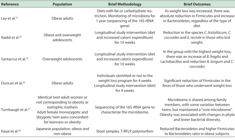
Data from clinical studies (table 1) that investigated changes in the composition of the intestinal microbiota in obesity have been, in general, in agreement with animal models; although these clinical studies are more heterogeneous, this could be attributed to the increased complexity of human lifestyles when compared to experimental animals.
Lipopolysaccharide – clinical features
Lipopolysaccharide glycolipids are large molecules formed by covalent bonds between lipid fractions and polysaccharides. They are found in the outer membrane of Gram-negative bacteria; they are also known as endotoxins due to their ability to trigger intense immune responses. The basic structure consists of lipid A associated with an antigenic group O which is probably the molecule responsible for triggering the inflammatory response and low-grade metabolic disorders.7
High fat diets appear to favor the uptake of lipopolysaccharides by the intestinal epithelium, but the exact mechanism remains under discussion.29,30 It has been shown
that chylomicron formation promotes absorption of LPS in human colorectal carcinoma model invitro. Chylomicron formation is associated with secretion of LPS by the cells.31
Other reports,32,33 however, showed that an increase
of about 10 times in the concentration of fatty acids can harm the integrity of the intestinal mucosal barrier. In animals, the endotoxemia that follows high-fat diet has been associated with reduced expression of genes that encode two proteins, namely zonula ocludens-1 and occludin, both important in keeping the selective permeability of the intestinal mucosa.34
An alternative route for LPS absorption has been described:35,36 internalization through intestinal microvilli
via a TLR4 dependent mechanism of protein and myeloid differentiation protein type 2; once inside enterocytes, LPS can be transported to the Golgi site of synthesis of chylomicrons.
Upon reaching the bloodstream, LPS is transported by a specific acute phase protein, the LPS binding protein and by lipoproteins into hepatocytes where it is cleared by excretion in bile.37,38 All classes of plasma lipoproteins can
sequester LPS and it seems to be dependent on the amount of phospholipids on their surface.39
Under normal conditions, high-density lipoprotein (HDL) cholesterol is the main acceptor of LPS. However,
Table 1 - Main studies evaluating changes in intestinal microbiota composition in humans
Reference Population Brief Methodology Brief Outcomes
Ley et al.15 Obese adults
Diets with fat or carbohydrate res-triction. Monitoring of microbiota for
1 year (sequencing of the 16S rRNA gene)
As weight loss was increased, there was absolute reduction in Firmicutes and increase
in Bacteroidetes, regardless of the type of diet.
Nadal et al.24 Obese and overweight
adolescents
Longitudinal study intervention (diet and increased caloric expenditure)
for 10 weeks
Reduction in the species C. histoliticum, C. coccoides and E. rectale in those who lost
weight.
Santacruz et al.25 Overweight adolescents
Longitudinal study intervention (diet and increased caloric expenditure)
for 10 weeks
In the group with the highest weight loss, there was an increase of B. fragilis and Lactobacillus and reduction B. longum and C.
coccoides
Duncan et al.26 Obese adults
Individuals ubmitted or not to the weight loss program for 4 weeks Longitudinal study intervention (diet)
for 4 weeks
Signiicant reduction of Firmicutes in the feces of those who underwent weight loss
Turnbaugh et al.27
Identical twin adult women or not corresponding to obesity or
eutrophic mothers Adult female monozygotic and dizygotic twin pairs concordant
for leanness or obesity
Sequencing of the 16S rRNA gene to characterize the microbiome.
Microbiome is shared among family members, with some variation between twins, but maintained a "core microbiome". Obesity was associated with changes in phyla
and lower bacterial diversity.
Kasai et al.28 Japanese population, obese and
non-obese Stool samples. T-RFLP polymorism
In the same study, Cani et al. examined the relationship of a diet rich in lipids and circulating LPS and noted that, after the ingestion of fat, LPS levels were approximately 1.4 times higher than observed in animals receiving standard diet, demonstrating a direct relationship between absorption of fat and endotoxin.29
The same authors endeavored to demonstrate an
in vivo relationship of cause and effect between systemic
exposure to slightly increased levels of endotoxin and development of weight gain, insulin resistance and low-grade inflammation, which some authors call “metabolic endotoxemia”. It seems that endotoxemia exacerbates and accelerates the pro-inflammatory, pro-diabetogenic stage promoted by fatty acids that, in turn, modulate activation of TLR4 by LPS.52
Another effect of LPS on adipose tissue is its influence upon adipocyte size, because chronic infusions of LPS were associated with reductions in the average size of adipocytes. The balance between hypertrophy and hyperplasia could influence individual risk of fat mass gain and development of insulin resistance and cardiovascular diseases.29,53
Another interesting fact is that the amount of LPS administered in the study performed by Cani et al.29 would
be enough to cause anorexia, which was not observed. Chronic exposure to endotoxin probably causes tolerance despite the already known anorexigenic mechanism of LPS. The reasons behind this observation are not completely understood but may be related to genotypic differences between species, hormonal factors such as leptin and ghrelin or biochemical characteristics of LPS.29
LPS inluences insulin signaling
The induction of the inflammatory response by LPS can establish an intersection with insulin signaling at various stages, and may even inhibit it. Insulin acts on target cells through binding to cell surface receptors inducing receptor autophosphorylation and activation of tyrosine kinase receptor activity that phosphorylates various substrates, such as members of insulin receptor family substrates (IRS), triggering its well known effects at the end of the cascade.54
It is also now well documented that phosphorylation of serine residues by the insulin receptor reduces ability of IRS-1 to associate with the receptor and thus inhibits the remainder of the cell signaling cascade. TNFα and IL-6 are responsible for the inhibitory phosphorylation of IRS-1.55
The key point in the integration of metabolic and immune pathways occurs at the level of the c-Jun N-terminal kinase Inflammatory signals lead to hyperactivation of the c-Jun N-terminal kinase, resulting in serine phosphorylation of IRS-1.56,57 However, other points of overlap between
immune and metabolic pathways occur at the levels of protein kinase C, the family of suppressors of cytokines such as SOCS-1, 3 and 6 and the induced nitric oxide synthase.57-61
in conditions of acute inflammation and infection, plasma HDL is reduced, while triglycerides and chylomicrons are increased. Thus, in this scenario, the LPS binding protein seems to transfer LPS preferentially to low-density lipoprotein (LDL cholesterol) and promote the formation of complexes of LPS with HDL and LDL.40,41 By increasing
and redistributing phospholipids between different lipoproteins, the immuno-stimulatory effect of LPS can be attenuated. This mechanism may represent part of the innate defense system against endotoxemia caused by gram-negative bacteria.42
At least three mechanisms have been proposed by which dietary lipid could promote greater absorption of LPS: (a) compositional changes in the intestinal microbiota, (b) increased availability of chylomicrons and (c) changes in the permeability of the intestinal epithelium. Therefore, a diet rich in lipids may contribute to greater absorption of LPS, a fundamental molecule for the process of chronic subclinical inflammation associated with many obesity-related diseases, especially atherosclerosis.
Endotoxemia and its importance in cellular metabolism
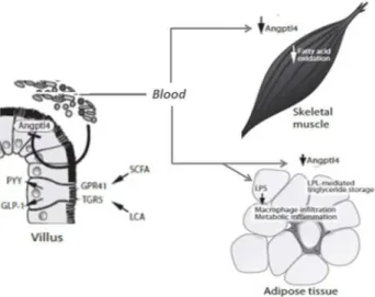
A new hypothesis, illustrated in Figure 2, has linked intestinal microbiota and metabolic homeostasis.47 Based
on evidence that obesity and type 2 diabetes are both associated with systemic low-grade inflammation48-51 in
the liver, adipose tissue and hypothalamus, Cani et al.29
suggested that bacterial LPS derived from gut bacteria could trigger inflammation that would lead to diabetes induced by high fat diet and obesity.
At the adipose tissue level, two other mechanisms also contribute to insulin resistance induced by LPS. First, activation of TLR4 in LPS-induced preadipocytes alters the expression of several cytokines, including TNFα and IL-6 by a paracrine route that inhibits insulin signaling in adipocytes.55 Moreover, LPS promotes the expression
of NF-kB and activation of the MAP kinase pathway in adipocytes, leading to target gene expression, such as glucose transporter stimulated by insulin-4 (GLUT-4), adiponectin, fatty acid synthase and perilipin. In adipocytes belonging to the 3T3-L1 cell line; LPS also promotes the expression of inducible nitric oxide synthase.62,63
It was observed that low doses of LPS induce a biphasic response in glucose uptake in non-obese individuals. In the early hours, there was an increase in insulin sensitivity, with subsequent reduction.64,65 This
biphasic effect on insulin sensitivity during experimental endotoxemia and sepsis may be due to expression of inducible nitric oxide synthase.66 LPS increases the uptake
of glucose by myocytes through inducible nitric oxide synthase induction. On the other hand, excessive production of nitric oxide (NO) causes a reduction in glucose uptake by muscle cells.67, 68 The effect of NO on insulin action may
be perturbed by release of TNFα and IL-6 induced by LPS. Furthermore, excessive production of NO may worsen insulin resistance by increasing the levels of circulating fatty acids, due to loss in lipoprotein lipase activity, which favors lipolysis.66
The behavior of blood glucose during sepsis demonstrates this biphasic effect, as shown by the evolution of hypoglycemia to hyperglycemia.64 LPS-induced
hypoglycemia is associated with a reduction in glucose synthesis. In addition, increased activity of NF-κB results in an increase in the expression of inducible nitric oxide synthase and reduced expression of genes that encode limiting enzymes for glucose synthesis such as glucose-6-phosphatase and phosphoenolpyruvate carboxykinase.69-71
Down-regulation of adiponectin receptor expression on surface of monocytes probably also contributes to the reduction in insulin sensitivity. The release of counter-regulatory hormones such as cortisol and growth hormone following the administration of low doses of LPS can contribute to reduction of peripheral and hepatic glucose uptake.65,72
Type 2 diabetes, hyperinsulinemia and endotoxemia
LPS stimulates insulin secretion and, reciprocally, chronic hyperinsulinemia causes reduction of LPS clearance by reducing the function of hepatic Kupffer cells.58,73 However, this mechanism that, in theory, would
protect against microorganisms of gastrointestinal tract reaching the blood stream, may become exaggerated in the presence of adiposity, hyperinsulinemia, high fat diet, and smoking.30,74,75 As result, LPS concentration in
plasma increases by about 50% after high-fat diet or when associated with smoking.30
Hyperinsulinemia with hyperglycemia may be intensified by endotoxemia, since they reduce jejunal motility and intestinal transit and may facilitate bacterial overgrowth and loss of mucosal integrity, which occurs quite frequently in patients with type 2 diabetes mellitus. Since hyperglycemia and hyperinsulinemia are usually accompanied by a diet rich in lipids and dyslipidemia, they can act as synergistic factors.76-78 It has been reported
that individuals with type 2 diabetes have serum levels of LPS that are 76% higher than those found in healthy individuals.79
Endotoxemia links dyslipidemia and cardiovascular disease
To our knowledge, the first study to demonstrate the relationship between LPS and cardiovascular risk was published in 1999 by Wiedermann et al,82 showing that
individuals with LPS levels above 50 pg/ml had a threefold higher risk of developing atherosclerosis than control subjects.
The positive association between increased levels of LPS and metabolic syndrome suggests a possible clinical utility of endotoxin in serum as a marker of low-grade inflammation and increased cardiovascular risk. This might add power to the classic markers of cardiovascular risk, by providing information about damage to tissues such as the liver endothelium. Through its connection to TLR4 in endothelial cells, monocytes and macrophages, LPS causes the release of pro-inflammatory cytokines, leading to severe endothelial dysfunction, atherosclerotic plaque formation, oxidation of LDL and thrombogenesis.83 TLRs are
not expressed in a constitutive manner in the endothelium, but their expression depends on mechanical and non-mechanical stimuli, which include blood flow disorders,84
oxidized LDL,85 endogenous nonlipid ligands,86 free fatty
acids,87,88 stress factors (heat shock proteins),87-89 advanced
glycosylation end products,89 as well as fibrinogen, heparan
sulfate and hyaluronic acid.90
By interacting with the endothelial surface of TLR4, LPS promotes direct endothelial damage by generation of reactive oxygen species.91 Furthermore, when stimulated
by LPS, endothelial cells release pro-inflammatory factors, chemotactic and cell adhesion molecules, which cause transmigration of monocytes,92,93 differentiation into
macrophages and atherosclerotic plaque formation. Among these cytokines, IL-8 is chemotactic for T lymphocytes, directed to the fibrous cap of atherosclerotic plaque;94,95
very low concentrations of endotoxin (<1ng / ml) can promote this process.92,93 An interesting aspect is
Immunity in obesity triggered by TLR
LPS, when not fully eliminated by hepatocytes, binds to TLR4 present on surface of Kupffer cells, preadipocytes and adipocytes, causing upregulation of CD1497 and expression of TLR2.79 Preadipocytes,
adipocytes and macrophages present in visceral adipose tissue appear to respond synergistically to inflammatory stimuli. Considerable potential has been assigned to pro-inflammatory visceral adipose tissue, when compared to non-visceral adipose tissue, because it is more readily mobilized during stress.98,99 Due to its features, visceral
adipose tissue seems to be decisive in determining the inflammatory response triggered by TLR activation.
The change in the content of protein CD36 which, in turn, regulates the uptake of plasma free fatty acids in adipose tissue, in muscle, and probably in the liver, might show an association between ectopic fat distribution and metabolic obesity-related disease.100,101
LPS and adipose tissue
Adipose tissue is composed of about 50-70% adipocytes, 20-40% of vascular stroma components, which include pre-adipocytes, fibroblasts, and mesenchymal stem cells, and 1-30% of infiltrating macrophages.102
Endotoxin in nanograms per milliliter levels induce the release of proinflammatory molecules of macrophages and pre-adipocytes, which in turn trigger insulin resistance in mature adipocytes.103,104 The trigger for the development
of insulin resistance occurs in adipocytes via activation of NF-κB and MAP Kinase signaling, reducing the activity of peroxisome proliferator-activated receptor gama and sensitivity to insulin. Moreover, LPS inhibits adiponectin expression in preadipocytes, whereas addition of LPS to cultures containing adipocytes alone did not adversely affect isulin mediated glucose uptake or adiponectin gene expression.63
Under normal conditions, adipocytes regulate lipid metabolic homeostasis, whereas macrophages are responsible for the inflammatory response. However, under special conditions, such as obesity and overeating, pre-adipocytes act as immune cells, displaying phagocytic and anti-microbial activities and differentiating into macrophages.105,106
The proinflammatory effect of LPS on preadipocytes can be partialy mediated by induction of TLR2 expression.63
Although TLR4 expression is constitutive in both preadipocytes and adipocytes, expression of TLR2 is induced by either LPS or TNFα and CD14. It seems that the TLR2 receptor is converted into a high molecular weight form, which may be due to recruitment of TLR4, with the formation of a complex. Alternatively, activation of TLR4 can induce the formation of intracellular effectors to form a complex with TLR2.103
Evidence supports the concept that fatty acids, more particularly lauric and palmitic acids, can promote insulin resistance and weight gain, being associated with low-grade inflammation through activation of TLR2 and TLR4.52 TLR4
activation mediated by fatty acids has been demonstrated in some cellular models.87,107 However, Erridge et al. in their
study attributed this activation to other molecules in their preparation and not to fatty acids.108
Thus, fatty acids appear to induce activation of TLR4 through formation of “lipid rafts” understood as membrane microdomains that help to bring together receptors, co-receptors, adapters and distal cascades of signaling molecules.52 Although saturated fatty acids have the ability
to activate this process, polyunsaturated fatty acids inhibit the dimerization and recruitment of TLR4.109
In type 2 diabetes mellitus, a condition in which we have a stimulated lipolysis, increased free fatty acids can promote activation of TLR in various tissues. In fact, it has been shown that high glucose levels and LDL oxidized particles, can strengthen expression and activation of TLR4, supporting the crucial role of fatty acids in the relationship between nutrition and immunity.110,111
Therapeutic prospects in the modulation of intestinal microbiota and microbiota-host mutualism
In animal models of insulin resistance (ob/ob mouse and diet-induced obesity), intestinal microbiota composition was modulated by administration of antibiotics. The combination of norfloxacin and ampicillin suppressed the number of aerobic and anaerobic cecal bacteria in mice with insulin resistance. After 2 weeks of antibiotic treatment, there was a significant improvement in fasting plasma glucose and oral glucose tolerance in ob/ob mice, independently of food intake or adiposity. A reduction in the amount of liver triglycerides and increased hepatic glycogen in animals treated with antibiotics was also observed. In addition, there was a decrease in LPS plasma levels and an increase in adiponectin levels, supporting the antidiabetic effects of treatment with antibiotics in ob/ob mouse.112
The use of prebiotics (oligosaccharides and derivatives of inulin, soluble fibers) may be justified by their ability to stimulate growth of beneficial bacteria such as Lactobacilli and Bifidobacteria in the intestines, as well as generation of fermentation products and short chain fatty acids with antinflammatory action by binding to leukocyte receptors, reducing appetite and inhibiting adhesion and infection of pathogens to intestinal epithelial cells.113
Although the use of prebiotics and probiotics has yielded encouraging results in experimental models of liver steatosis, results in human studies of obesity and cardiovascular disease are not consistent.
Recently, Plovier et al.114 observed improved
Akkermansia muciniphila either as a purified membrane protein or in pasteurized form, with better results when pasteurization was performed.
■
CONCLUSIONGut microbiota has an enormous potential to become one of the most important features in the fight against obesity, type 2 diabetes and cardiovascular disease worldwide. However, much remains to be investigated in this fascinating field of science. As further studies are conducted, important new discoveries will be added to our scant current knowledge, so that in near future, we can have more effective ways to modulate the microbiome with possible beneficial effects on human health.
■
AUTHORS CONTRIBUTIONSVLSJ, FAML (literature review and writing of the manuscript); RMA and EB (revised the manuscript); MGCS, CMLB and PAM (literature review); LGKA (conceived the review, revised the manuscript and edited the final version).
■
CONFLICT OF INTERESTThe authors declare no conflict of interest.
■
FUNDINGThis study was supported by grants from the National Council for Scientific and Technologic Development (CNPq) and Carlos Chagas Filho Foundation for Research Support in the State of Rio de Janeiro (FAPERJ).
REVISÃO: OBESIDADE E MICROBIOTA INTESTINAL - O QUE SABEMOS ATÉ AGORA?
Na história da medicina apenas recentemente a obesidade foi reconhecida como uma doença. Sabemos agora que é uma doença pandêmica, explicada em parte pelo chamado estilo de vida ocidental e relacionado a múltiplas outras comorbidades em vários sistemas. O referido estilo de vida inclui comer grandes porções, ricas em gorduras saturadas e açúcares refinados, e hábitos sedentários.
Nos últimos anos, a microbiota intestinal foi associada aos aspectos fisiopatológicos envolvidos na obesidade. De estudos com animais livres de bactérias no trato digestivo, conhecidos como “animais sem germes”, a relevância da microbiota intestinal na regulação da gordura corporal tornou-se evidente e sua importância também se estendeu à fisiopatologia de doenças como diabetes mellitus e doença cardíaca coronária. A caracterização dos
receptores “Toll-like” levou à descoberta de mecanismos que ligam o sistema imunológico a algumas vias metabólicas e abriram novas avenidas de um mundo anteriormente desconhecido para as ciências biológicas.
O aumento do conhecimento sobre as interações entre a microbiota intestinal e o hospedeiro certamente pode revelar, em um futuro não muito distante, novas perspectivas terapêuticas para a obesidade e suas doenças relacionadas.
PALAVRAS-CHAVE: obesidade; intestino; Microbiota
■
REFERENCES1. Marco Antônio de Carvalho Filho JRP, Eduardo Rochete Ropelle, Dennys
Esper Cintra. Obesidade e diabetes: da origem ao caos. In. Obesidade e Diabetes: Fisiopatologia e Sinalização Celular. 1 ed. Dennys E. Cintra ERRJRP, editor. São Paulo: SARVIER; 2011 405 p.
2. C. B. The Genetics of Obesity. In: Bouchard C, ed. Genetics of obesity: overview and research direction. Boca Raton; 1994:223–2331994. 3. R. P. Genetics and common obesities: background, current status,
strategies, and future prospects. In: Wadden T, Stunkard AJ, eds. Handbook for Obesity Treatment. New York, NY: Guilford Press; 2002:73–94.2002.
4. Neel JV. Diabetes mellitus: a “thrifty” genotype rendered detrimental by “progress”? Am J Hum Genet. 1962;14:353-62.
5. Global Strategy on Diet, Physical activity and Health. World health Organization retrieved from www.who.int/dietphysicalactivity/ factsheet_adults/en/index.html.
6. Snyder EE, Walts B, Perusse L, Chagnon YC, Weisnagel SJ, Rankinen
T, et al. The human obesity gene map: the 2003 update. Obes Res. 2004;12(3):369-439. DOI:10.1038/oby.2004.47.
7. Beutler B, Hoebe K, Du X, Ulevitch RJ. How we detect microbes and
respond to them: the Toll-like receptors and their transducers. J Leukoc Biol. 2003;74(4):479-85. DOI:10.1189/jlb.0203082.
8. Manco M, Putignani L, Bottazzo GF. Gut microbiota, lipopolysaccharides, and innate immunity in the pathogenesis of obesity and cardiovascular
risk. Endocr Rev. 2010;31(6):817-44. DOI:10.1210/er.2009-0030.
9. Bettelheim KA, Breadon A, Faiers MC, O’Farrell SM, Shooter RA. Origin of O serotypes of Escherichia coli in babies after normal delivery. J Hyg
(Lond). 1974;72(1):67-70.
10. Adlerberth I, Wold AE. Establishment of the gut microbiota in Western
infants. Acta Paediatr. 2009;98(2):229-38.
DOI:10.1111/j.1651-2227.2008.01060.x.
11. Hooper LV, Gordon JI. Commensal host-bacterial relationships in the
gut. Science. 2001;292(5519):1115-8. DOI:10.1126/science.1058709. 12. Guarner F, Malagelada JR. Gut flora in health and disease. Lancet.
2003;361(9356):512-9. DOI:10.1016/S0140-6736(03)12489-0.
13. Gill SR, Pop M, Deboy RT, Eckburg PB, Turnbaugh PJ, Samuel BS, et al. Metagenomic analysis of the human distal gut microbiome. Science.
2006;312(5778):1355-9. DOI:10.1126/science.1124234.
14. Bergman EN. Energy contributions of volatile fatty acids from the
gastrointestinal tract in various species. Physiol Rev.
1990;70(2):567-90.
15. Ley RE, Turnbaugh PJ, Klein S, Gordon JI. Microbial ecology: human gut microbes associated with obesity. Nature. 2006;444(7122):1022-3. DOI:10.1038/nature4441022a.
16. Backhed F, Ding H, Wang T, Hooper LV, Koh GY, Nagy A, et al. The gut microbiota as an environmental factor that regulates fat storage.
Proc Natl Acad Sci U S A. 2004;101(44):15718-23. DOI:10.1073/
pnas.0407076101.
17. Backhed F, Manchester JK, Semenkovich CF, Gordon JI. Mechanisms underlying the resistance to diet-induced obesity in germ-free
mice. Proc Natl Acad Sci U S A. 2007;104(3):979-84. DOI:10.1073/
18. Fleissner CK, Huebel N, Abd El-Bary MM, Loh G, Klaus S, Blaut M. Absence of intestinal microbiota does not protect mice from
diet-induced obesity. Br J Nutr. 2010;104(6):919-29. DOI:10.1017/
S0007114510001303.
19. Turnbaugh PJ, Ley RE, Mahowald MA, Magrini V, Mardis ER, Gordon JI. An obesity-associated gut microbiome with increased capacity for
energy harvest. Nature. 2006;444(7122):1027-31. DOI:10.1038/
nature05414.
20. Yoshida K, Shimizugawa T, Ono M, Furukawa H. Angiopoietin-like protein 4 is a potent hyperlipidemia-inducing factor in mice and
inhibitor of lipoprotein lipase. J Lipid Res. 2002;43(11):1770-2. DOI:10.1194/jlr.C200010-JLR200.
21. Xue B, Kahn BB. AMPK integrates nutrient and hormonal signals to regulate food intake and energy balance through effects in the
hypothalamus and peripheral tissues. J Physiol. 2006;574:73-83. DOI:10.1113/jphysiol.2006.113217
22. Cani PD, Joly E, Horsmans Y, Delzenne NM. Oligofructose promotes
satiety in healthy human: a pilot study. Eur J Clin Nutr. 2006;60(5):567-72. DOI:10.1038/sj.ejcn.1602350.
23. Cani PD, Neyrinck AM, Maton N, Delzenne NM. Oligofructose promotes
satiety in rats fed a high-fat diet: involvement of glucagon-like
Peptide-1. Obes Res. 2005;13(6):1000-7. DOI:10.1038/oby.2005.117. 24. Nadal I, Santacruz A, Marcos A, Warnberg J, Garagorri JM, Moreno LA, et al. Shifts in clostridia, bacteroides and immunoglobulin-coating fecal bacteria associated with weight loss in obese adolescents. Int J Obes.
2009;33(7):758-67. DOI:10.1038/ijo.2008.260.
25. Santacruz A, Marcos A, Warnberg J, Marti A, Martin-Matillas M, Campoy C, et al. Interplay between weight loss and gut microbiota composition
in overweight adolescents. Obesity (Silver Spring).
2009;17(10):1906-15. Interplay between weight loss and gut microbiota composition in overweight adolescents.
26. Duncan SH, Lobley GE, Holtrop G, Ince J, Johnstone AM, Louis P, et al. Human colonic microbiota associated with diet, obesity and weight
loss. Int J Obes. 2008;32(11):1720-4. DOI:10.1038/ijo.2008.155.
27. Turnbaugh PJ, Hamady M, Yatsunenko T, Cantarel BL, Duncan A, Ley RE, et al. A core gut microbiome in obese and lean twins. Nature.
2009;457(7228):480-4. DOI:10.1038/nature07540.
28. Kasai C, Sugimoto K, Moritani I, Tanaka J, Oya Y, Inoue H, et al. Comparison of the gut microbiota composition between obese and non-obese individuals in a Japanese population, as analyzed by terminal restriction fragment length polymorphism and next-generation sequencing. BMC Gastroenterol. 2015;15:100. DOI:10.1186/s12876-015-0330-2.
29. Cani PD, Amar J, Iglesias MA, Poggi M, Knauf C, Bastelica D, et al. Metabolic endotoxemia initiates obesity and insulin resistance.
Diabetes. 2007;56(7):1761-72. DOI:10.2337/db06-1491.
30. Erridge C, Attina T, Spickett CM, Webb DJ. A high-fat meal induces
low-grade endotoxemia: evidence of a novel mechanism of postprandial inflammation. Am J Clin Nutr. 2007;86(5):1286-92.
31. Ghoshal S, Witta J, Zhong J, de Villiers W, Eckhardt E. Chylomicrons promote intestinal absorption of lipopolysaccharides. J Lipid Res.
2009;50(1):90-7. DOI:10.1194/jlr.M800156-JLR200.
32. Brun P, Castagliuolo I, Di Leo V, Buda A, Pinzani M, Palu G, et al.
Increased intestinal permeability in obese mice: new evidence in the
pathogenesis of nonalcoholic steatohepatitis. Am J Physiol Gastrointest
Liver Physiol. 2007;292(2):G518-25. DOI:10.1152/ajpgi.00024.2006.
33. Velasquez OR, Henninger K, Fowler M, Tso P, Crissinger KD. Oleic acid-induced mucosal injury in developing piglet intestine. Am J Physiol.
1993;264(3 Pt 1):G576-82. PubMed PMID: 8460708.
34. Cani PD, Bibiloni R, Knauf C, Waget A, Neyrinck AM, Delzenne NM, et al. Changes in gut microbiota control metabolic endotoxemia-induced
inflammation in high-fat diet-induced obesity and diabetes in mice. Diabetes. 2008;57(6):1470-81. DOI:10.2337/db07-1403.
35. Neal MD, Leaphart C, Levy R, Prince J, Billiar TR, Watkins S, et al. Enterocyte TLR4 mediates phagocytosis and translocation of bacteria
across the intestinal barrier. J Immunol. 2006;176(5):3070-9. DOI:10.4049/jimmunol.176.5.3070.
36. Hornef MW, Frisan T, Vandewalle A, Normark S, Richter-Dahlfors A. Toll-like receptor 4 resides in the Golgi apparatus and colocalizes with internalized lipopolysaccharide in intestinal epithelial cells. J Exp Med.
2002;195(5):559-70. DOI:10.1084/jem.20011788.
37. Munford RS, Andersen JM, Dietschy JM. Sites of tissue binding and uptake in vivo of bacterial lipopolysaccharide-high density lipoprotein
complexes: studies in the rat and squirrel monkey. J Clin Invest. 1981;68(6):1503-13. DOI: 10.1172/JCI110404.
38. Read TE, Harris HW, Grunfeld C, Feingold KR, Calhoun MC, Kane JP, et al. Chylomicrons enhance endotoxin excretion in bile. Infect Immun.
1993;61(8):3496-502.
39. Levels JH, Abraham PR, van den Ende A, van Deventer SJ. Distribution and kinetics of lipoprotein-bound endotoxin. Infect Immun. 2001
May;69(5):2821-8. DOI:10.1128/IAI.69.5.2821-2828.2001.
40. Levels JH, Marquart JA, Abraham PR, van den Ende AE, Molhuizen HO, van Deventer SJ, et al. Lipopolysaccharide is transferred from high-density to low-density lipoproteins by lipopolysaccharide-binding protein and phospholipid transfer protein. Infect Immun.
2005;73(4):2321-6. DOI:10.1128/IAI.73.4.2321-2326.2005.
41. Kitchens RL, Thompson PA, Munford RS, O’Keefe GE. Acute
inflammation and infection maintain circulating phospholipid levels
and enhance lipopolysaccharide binding to plasma lipoproteins. J Lipid
Res. 2003;44(12):2339-48. DOI:10.1194/jlr..M300228-JLR200.
42. Levine DM, Parker TS, Donnelly TM, Walsh A, Rubin AL. In vivo protection against endotoxin by plasma high density lipoprotein.
Proc Natl Acad Sci U S A. 1993;90(24):12040-4. DOI:10.1073/
pnas.90.24.12040.
43. Mowat AM. Anatomical basis of tolerance and immunity to intestinal
antigens. Nat Rev Immunol. 2003;3(4):331-41. DOI:10.1038/nri1057. 44. Cario E. Bacterial interactions with cells of the intestinal mucosa:
Toll-like receptors and NOD2. Gut. 2005;54(8):1182-93. DOI:10.1136/
gut.2004.062794.
45. Strobel S, Mowat AM. Oral tolerance and allergic responses to
food proteins. Curr Opin Allergy Clin Immunol. 2006;6(3):207-13. DOI:10.1097/01.all.0000225162.98391.81.
46. Weiner HL, da Cunha AP, Quintana F, Wu H. Oral tolerance.
Immunol Rev. 2011;241(1):241-59.
DOI:10.1111/j.1600-065X.2011.01017.x.
47. Bäckhed F. Programming of host metabolism by the gut microbiota.
Annals of Nutrition and Metabolism. 2011;58(Suppl2):44-52. DOI:10.1159/000328042
48. Wellen KE, Hotamisligil GS. Inflammation, stress, and diabetes. J Clin
Invest. 2005;115(5):1111-9. DOI: 10.1172/JCI200525102.
49. Caricilli AM, Nascimento PH, Pauli JR, Tsukumo DM, Velloso LA, Carvalheira JB, et al. Inhibition of toll-like receptor 2 expression improves insulin sensitivity and signaling in muscle and white adipose tissue of mice fed a high-fat diet. J Endocrinol. 2008;199(3):399-406.
DOI:10.1677/JOE-08-0354.
50. Carvalho-Filho MA, Ueno M, Hirabara SM, Seabra AB, Carvalheira JB, de Oliveira MG, et al. S-nitrosation of the insulin receptor, insulin
receptor substrate 1, and protein kinase B/Akt: a novel mechanism
of insulin resistance. Diabetes. 2005;54(4):959-67. DOI:10.2337/ diabetes.54.4.959.
51. Tsukumo DM, Carvalho-Filho MA, Carvalheira JB, Prada PO, Hirabara SM, Schenka AA, et al. Loss-of-function mutation in Toll-like receptor 4 prevents diet-induced obesity and insulin resistance. Diabetes.
2007;56(8):1986-98. DOI:10.2337/db06-1595.
52. Lee JY, Zhao L, Hwang DH. Modulation of pattern recognition
receptor-mediated inflammation and risk of chronic diseases by dietary fatty acids. Nutr Rev. 2010;68(1):38-61.
DOI:10.1111/j.1753-4887.2009.00259.x.
53. Virtue S, Vidal-Puig A. It’s not how fat you are, it’s what you do with it that counts. PLoS Biol. 2008;6(9):e237. DOI:10.1371/journal. pbio.0060237.
54. Saltiel AR, Pessin JE. Insulin signaling pathways in time and
space. Trends Cell Biol. 2002;12(2):65-71.
55. Hotamisligil GS, Peraldi P, Budavari A, Ellis R, White MF, Spiegelman BM. IRS-1-mediated inhibition of insulin receptor tyrosine kinase activity in TNF-alpha- and obesity-induced insulin resistance. Science.
1996;271(5249):665-8. DOI:10.1126/science.271.5249.665.
56. Nakatani Y, Kaneto H, Kawamori D, Hatazaki M, Miyatsuka T, Matsuoka TA, et al. Modulation of the JNK pathway in liver affects insulin
resistance status. J Biol Chem. 2004;279(44):45803-9. DOI:10.1074/
jbc.M406963200.
57. Gao Z, Hwang D, Bataille F, Lefevre M, York D, Quon MJ, et al. Serine phosphorylation of insulin receptor substrate 1 by inhibitor kappa B
kinase complex. J Biol Chem. 2002;277(50):48115-21. DOI:10.1074/
jbc.M209459200.
58. Cornell RP. Mechanisms of acute hyperinsulinemia after Kupffer cell
phagocytosis. Am J Physiol. 1980;238(3):E276-83.
59. Rui L, Yuan M, Frantz D, Shoelson S, White MF. SOCS-1 and SOCS-3 block insulin signaling by ubiquitin-mediated degradation of IRS1 and IRS2.
J Biol Chem. 2002;277(44):42394-8. DOI:10.1074/jbc.C200444200.
60. Mooney RA, Senn J, Cameron S, Inamdar N, Boivin LM, Shang Y, et al. Suppressors of cytokine signaling-1 and -6 associate with and inhibit the insulin receptor. A potential mechanism for cytokine-mediated
insulin resistance. J Biol Chem. 2001;276(28):25889-93. DOI:10.1074/
jbc.M010579200.
61. Perreault M, Marette A. Targeted disruption of inducible nitric oxide synthase protects against obesity-linked insulin resistance in muscle.
Nat Med. 2001;7(10):1138-43. DOI:10.1038/nm1001-1138.
62. Song MJ, Kim KH, Yoon JM, Kim JB. Activation of Toll-like receptor 4 is associated with insulin resistance in adipocytes. Biochem Biophys
Res Commun. 2006;346(3):739-45. DOI:10.1016/j.bbrc.2006.05.170.
63. Chung S, Lapoint K, Martinez K, Kennedy A, Boysen Sandberg M, McIntosh MK. Preadipocytes mediate lipopolysaccharide-induced
inflammation and insulin resistance in primary cultures of newly differentiated human adipocytes. Endocrinology. 2006;147(11):5340-51. DOI:10.1210/en.2006-0536.
64. van der Crabben SN, Blumer RM, Stegenga ME, Ackermans MT, Endert E, Tanck MW, et al. Early endotoxemia increases peripheral and hepatic insulin sensitivity in healthy humans. J Clin Endocrinol Metab.
2009;94(2):463-8. DOI:10.1210/jc.2008-0761.
65. Anderson PD, Mehta NN, Wolfe ML, Hinkle CC, Pruscino L, Comiskey LL, et al. Innate immunity modulates adipokines in humans. J Clin
Endocrinol Metab. 2007;92(6):2272-9. DOI:10.1210/jc.2006-2545. 66. Kapur S, Picard F, Perreault M, Deshaies Y, Marette A. Nitric oxide: a
new player in the modulation of energy metabolism. Int J Obes Relat
Metab Disord. 2000;24 Suppl 4:S36-40.
67. Kapur S, Bedard S, Marcotte B, Cote CH, Marette A. Expression of
nitric oxide synthase in skeletal muscle: a novel role for nitric oxide as a modulator of insulin action. Diabetes. 1997;46(11):1691-700. DOI:10.2337/diabetes.46.11.1691.
68. Bedard S, Marcotte B, Marette A. Cytokines modulate glucose transport in skeletal muscle by inducing the expression of inducible nitric
oxide synthase. Biochem J. 1997;325 ( Pt 2):487-93. DOI:10.1042/
bj3250487.
69. Titheradge MA, Knowles RG, Smith FS, Horton RA, Ceppi ED. Mechanism
of inhibition of hepatic gluconeogenesis by bacterial endotoxin: a role for nitric oxide? Biochem Soc Trans. 1995;23(4):1002-8.
70. Maitra SR, Gestring ML, El-Maghrabi MR, Lang CH, Henry MC. Endotoxin-induced alterations in hepatic glucose-6-phosphatase
activity and gene expression. Mol Cell Biochem. 1999;196(1-2):79-83. DOI:10.1007/978-1-4615-5097-6_9.
71. Spitzer JJ, Bagby GJ, Meszaros K, Lang CH. Alterations in lipid and carbohydrate metabolism in sepsis. JPEN J Parenter Enteral Nutr.
1988;12(6 Suppl):53S-8S.
72. Agwunobi AO, Reid C, Maycock P, Little RA, Carlson GL. Insulin resistance and substrate utilization in human endotoxemia. J Clin
Endocrinol Metab. 2000;85(10):3770-8. DOI:10.1210/jc.85.10.3770.
73. Walrand S, Guillet C, Boirie Y, Vasson MP. In vivo evidences that insulin regulates human polymorphonuclear neutrophil functions. J Leukoc
Biol. 2004;76(6):1104-10. DOI:10.1189/jlb.0104050.
74. Muzio M, Polentarutti N, Bosisio D, Manoj Kumar PP, Mantovani A. Toll-like receptor family and signalling pathway. Biochem Soc Trans.
2000;28(5):563-6. DOI:10.1042/bst0280563.
75. Hasday JD, Bascom R, Costa JJ, Fitzgerald T, Dubin W. Bacterial endotoxin
is an active component of cigarette smoke. Chest. 1999;115(3):829-35. DOI:10.1378/chest.115.3.829.
76. Byrne MM, Pluntke K, Wank U, Schirra J, Arnold R, Goke B, et al.
Inhibitory effects of hyperglycaemia on fed jejunal motility: potential
role of hyperinsulinaemia. Eur J Clin Invest. 1998;28(1):72-8.
DOI:10.1046/j.1365-2362.1998.00240.x.
77. Cuoco L, Montalto M, Jorizzo RA, Santarelli L, Arancio F, Cammarota G, et al. Eradication of small intestinal bacterial overgrowth and oro-cecal
transit in diabetics. Hepatogastroenterology. 2002;49(48):1582-6.
78. Yang SQ, Lin HZ, Lane MD, Clemens M, Diehl AM. Obesity increases
sensitivity to endotoxin liver injury: implications for the pathogenesis of steatohepatitis. Proc Natl Acad Sci U S A. 1997;94(6):2557-62. DOI:10.1073/pnas.94.6.2557.
79. Creely SJ, McTernan PG, Kusminski CM, Fisher f M, Da Silva NF, Khanolkar M, et al. Lipopolysaccharide activates an innate immune system response in human adipose tissue in obesity and type 2
diabetes. Am J Physiol Endocrinol Metab. 2007;292(3):E740-7. DOI:10.1152/ajpendo.00302.2006.
80. Chinetti G, Fruchart JC, Staels B. Peroxisome proliferator-activated
receptors (PPARs): nuclear receptors at the crossroads between lipid metabolism and inflammation. Inflamm Res. 2000;49(10):497-505. DOI:10.1007/s000110050622.
81. Festa A, D’Agostino R, Jr., Tracy RP, Haffner SM. Insulin Resistance Atherosclerosis S. Elevated levels of acute-phase proteins and plasminogen activator inhibitor-1 predict the development of type
2 diabetes: the insulin resistance atherosclerosis study. Diabetes. 2002;51(4):1131-7. DOI:10.2337/diabetes.51.4.1131.
82. Wiedermann CJ, Kiechl S, Dunzendorfer S, Schratzberger P, Egger G, Oberhollenzer F, et al. Association of endotoxemia with carotid
atherosclerosis and cardiovascular disease: prospective results from the Bruneck Study. J Am Coll Cardiol. 1999;34(7):1975-81. DOI:10.1016/S0735-1097(99)00448-9.
83. Liao W. Endotoxin: possible roles in initiation and development of atherosclerosis. J Lab Clin Med. 1996;128(5):452-60. DOI:10.1016/
S0022-2143(96)90042-6.
84. Partridge J, Carlsen H, Enesa K, Chaudhury H, Zakkar M, Luong L, et al. Laminar shear stress acts as a switch to regulate divergent functions
of NF-kappaB in endothelial cells. FASEB J. 2007;21(13):3553-61. DOI:10.1096/fj.06-8059com.
85. Boullier A, Friedman P, Harkewicz R, Hartvigsen K, Green SR, Almazan F, et al. Phosphocholine as a pattern recognition ligand for CD36. J Lipid
Res. 2005;46(5):969-76. DOI:10.1194/jlr.M400496-JLR200.
86. Miller YI, Viriyakosol S, Binder CJ, Feramisco JR, Kirkland TN, Witztum
JL. Minimally modified LDL binds to CD14, induces macrophage
spreading via TLR4/MD-2, and inhibits phagocytosis of apoptotic cells.
J Biol Chem. 2003;278(3):1561-8. DOI:10.1074/jbc.M209634200.
87. Shi H, Kokoeva MV, Inouye K, Tzameli I, Yin H, Flier JS. TLR4 links innate immunity and fatty acid-induced insulin resistance. J Clin Invest.
2006;116(11):3015-25. DOI:10.1172/JCI28898.
88. Suganami T, Tanimoto-Koyama K, Nishida J, Itoh M, Yuan X, Mizuarai S, et al. Role of the Toll-like receptor 4/NF-kappaB pathway in
saturated fatty acid-induced inflammatory changes in the interaction
between adipocytes and macrophages. Arterioscler Thromb Vasc Biol.
2007;27(1):84-91. DOI:10.1161/01.ATV.0000251608.09329.9a. 89. Lin L, Park S, Lakatta EG. RAGE signaling in inflammation and arterial
aging. Front Biosci (Landmark Ed). 2009;14:1403-13.
90. Termeer C, Benedix F, Sleeman J, Fieber C, Voith U, Ahrens T, et al. Oligosaccharides of Hyaluronan activate dendritic cells via
toll-like receptor 4. J Exp Med. 2002;195(1):99-111. DOI:10.1084/
jem.20001858.
91. Gibbs LS, Del Vecchio PJ, Shaffer JB. Mn and Cu/Zn SOD expression in cells from LPS-sensitive and LPS-resistant mice. Free Radic Biol Med.
92. Stoll LL, Denning GM, Li WG, Rice JB, Harrelson AL, Romig SA, et
al. Regulation of endotoxin-induced proinflammatory activation in human coronary artery cells: expression of functional
membrane-bound CD14 by human coronary artery smooth muscle cells. J
Immunol. 2004;173(2):1336-43. DOI:https://doi.org/10.4049/
jimmunol.173.2.1336.
93. Rice JB, Stoll LL, Li WG, Denning GM, Weydert J, Charipar E, et al.
Low-level endotoxin induces potent inflammatory activation of human blood vessels: inhibition by statins. Arterioscler Thromb Vasc Biol. 2003;23(9):1576-82. DOI:10.1161/01.ATV.0000081741.38087.F9.
94. Gerszten RE, Garcia-Zepeda EA, Lim YC, Yoshida M, Ding HA,
Gimbrone MA, Jr., et al. MCP-1 and IL-8 trigger firm adhesion of monocytes to vascular endothelium under flow conditions. Nature. 1999;398(6729):718-23.
95. Larsen CG, Anderson AO, Appella E, Oppenheim JJ, Matsushima K. The neutrophil-activating protein (NAP-1) is also chemotactic for T
lymphocytes. Science. 1989;243(4897):1464-6.
96. Pahan K, Sheikh FG, Namboodiri AM, Singh I. Lovastatin and phenylacetate inhibit the induction of nitric oxide synthase and cytokines in rat primary astrocytes, microglia, and macrophages. J
Clin Invest. 1997;100(11):2671-9. DOI: 10.1172/JCI119812.
97. Fogelstrand L, Hulthe J, Hulten LM, Wiklund O, Fagerberg B. Monocytic expression of CD14 and CD18, circulating adhesion molecules and
inflammatory markers in women with diabetes mellitus and impaired glucose tolerance. Diabetologia. 2004;47(11):1948-52. DOI:10.1007/
s00125-004-1553-x.
98. Alessi MC, Peiretti F, Morange P, Henry M, Nalbone G, Juhan-Vague I. Production of plasminogen activator inhibitor 1 by human adipose
tissue: possible link between visceral fat accumulation and vascular
disease. Diabetes. 1997;46(5):860-7. DOI:10.2337/diabetes.46.5.860. 99. Thomas EL, Saeed N, Hajnal JV, Brynes A, Goldstone AP, Frost G,
et al. Magnetic resonance imaging of total body fat. J Appl Physiol.
1998;85(5):1778-85.
100. Gastaldelli A, Miyazaki Y, Pettiti M, Matsuda M, Mahankali S, Santini E, et al. Metabolic effects of visceral fat accumulation in type 2 diabetes.
J Clin Endocrinol Metab. 2002;87(11):5098-103. DOI:10.1210/
jc.2002-020696.
101. Goldberg IJ, Eckel RH, Abumrad NA. Regulation of fatty acid uptake
into tissues: lipoprotein lipase- and CD36-mediated pathways. J Lipid Res. 2009;50(Suppl):S86-90. DOI:10.1194/jlr.R800085-JLR200.
102. Hauner H. Secretory factors from human adipose tissue and their
functional role. Proc Nutr Soc. 2005 May;64(2):163-9. DOI:10.1079/
PNS2005428.
103. Lin Y, Lee H, Berg AH, Lisanti MP, Shapiro L, Scherer PE. The lipopolysaccharide-activated toll-like receptor (TLR)-4 induces synthesis of the closely related receptor TLR-2 in adipocytes. J Biol
Chem. 2000;275(32):24255-63. DOI:10.1074/jbc.M002137200.
104. Berg AH, Lin Y, Lisanti MP, Scherer PE. Adipocyte differentiation induces dynamic changes in NF-kappaB expression and activity. Am
J Physiol Endocrinol Metab. 2004;287(6):E1178-88. DOI:10.1152/
ajpendo.00002.2004.
105. Cousin B, Munoz O, Andre M, Fontanilles AM, Dani C, Cousin JL, et al. A role for preadipocytes as macrophage-like cells. FASEB J. 1999
Feb;13(2):305-12.
106. Charriere G, Cousin B, Arnaud E, Andre M, Bacou F, Penicaud L, et al. Preadipocyte conversion to macrophage. Evidence of plasticity. J Biol
Chem. 2003;278(11):9850-5. DOI:10.1074/jbc.M210811200.
107. Lee JY, Zhao L, Youn HS, Weatherill AR, Tapping R, Feng L, et al. Saturated fatty acid activates but polyunsaturated fatty acid inhibits Toll-like receptor 2 dimerized with Toll-like receptor 6 or 1. J Biol Chem.
2004;279(17):16971-9. DOI:10.1074/jbc.M312990200.
108. Erridge C, Samani NJ. Saturated fatty acids do not directly stimulate Toll-like receptor signaling. Arterioscler Thromb Vasc Biol.
2009;29(11):1944-9. DOI:10.1161/ATVBAHA.109.194050.
109. Wong SW, Kwon MJ, Choi AM, Kim HP, Nakahira K, Hwang DH. Fatty acids modulate Toll-like receptor 4 activation through regulation of receptor dimerization and recruitment into lipid rafts in a reactive
oxygen species-dependent manner. J Biol Chem. 2009;284(40):27384-92. DOI:10.1074/jbc.M109.044065.
110. Miller YI, Viriyakosol S, Worrall DS, Boullier A, Butler S, Witztum JL. Toll-like receptor 4-dependent and -independent cytokine secretion induced by minimally oxidized low-density lipoprotein in
macrophages. Arterioscler Thromb Vasc Biol. 2005;25(6):1213-9. DOI:10.1161/01.ATV.0000159891.73193.31.
111. Dasu MR, Devaraj S, Zhao L, Hwang DH, Jialal I. High glucose induces
toll-like receptor expression in human monocytes: mechanism of
activation. Diabetes. 2008;57(11):3090-8. DOI:10.2337/db08-0564. 112. Membrez M, Blancher F, Jaquet M, Bibiloni R, Cani PD, Burcelin RG,
et al. Gut microbiota modulation with norfloxacin and ampicillin
enhances glucose tolerance in mice. FASEB J. 2008;22(7):2416-26. DOI:10.1096/fj.07-102723.
113. Roberfroid M. Prebiotics: the concept revisited. J Nutr. 2007;137(3 Suppl 2):830S-7S.
114. Plovier H, Everard A, Druart C, Depommier C, Van Hul M, Geurts L,
et al. A purified membrane protein from Akkermansia muciniphila or
the pasteurized bacterium improves metabolism in obese and diabetic