Article
ISSN 0102-695X http://dx.doi.org/10.1590/S0102-695X2011005000191 Received 31 Aug 2010 Accepted 3 Mar 2011 Available online 14 Oct 2011
elicited by an
Eugenia punicifolia
extract in
chromaffin cells
Ricardo de Pascual,
1Inés Colmena,
1Cristobal de los Rios,
1,2Juliana M. Rosa,
1Paulo E. Correa-Leite,
3Kátia G.
Lima-Araújo,
4Vitor F. Ferreira,
5David R. Rocha,
5Daniel T. G.
Gonzaga,
5Antonio G. García,
1,2Wilson C. Santos,
1,6Luis
Gandía
*,11Instituto Teófilo Hernando, Departamento de Farmacología, Facultad de Medicina,
Universidad Autónoma de Madrid, Spain,
2Servicio de Farmacología Clínica, Hospital Universitario de la Princesa, Madrid,
Spain,
3Departamento de Biologia Celular e Molecular, Universidade Federal Fluminense,
Brazil,
4Departamento de Bromatologia, Faculdade de Farmácia, Universidade Federal
Fluminense, Brazil,
5Programa de Pós-graduação em Química Orgânica, Instituto de Química,
Universidade Federal Fluminense, Brazil,
6Departamento de Farmácia e Administração Farmacêutica, Faculdade de
Farmácia, Universidade Federal Fluminense, Brazil.
Abstract: Plant extracts of Eugenia punicifolia (Kunth) DC., Myrtaceae, are used in Amazon region of Brazil to treat diarrhea and stomach disturbances, and as hypoglycemic medicine. We have recently shown that an aqueous extract of E. punicifolia augmented cholinergic neurotransmission in a rat phrenic nerve-diaphragm preparation. In this study, we investigated the effects of an E. punicifolia dichloromethane extract (EPEX) in a neuronal model of cholinergic neurotransmission, the bovine adrenal chromaffin cell. EPEX augmented the release of catecholamine triggered by acetylcholine (ACh) pulses but did not enhance ACh-evoked inward currents, which were inhibited by 30%. Since EPEX did not cause a blockade of acetylcholinesterase or butyrylcholinesterase, it seems that EPEX is not directly activating the cholinergic system. EPEX also augmented K+-elicited
secretion without enhancing the whole-cell inward calcium current. This novel and potent effect of EPEX in enhancing exocytosis might help to identify the active component responsible for augmenting exocytosis. When elucidated, the molecular structure of this active principle could serve as a template to synthesise novel compounds to regulate the exocytotic release of neurotransmitters.
Keywords:
calcium channels catecholamine release chromaffin cells
Eugenia punicifolia
nicotinic receptors
Introduction
Historically, there is a long tradition investigating
the pharmacological proi le of plant extracts and looking
for new active compounds with potential therapeutic interest. This strategy has given rise to many new compounds that lead to the synthesis of new chemical entities to treat several diseases (Marcaurelle & Johannes, 2008). Our interest is in determining natural product extracts with pharmacological activity, and we recently became interested in studying the cholinergic effects of Eugenia punicifolia (Kunt) DC., Myrtaceae, a plant that grows in the Amazonia forests of Brazil. We studied the
effects of a 5% (w/v) aqueous extract of E. punicifolia on cholinergic neurotransmission at the muscle endplate (rat phrenic nerve-diaphragm preparation), and we found that this extract augmented neurotransmission mediated by nicotinic receptors for acetylcholine (nAChR) (Grangeiro et al., 2006).
butyrylcholinesterase (BuChE) (Marco-Contelles et al., 2006; Marco & do Carmo Carreiras, 2006). For the last decade, we have been designing and synthesising
novel hybrid compounds with multi-target proiles,
such as inhibiting AChE and/or BuChE, the modulation of nAChR and neuronal calcium (Ca2+) signalling, or neuroprotection against different neurotoxic stimuli (de los Rios et al., 2002; Leon et al., 2005; Orozco et al., 2006; de los Rios et al., 2010). In this context, we hypothesised that the E. punicifolia extract that enhances cholinergic neurotransmission in the rat diaphragm (Grangeiro et al., 2006) could also augment nAChR-mediated catecholamine release responses in
adrenal medullary chromafin cells. This could further
stimulate investigations identifying the possible active component(s) in the extract and eventually serve as a starting point for a program synthesising new compounds aimed at optimising the natural active compound.
In this study, we have chosen the chromafin cell
because it is considered an excellent model for studies on basic mechanisms of neuronal communication, Ca2+ signalling, and exocytosis linked to nAChR (Garcia et al., 2006; Livett et al., 2006). We report here that an E. punicifolia extract (EPEX) augmented the release of
catecholamines from bovine chromafin cells stimulated
with either acetylcholine (ACh) or with high K+ concentrations. In elucidating the mechanisms involved in such augmentation, we also investigated the effects of EPEX on ACh-elicited inward currents (IACh), Na+ channel currents (INa), Ca2+ channel currents (I
Ca) and cytosolic Ca2+ concentrations ([Ca2+]
c). Furthermore, we also investigated EPEX effects on the activity of both
puriied AChE and BuChE enzymes.
Material and Methods
Plant material and preparation of crude extracts
Eugenia punicifolia (Kunth) DC., Myrtaceae, was kindly supplied by Centro de Instrução de Guerra na
Selva (Manaus-AM, Brazil) and was identiied at Museu
Nacional, UFRJ (Brazil), where voucher specimens were
kept for future reference. The oficial authorisation to
investigate this plant was given by Instituto Brasileiro de Meio Ambiente e Recursos Renováveis, Brazil, and it was registered under the number 16602-1. The plant was successively extracted at room temperature with solvents of increasing polarity beginning with n-hexane,
then dichloromethane and inally methanol. The extracts
were concentrated under reduced pressure to yield oily (hexane extract) or solid (dichloromethane and methanol extracts) residues, and they were stored at 4 °C. Stock solutions were prepared at 1 mg/mL in 10-2 M DMSO. Appropriate dilutions of the solutions were made for each experiment. All experiments in this study used the
dichloromethane extract (EPEX). Appropriate controls
with this solvent were made and no signiicant effects
were observed in the presence of DMSO in any of the experimental protocols (see below) used in this study.
Measurement of AChE activity
The effect of EPEX on acetylcholinesterase (AChE) activity was determined following the method of Ellman et al. (Ellman et al., 1961) using AChE from Electrophorus electricus (Electric eel; eeAChE) and acetylthiocholine iodide (0.35 mM) as substrate. The
reaction took place in a inal volume of 3 mL and consisted
of a phosphate buffer solution at pH 8.0 containing 0.035 U of eeAChE and 0.35 mM of 5,5’-dithiobis-2-nitrobenzoic acid (DTNB), at 25 °C. The yellow anion 5-thio-2-nitrobenzoic acid is produced as the enzymatic reaction proceeds. After a 10-min preincubation of the enzyme with different EPEX concentrations, the substrate was added and the enzymatic reaction was allowed to proceed for 15 additional min. A sample without EPEX was always present as a negative control to determine 100% of enzymatic activity, along with a sample containing 100 nM tacrine as a reference compound (Guillou et al., 2000). After the 15-min incubation period, the production of colour as an indication of enzymatic activity was evaluated by measuring absorbance at 410 nm in a spectrophotometer plate reader (iEMS Reader MF, Labsystems).
Measurement of BuChE activity
The effect of EPEX on butyrylcholinesterase (BuChE) was also determined following the method of Ellman et al. (Ellman et al., 1961) using BuChE from horse serum (eqBuChE) and butyrylthiocholine iodide (0.5 mM) as the substrate. The reaction took place at a
inal volume of 3 mL and consisted of a phosphate buffer
Isolation and culture of adrenal medulla chromafin
cells
Bovine adrenal glands were obtained from
a local slaughterhouse. Adrenal medulla chromafin
cells were isolated following standard methods (Livett,
1984) with some modiications (Moro et al., 1990).
Cells were suspended in DMEM supplemented with 5% foetal calf serum, 10 µM cytosine arabinoside, 10 µM
luorodeoxyuridine, 50 IU/mL penicillin, and 50 µg/mL
streptomycin. For the patch-clamp studies, cells were plated on 1-cm diameter glass coverslips at a low density (5x10⁴ cells per coverslip). For the catecholamine release measurements, cells were plated on 5-cm diameter Petri dishes at 5x10⁶ cells per dish. To study changes in cytosolic Ca2+ levels ([Ca2+]
c), cells were plated at a density of 2x10⁵ cells/well in 96-well black dishes.
On-line measurements of catecholamine release
Bovine chromafin cells were scrapped off
carefully from the bottom of the Petri dishes with a rubber policeman and centrifuged at 800 rpm for 10 min. The cell pellet was resuspended in a 200 µL Krebs-HEPES solution at pH 7.4 (in mM: 144 NaCl, 5.9 KCl, 1.2 MgCl2, 11 glucose, 10 N-(2-hydroxyethyl) piperazine-N ´-(2-ethanesulfonic acid) (HEPES)). Cells were placed in a microchamber for their superfusion at the rate of 2 mL/ min at 37 °C in the Krebs-HEPES solution. Under these
conditions, the cell superfusion luid emanating from the
microchamber was measured with a thermistor probe and showed a temperature of 35 °C. For the detection
of released catecholamines, the liquid lowing from the
superfusion chamber reached an electrochemical detector (model CH-9100; Metrohm AG, Herisau, Switzerland) equipped with a glassy carbon working electrode, an Ag/ AgCl reference electrode and a gold auxiliary electrode. Catecholamines are oxidised at +0.65 V and the oxidation current was recorded on a PC computer (Borges et al., 1986).
Cells were stimulated to secrete with short pulses (5 s) of a Krebs-HEPES solution containing either 35 or 70 mM K+ or 100 μM acetylcholine, in the absence or presence of different extract concentrations (See Results). Solutions were rapidly exchanged through electrovalves operated by a computer.
Patch-clamp current measurements and analysis
Inward currents through voltage-gated Ca2+ channels (IBa), voltage-gated Na+ (I
Na), and nAChR (IACh) were recorded using the whole-cell coniguration of the patch-clamp technique (Hamill et al., 1981).
Whole-cell recordings were made with ire-polished electrodes (resistance 2-5 MΩ when illed with the
standard intracellular solutions) that were mounted on
the headstage of an EPC-9 patch-clamp ampliier (Heka
Electronic, Lambrecht, Germany), allowing cancellation of capacitative transients and compensation of series resistance. Data were acquired with a sample frequency of 20 kHz by using PULSE 8.74 software (Heka Elektronik, Lambrecht, Germany). Linear leak and capacitative components were substracted by using a P/4 protocol and series resistance was compensated by 80%. The data analysis was performed with Igor Pro (Wavemetrics, Lake Oswego, OR) and PULSE programs (Heka Elektronik).
Coverslips containing the cells were placed on an experimental chamber mounted on the stage of a Nikon Diaphot inverted microscope. Cells were continuously superfused with a control Tyrode solution at pH 7.4 containing (in mM): 137 NaCl, 1 MgCl2, 2 CaCl2, 10 HEPES/NaOH. Once the patch membrane was ruptured
and the whole-cell coniguration of the patch-clamp
technique had been established, the cell was locally, rapidly and constantly superfused with an extracellular solution of similar composition to the chamber solution, but containing nominally 0 mM Ca2+ to measure I
Na, 2 mM Ca2+ to measure I
ACh, or 10 mM Ba
2+ (instead of Ca2+)
to measure IBa (see Results for speciic experimental protocols). Cells were internally dialysed with an intracellular solution containing (in mM): 100 CsCl, 14 EGTA, 20 TEA.Cl, 10 NaCl, 5 Mg-ATP, 0.3 Na-GTP, and
20 HEPES⁄CsOH (pH 7.3). The external solutions were
rapidly exchanged using electronically driven miniature solenoid valves coupled to a multi-barrel concentration-clamp device, the common outlet of which was placed
within 100 μm of the cell to be patched. The low rate
was 1 mL/min and regulated by gravity.
Cells were held at -80 mV; Na+ currents were generated by 15 ms depolarizing pulses to -10 mV, Ba2+ currents were generated by 50 ms depolarizing pulses at 0 or +10 mV; nAChR currents were generated by
the application of 250 ms ACh pulses (100 μM). All
experiments were performed at room temperature (24±2 °C) on cells from 2 to 4 days after culture.
Measurement of [Ca2+] c
Cells were plated at a density of 2x10⁵ cells per well into 96-well plates, and the experiments were performed 48 h later. Cells were loaded with a Krebs-HEPES solution at pH 7.4 (in mM: 144 NaCl, 5.9 KCl, 1.2 MgCl2, 2 CaCl2, 11 D-glucose, and 10 HEPES)
containing 10 µM luo-4-AM and 0.2% pluronic acid.
The cells were incubated in this solution for 45 min at 37 °C in the dark. After this incubation period, cells were washed twice with the Krebs-HEPES solution at
room temperature in the dark. Changes in luorescence
(excitation 485 nm, emission 520 nm) were measured
Offenburg, Germany). Basal levels of luorescence
were monitored before adding the stimulation solution
(containing 100 μM acetylcholine) with an automatic
dispenser. After stimulation of the cells, changes in
luorescence were measured for 40 s. To normalise luo-4
signals, responses from each well were calibrated by
measuring maximum and minimum luorescence values.
At the end of each experiment, 3% Triton X-100 (Fmax) was added followed by 0.2 M MnCl2 (Fmin). Data were calculated as a percentage of Fmax-Fmin.
Reagents
Collagenase type A from Clostridium histolyticum was purchased from Boehringer-Mannheim
(Madrid, Spain). Dulbecco’s modiied Eagle’s medium
(DMEM), bovine serum albumin fraction V, foetal calf serum and antibiotics were purchased from Gibco (Madrid, Spain). Collagenase type I from Clostridium histolyticum, 3-aminobenzoic acid ethyl ester, and acetylcholine chloride came from Sigma Chemical Co. (St Louis, MO). The agonist (ACh) was freshly prepared in cold water and diluted to the desired concentration in the extracellular solution.
Data analysis
The results are presented as mean±SE. The
measurements of peak current and charge that low
through the channel after its activations were calculated
with the program Clampit 5.03. IC50 values were
estimated through non-linear regression analysis using Origin software (OriginLab Corporation, Northampton, USA). Comparisons between means of group data were performed by one-way analysis of variance (ANOVA) followed by the Duncan post-hoc test when appropriate. p≤0.05 was taken as the limit of signiicance.
Results
Effects of EPEX on catecholamine release elicited by
repeated ACh pulsing in chromafin cells
Because EPEX was previously shown to enhance neurotransmission at the muscle endplate (rat phrenic nerve-diaphragm preparation) (Grangeiro et al., 2006),
we irst explored whether the extract had the capability
of augmenting the exocytotic release of catecholamines. Thus, an electrochemical detector working under the amperometric mode was used to measure the real-time
rate of catecholamine release from bovine chromafin
cell populations (around 5 million cells per experiment). These cells were trapped in a microchamber and fast-perfused with a Krebs-HEPES solution containing 2 mM Ca2+. In each experiment, cells were initially perfused for
a 5 to 10-min period to allow the stabilisation of basal secretion (around 10 nA). Accumulated experience in our laboratory shows that these cells give highly reproducible amperometric secretory responses upon repeated pulses applied at regular intervals, using solutions containing supramaximal concentrations of ACh (Santos et al., 2001; Cuchillo-Ibáñez et al., 2002; Tapia et al., 2009).
We used 3-s pulses of solutions containing 100 μM ACh
(100 ACh), which in a previous study were found to fully deplete the ready-releasable vesicle pools under these experimental conditions and produce a maximal peak secretory response (Cuchillo-Ibáñez et al., 2002).
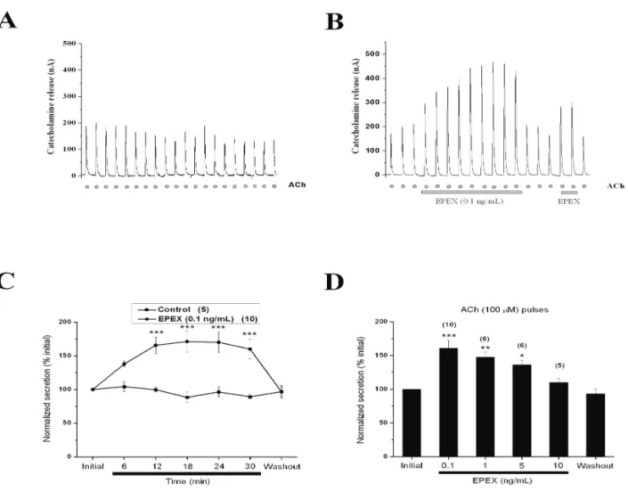
Figure 1A shows a prototypical experiment using repeated 3-s 100 ACh pulses given at 5-min intervals. Note that the peak of the initial secretory spike of 200 nA was maintained fairly constant throughout the experiment. Using this protocol, we surprisingly found that at the low concentration of 0.1 ng/mL, EPEX gradually augmented the ACh-evoked, catecholamine release response, reaching an amplitude 2.5 fold higher than the initial response (Figure 1B). As shown in Figure 1B, basal catecholamine release was not affected by EPEX perfusion; however, upon EPEX washout, the response quickly returned to initial levels and was enhanced once more upon reperfusion of the extract. Pooled data plotted in Figure 1C shows EPEX causing a gradual increase in catecholamine secretion, which was maintained for 15 min and reversed upon EPEX washout.
However, we did not ind an obvious
concentration-dependence for the secretion-enhancing effects of EPEX that seemed to augment this response even more at 0.1 ng/mL than at 1 and 5 ng/mL, although there were no statistical differences between the three concentrations (Figure 1D).
Effects of EPEX on ACh-elicited inward currents
In trying to elucidate the mechanism involved in the enhancement of ACh-evoked catecholamine release elicited by EPEX, we explored the possibility that the extract was modifying the ACh-elicited inward currents (IACh). Thus, we performed experiments in
voltage-clamped cells using the whole-cell coniguration of the
patch-clamp technique.
2001). We performed experiments on the basis of the
following previous indings: i) augmentation of synaptic
transmission at the muscle endplate elicited by EPEX could be due to inhibition of AChE and BuChE (Grangeiro et al., 2006); ii) drugs inhibiting AChE and BuChE have
proven to have beneicial effects in Alzheimer’s disease
patients (Schmidt et al., 2008; van Marum, 2008) and iii) inhibition of AChE and BuChE could explain the EPEX-elicited augmentation of ACh-evoked catecholamine release responses.
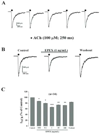
Figure 3 shows the concentration response curves for enzyme inhibition by tacrine, a well-known inhibitor of both AChE and BuChE (Summers et al., 1986). As expected, tacrine inhibited both enzymes in a concentration-dependent manner, with an IC50 for BuChE at 2.0 nM, and 25 nM for AChE. In contrast, the concentrations of EPEX that augmented ACh-evoked 63% of the initial current, and higher concentrations
(10-100 ng/mL) did not produce additional blockade. EPEX washout produced a partial current recovery.
We also tested whether EPEX could affect other ionic channels by measuring the TTX-sensitive sodium current (INa) in voltage-clamped (-80 mV) cells. We found no effect of EPEX on such current at 0.1 and 1 ng/ mL (data not shown).
Effects of EPEX on the activities of AChE and BuChE
Since the effects of EPEX in enhancing ACh-stimulated catecholamine release did not seem to be associated with a direct action on nAChR, we wanted to know whether EPEX affected the activities of two enzymes involved in the rapid degradation of ACh at synaptic sites, AChE and BuChE (Kutty, 1980; Buchwald,
Figure 1. EPEX augments the release of catecholamines triggered by acetylcholine (ACh) pulses. Chromafin cells (5x10⁶) were
trapped in a microchamber and perfused with Krebs-HEPES solution. Once stabilised, the basal catecholamine release was between
10 and 20 nA. (A) Representative experiment in which cells were stimulated with a solution containing 100 μM ACh for 5 s at 3-min
secretion (0.1-1 ng/mL, Figure 1 B and D) or even 1000-fold higher concentrations of EPEX, did not affect the activities of AChE or BuChE.
Figure 2. EPEX causes a concentration-dependent mild inhibition of the inward whole-cell current generated by ACh pulses. Cells were voltage-clamped at -80 mV. Upon breaking
into the cell, ACh pulses (100 μM, 250 ms) were sequentially
applied to each individual cell at 60 s intervals. (A) An example cell stimulated with ACh pulses that produced inward IACh. (B) An example cell showing the IACh traces obtained before (initial), 3 min after perfusion with EPEX, and 3 min after extract washout. (C) Pooled results obtained in cells that were sequentially stimulated with ACh pulses before (initial IACh) and 3 min after perfusion with increasing concentrations of EPEX (bottom horizontal bar). Each concentration was tested in a different cell. Data were normalised as the percent of IACh amplitude obtained initially and are represented as mean±SE
of 34 cells from ive different cultures. *p<0.05, **p<0.01, and ***p<0.001, with respect to initial IACh amplitude.
Effects of EPEX on catecholamine release responses triggered by repeated application of high-K+ pulses
The sequence of events leading to the coupling between ACh stimulation and the secretory response (Douglas, 1968) include cell depolarization, opening of voltage-activated Ca2+ channels (VACC) and exocytosis (Garcia et al., 2006). Because EPEX did not seem to augment secretion through a cholinergic-mediated pathway, we explored the possibility that the extract could act on VACC to enhance secretion. We therefore
triggered secretion with direct depolarization of the cell membrane with a solution containing 70 mM K+ (70K+). At this concentration, the bovine chromafin cell membrane potential depolarizes to near 0 mV (Orozco et al., 2006) and opens all VACC (Garcia et al., 2006).
Figure 3. EPEX did not affect the activities of acetylcholinesterase (AChE) and butyrylcholinesterase (BuChE). Purified AChE from Electrophorus electricus or BuChE from horse serum were incubated with EPEX at the concentrations indicated on the abscissa. Tacrine was used as a positive control. In each individual experiment, the maximal enzyme activities (100%) were determined in the presence of vehicle, and enzyme activities were determined in the presence of EPEX or tacrine, expressed as the percent of maximal activity. Data are represented as mean±SE from eight wells from at least two different experiments.
Effects of EPEX on inward currents through Na+ and
Ca2+ channels
High K+ concentrations will induce a cell membrane depolarization allowing the opening of voltage-gated Na+ channels and VACC to enhance Ca2+ entry, [Ca2+]
c and catecholamine secretion. Thus, EPEX-elicited enhanced secretion could be due to augmentation of Ca2+ entry through VACC. The most direct way of testing this possibility was measuring the whole-cell inward currents through VACC. Thus, we performed experiments aimed to test the possible effects of EPEX on voltage-gated Na+ and Ca2+ channels by recording whole-cell inward currents through these channels.
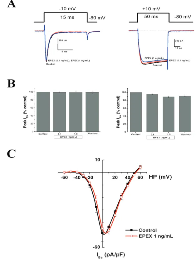
Figure 5A shows original recordings of INa (left panel) and IBa (right panel) elicited by 15 or 50 ms
depolarizing pulses (see protocol on top or the original recordings), in the absence and/or the presence of EPEX (0.1 and 1 ng/mL). The peak Na+ current density in cells from different batches was 42.15±6.65 pA/pF (n=8) and the peak Ba2+ current density in cells from different batches was 45.63±4,13 pA/pF (n=15). Averaged results obtained with these experimental protocols showed that INa was not signiicantly modiied in the presence of EPEX (left panel in Figure 5B), and only a mild (around 10%),
not signiicant, decrease of IBa at 0.1-1 ng/mL EPEX was observed. Figure 5C shows current-voltage curves generated by increasing voltage-step depolarizations. Peak current was reached at 0-+10 mV and was reduced by 5-10% in the presence of 1 ng/mL EPEX; however, no shift of the current-voltage (I-V) curve was found during cell treatment with the extract.
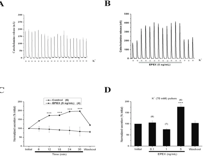
Figure 4. EPEX augmented the release of catecholamines from cells repeatedly stimulated with K+-enriched solutions. Cells
were initially perfused with Krebs-Hepes solution and, once baseline secretion was stable, cells were challenged for 5 s every 3 min with a solution containing 70 mM K+ (with low Na+) (dots at the bottom). (A) An example cell showing initial
secretion spikes of 200 nA. This spike amplitude was maintained during the first five initial pulses and declined by 20% at the end of the experiment. (B) An example cell perfused with EPEX during the period indicated by the bottom horizontal bar. (C) Pooled data obtained in different experiments made with the protocols shown in panels A (control) and B (EPEX-treated cells). (D) The effects of increasing concentrations of EPEX on K+-evoked secretion. Data in panels C and D are normalised
Effects of EPEX on the elevations of cytosolic calcium concentrations ([Ca2+]
c ) elicited by ACh
In bovine chromafin cells, ACh pulses cause
depolarisation and action potentials that give rise to transient [Ca2+]c elevations (de Diego et al., 2008). Such Ca2+ signals are the result of Ca2+ entry through nAChR via VACC, or from Ca2+ release from the endoplasmic reticulum (Garcia et al., 2006). Thus, we tested whether EPEX could alter this ACh-elicited Ca2+ signal to explain its ability to augment depolarization-evoked secretion.
Fluo-4-loaded cells were challenged with ACh pulses in the absence or the presence of increasing concentrations of EPEX. Figure 6A shows two
luorescence records indicating [Ca2+]
c elevations elicited by ACh (bottom black horizontal bar), before (left) and during the application of 0.1 ng/mL EPEX, as indicated by the bottom horizontal grey bar. EPEX slightly augmented the rate and amplitude of the ACh-evoked [Ca2+]
c elevation. Pooled results are given in Figure 6A, showing that at 0.1 and 0.3 ng/mL, EPEX augmented the [Ca2+]
c elevation by 7 and 12%, respectively (not
signiicant). At greater concentrations, the extract did not
change the ACh-evoked [Ca2+]
c elevation.
Discussion
In this study, we found that an extract of the plant E. punicifolia (EPEX) caused an augmentation of catecholamine release from bovine adrenal medullary cells challenged with ACh (Figure 1) or high K+ (Figure 4). Both type of stimuli are widely used to trigger secretion (both induce cell membrane depolarization and subsequently Ca2+ entry through voltage-gated Ca2+ channels, but they differ in the fact that ACh-induced catecholamine secretion implies the activation of nAChR and indirectly depolarize cell membrane, while high K+ serves to directly depolarise cell membrane (Garcia et al., 2006). We also used electrophysiological and Ca2+ imaging techniques to determine the mechanisms underlying this potentiation.
Augmentation of ACh-evoked secretion could be due to a direct action of EPEX on nAChR by behaving
as an allosteric modulator of α7 nAChR (Faghih et al., 2007) that have been identiied in bovine chromafin
cells (Quik et al., 1987; Geertsen et al., 1988; Criado et al., 1997; Lopez et al., 1998; Fuentealba et al., 2004). However, this does not seem to be the case because i) EPEX did not augment IACh, but it mildly inhibited IACh at concentrations that augmented exocytosis, suggesting that the potentiating effects of EPEX was exerted beyond the nAChR (Figure 2) and ii) EPEX also augmented the K+-evoked secretion (Figure 4), a stimulus that directly depolarises the cells without intervention of nAChR.
In chromafin cells, the L-subtype VDCC
mediates the secretion elicited by K+ depolarization, and this response is augmented by the L-type Ca2+ channel activator Bayk 8644 (Garcia et al., 1984). Thus, we considered the possibility that, similar to Bayk 8644, EPEX was acting by enhancing Ca2+ entry through L-channels; however, EPEX mildly reduced ICa (Figure 5). Although EPEX slightly increased the [Ca2+]
c elevations induced by ACh, this was not signiicant (Figure 6). One possibility is that EPEX could augment
the quantal content of individual chromafin vesicles and/
or of the single secretory spike burst, by actin at some of the initial steps of exocytosis. This will be tested in future experiments on quantal catecholamine release at
the single cell level, using a carbon ibre microelectrode
(Miranda-Ferreira et al., 2008; Miranda-Ferreira et al., 2009)
Although we have not yet uncovered the mechanism of action of EPEX, this study has served to unravel an entirely new biological activity for this plant extract, augmentation of exocytosis. In Brazil, E. punicifolia extracts are employed in Amazon region to treat diarrhea and stomach disturbances, and as hypoglycemic medicine (Brito et al., 2007; Bopp et al., 2009). The mechanism underlying pharmacological properties of the genus Eugenia are partially attributed to
lavonoids (myricitrin, quercetin, and quercetrin), steroids,
terpenoids, tanines, and anthraquinones (Consolini & Sarubbio, 2002). We keep doing experiments on the dichloromethane extract used in the presents tudy to gain further information on the composition and to identify the component/s that could be responsible of the observed effects on the cholinergic neurotransmission.
Its use for the treatment of diabetes could be linked to the augmentation of the exocytotic release of insulin in response to hyperglycemia. Also, it is interesting that hypoglycaemic drugs block a type of ATP-dependent potassium channel, and we cannot rule out that a ligand present in the E. punicifolia extract may bind to these potassium channels to elicit an effect. Unfortunately, this
type of channel is not present in bovine chromafin cells
and hence we should not explore such possibility. Related to the possibility that EPEX could be acting on other K+ channel types to regulate the catecholamine secretory response, our laboratory found that SK (small conductance calcium-activated K+ channels) and BK (big conductance calcium-activated K+ channels) were involved in the modulation of exocytosis, due to an indirect effect, by Ca2+ entering through VDCCs (Uceda et al., 1994; Lara et al.,
1995). However, we did not ind any effect of EPEX on
such currents (not shown).
We have discovered a simple and eficient
submilligram concentrations are suficient to analyse
exocytotic release of catecholamines from bovine
chromafin cells, testing different fractions of puriied
extracts can be performed.
In addition to the identiication of the active compound in EPEX, our indings have also interested
us to search for new biological targets and ligands with potential therapeutic interest in neurodegenerative
diseases, particularly in Alzheimer’s disease (Raii &
Aisen, 2009). A compound that augments exocytosis might also enhance neurotransmission, synaptic plasticity and neuronal survival. If the molecular structure of the active compound present in EPEX could eventually be
identiied, it could become a prototype for the design
and synthesis of new compounds capable of improving synaptic plasticity and neuronal communication.
In conclusion, we have discovered that an Eugenia punicifolia extract augments the exocytotic
catecholamine release from bovine adrenal chromafin
cells stimulated with ACh or K+. Using catecholamine release as a biological probe in subsequent studies, could facilitate the isolation of the active compound and the elucidation of its molecular structure. This could be used as a template to synthesise new derivatives with therapeutic potential as cognition enhancers in Alzheimer’s disease patients.
Acknowledgements
This work was partially supported by grants from the Ministerio de Ciencia e Innovación (SAF2010-21795 to AGG, SAF2010-18837 to LG, and
SAB2006-Figure 6. EPEX caused a mild augmentation of cytosolic calcium concentration ([Ca2+]
c) elevations elicited by ACh. Cells
loaded with Fluo-4 in a 96-well black plate were challenged with ACh in the absence (control) and the presence of increasing concentrations of EPEX. (A) Original [Ca2+]
c traces evoked by ACh in untreated cells (control, left) and cells incubated with
0123 to WCS), Comunidad Autónoma de Madrid (S-SAL-0275-2006), and CEAL-SANTANDER (to LG). We thank the Fundación Teófilo Hernando (Spain) and FAPERJ (Brasil) for funding and continued support. Authors are also indebted to Centro de Instrução de Guerra na Selva, Manaus-AM, Brasil, for the provision of plants. WCS, KGLA and VFF are members of the INCT de Processos Redox em Biomedicina funded by CNPq/FAPESP (Brazil).
References
Bopp A, De Bona KS, Belle LP, Moresco RN, Moretto MB 2009. Syzygium cumini inhibits adenosine deaminase activity and reduces glucose levels in hyperglycemic patients. Fundam Clin Pharmacol 23: 501-507. Borges R, Sala F, Garcia AG 1986. Continuous monitoring of
catecholamine release from perfused cat adrenals. J Neurosci Methods 16: 289-300.
Brito FA, Lima LA, Ramos MF, Nakamura MJ, Cavalher-Machado SC, Siani AC, Henriques MGMO, Sampaio ALF 2007. Pharmacological study of anti-allergic activity of Syzygium cumini (L.) Skeels. Braz J Med Biol Res 40: 105-115.
Buchwald P 2001. Structure-metabolism relationships: steric effects and the enzymatic hydrolysis of carboxylic esters. Mini Rev Med Chem 1: 101-111.
Consolini AE, Sarubbio MG 2002. Pharmacological effects of
Eugenia unilora (Myrtaceae) aqueous crude extract on rat's heart. J Ethnopharmacol 81: 57-63.
Criado M, Dominguez del Toro E, Carrasco-Serrano C, Smillie FI, Juiz JM, Viniegra S, Ballesta JJ 1997. Differential expression of alpha-bungarotoxin-sensitive neuronal
nicotinic receptors in adrenergic chromafin cells: a
role for transcription factor Egr-1. J Neurosci 17: 6554-6564.
Cuchillo-Ibáñez I, Olivares R, Aldea M, Villarroya M, Arroyo G, Fuentealba J, García AG, Albillos A 2002. Acetylcholine and potassium elicit different patterns
of exocytosis in chromafin cells when the intracellular
calcium handling is disturbed. Plugers Arch 444: 133-142.
de Diego AM, Arnaiz-Cot JJ, Hernandez-Guijo JM, Gandia L, Garcia AG 2008. Differential variations in Ca2+ entry,
cytosolic Ca2+ and membrane capacitance upon steady
or action potential depolarizing stimulation of bovine
chromafin cells. Acta Physiol (Oxf) 194: 97-109. de los Rios C, Egea J, Marco-Contelles J, Leon R, Samadi A,
Iriepa I, Moraleda I, Galvez E, Garcia AG, Lopez MG, Villarroya M, Romero A 2010. Synthesis, inhibitory
activity of cholinesterases, and neuroprotective proile
of novel 1,8-naphthyridine derivatives. J Med Chem 53: 5129-5143.
de los Rios C, Marco JL, Carreiras MD, Chinchon PM, Garcia AG, Villarroya M 2002. Novel tacrine derivatives that
block neuronal calcium channels. Bioorg Med Chem 10: 2077-2088.
Douglas WW 1968. Stimulus-secretion coupling: the concept
and clues from chromafin and other cells. Br J Pharmacol 34: 453-474.
Ellman GL, Courtney KD, Andres V, Jr., Feather-Stone RM 1961. A new and rapid colorimetric determination of acetylcholinesterase activity. Biochem Pharmacol 7: 88-95.
Faghih R, Gfesser GA, Gopalakrishnan M 2007. Advances in the discovery of novel positive allosteric modulators of the alpha7 nicotinic acetylcholine receptor. Recent Pat CNS Drug Discov 2: 99-106.
Fuentealba J, Olivares R, Ales E, Tapia L, Rojo J, Arroyo G, Aldea M, Criado M, Gandi L, Garci AG 2004. A choline-evoked [Ca2+]
c signal causes catecholamine release and
hyperpolarization of chromafin cells. Faseb J 18: 1468-1470.
Garcia AG, De-Diego AM, Gandia L, Borges R, Garcia-Sancho J 2006. Calcium signaling and exocytosis in
adrenal chromafin cells. Physiol Rev 86: 1093-1131. Garcia AG, Sala F, Reig JA, Viniegra S, Frias J, Fonteriz R,
Gandía L 1984. Dihydropyridine BAY-K-8644 activates
chromafin cell calcium channels. Nature 309: 69-71. Geertsen S, Afar R, Trifaro JM, Quik M 1988. Regulation of
alpha-bungarotoxin sites in chromafin cells in culture
by nicotinic receptor ligands, K+, and cAMP. Mol Pharmacol 34: 549-556.
Grangeiro MS, Calheiros-Lima AP, Martins MF, Arruda LF, Garcez-do-Carmo L, Santos WC 2006. Pharmacological effects of Eugenia punicifolia (Myrtaceae) in cholinergic nicotinic neurotransmission. J Ethnopharmacol 108: 26-30.
Guillou C, Mary A, Renko DZ, Gras E, Thal C 2000. Potent acetylcholinesterase inhibitors: design, synthesis and structure-activity relationships of alkylene linked bis-galanthamine and bis-galanthamine-galanthaminium salts.
Bioorg Med Chem Lett 10: 637-639.
Hamill OP, Marty A, Neher E, Sakmann B, Sigworth FJ 1981. Improved patch-clamp techniques for high-resolution current recording from cells and cell-free membrane patches. Plugers Arch 391: 85-100.
Kutty KM 1980. Biological function of cholinesterase. Clin Biochem 13: 239-243.
Lara B, Zapater P, Montiel C, de la Fuente MT, Martinez-Sierra R, Ballesta JJ, Gandía L, García AG 1995. Density of apamin-sensitive Ca(2+)-dependent K+ channels in
bovine chromafin cells: relevance to secretion. Biochem Pharmacol 49: 1459-1468.
Leon R, Marco-Contelles J, Garcia AG, Villarroya M 2005. Synthesis, acetylcholinesterase inhibition and neuroprotective activity of new tacrine analogues.
Bioorg Med Chem 13: 1167-1175.
Livett BG, Sandall DW, Keays D, Down J, Gayler KR, Satkunanathan N, Khalil Z 2006. Therapeutic applications of conotoxins that target the neuronal nicotinic acetylcholine receptor. Toxicon 48: 810-829. Lopez MG, Montiel C, Herrero CJ, Garcia-Palomero E,
Mayorgas I, Hernandez-Guijo JM, Villarroya M, Olivares R, Gandía L, McIntosh JM, Oliveira BM, García AG 1998. Unmasking the functions of the
chromafin cell alpha7 nicotinic receptor by using short
pulses of acetylcholine and selective blockers. Proc Natl Acad Sci U S A 95: 14184-14189.
Marcaurelle LA, Johannes CW 2008. Application of natural product-inspired diversity-oriented synthesis to drug discovery. Prog Drug Res 66: 187-216.
Marco-Contelles J, do Carmo Carreiras M, Rodriguez C, Villarroya M, Garcia AG 2006. Synthesis and pharmacology of galantamine. Chem Rev 106: 116-133.
Marco L, do Carmo Carreiras M 2006. Galanthamine, a natural product for the treatment of Alzheimer's disease. Recent Pat CNS Drug Discov 1: 105-111.
Miranda-Ferreira R, de Pascual R, Caricati-Neto A, Gandia L, Jurkiewicz A, Garcia AG 2009. Role of the endoplasmic reticulum and mitochondria on quantal catecholamine release from chromaffin cells of control and hypertensive rats. J Pharmacol Exp Ther 329: 231-240.
Miranda-Ferreira R, de Pascual R, de Diego AM, Caricati-Neto A, Gandia L, Jurkiewicz A, García AG 2008. Single-vesicle catecholamine release has greater quantal
content and faster kinetics in chromafin cells from
hypertensive, as compared with normotensive, rats. J Pharmacol Exp Ther 324: 685-693.
Moro MA, López MG, Gandía L, Michelena P, García AG 1990. Separation and culture of living adrenaline- and noradrenaline-containing cells from bovine adrenal medullae. Anal Biochem 185: 243-248.
Orozco C, Garcia-de-Diego AM, Arias E, Hernandez-Guijo JM, Garcia AG, Villarroya M, López MG 2006. Depolarization preconditioning produces cytoprotection
against veratridine-induced chromafin cell death. Eur J
Pharmacol 553: 28-38.
Quik M, Geertsen S, Trifaro JM 1987. Marked up-regulation of
the beta-bungarotoxin site in adrenal chromafin cells by speciic nicotinic antagonists. Mol Pharmacol 31: 385-391.
Raii MS, Aisen PS 2009. Recent developments in Alzheimer's
disease therapeutics. BMC Med 7: 7.
Santos WC, Hernandez-Guijo JM, Ruiz-Nuno A, Olivares R, Jurkiewicz A, Gandia L, García AG 2001. Blockade by
agmatine of catecholamine release from chromafin cells
is unrelated to imidazoline receptors. Eur J Pharmacol 417: 99-109.
Schmidt R, Neff F, Lampl C, Benke T, Anditsch M, Bancher C, Dal-Bianco P, Reisecker F, Marksteiner J, Rainer M, Kepeller P, Dodel R 2008. Therapy of Alzheimer's disease: current status and future development.
Neuropsychiatr 22: 153-171.
Summers WK, Majovski LV, Marsh GM, Tachiki K, Kling A 1986. Oral tetrahydroaminoacridine in long-term treatment of senile dementia, Alzheimer type. N Engl J Med 315: 1241-1245.
Tapia L, Garcia-Eguiagaray J, Garcia AG, Gandia L 2009.
Preconditioning stimuli that augment chromafin cell
secretion. Am J Physiol Cell Physiol 296: C792-800. Uceda G, Artalejo AR, de la Fuente MT, Lopez MG, Albillos A,
Michelena P, Garcia AG, Montiel C 1994. Modulation by L-type Ca2+ channels and apamin-sensitive K+
channels of muscarinic responses in cat chromafin
cells. Am J Physiol 266: C1432-1439.
van Marum RJ 2008. Current and future therapy in Alzheimer's disease. Fundam Clin Pharmacol 22: 265-274.
*Correspondence
Luis Gandía
Instituto Teóilo Hernando de I+D del Medicamento, Facultad
de Medicina, Universidad Autónoma de Madrid Arzobispo Morcillo, 4; 28029 Madrid, Spain [email protected]