Article
ISSN 0102-695X http://dx.doi.org/10.1590/S0102-695X2011005000184 Received 20 Jan 2011 Accepted 12 Jul 2011 Available online 7 Oct 2011
of lichen
Toninia candida
Nedeljko T. Manojlovic,
*,1Perica J. Vasiljevic,
2Pavle Z.
Maskovic
31Department of Pharmacy, Medical Faculty, University of Kragujevac, Serbia,
2Department of Biology, Faculty of Science, University of Nis, Serbia,
3Faculty of Agronomy, University of Kragujevac, Serbia.
Abstract: In the present investigation, methanol, chloroform and petrol ether extracts from the lichen Toninia candida (Weber) Th. Fr, Catillariaceae, were assayed for their antioxidant activity. The phenolic composition of the extracts was determined by HPLC-UV analysis. The predominant phenolic compound in all the extracts was depsidone, norstictic acid. All the tested extracts of T. candida
contain, besides norstictic acid, atranorin, stictic, protocetraric and usnic acid, but in different amounts and relations. The lichen extracts showed comparable and strong antioxidant activity, exhibited higher DPPH and hydroxyl radical scavengings, chelating activity and inhibitory activity towards lipid peroxidation. This is the first report of chemical composition and antioxidant antimicrobial activity of the lichen
Toninia candida.
Keywords:
antioxidant activity chemical composition HPLC
Toninia candida
Introduction
Lichens are valuable plant resources and are used as food, fodder, medicine, dyes perfume, spice, and for miscellaneous purposes throughout the world. More than one thousand primary and secondary metabolites with identii ed structures are currently known in lichens (Shukla et al., 2010). The use of lichens in medicine is based on the fact that they contain unique and varied biologically active substances. Lichen metabolites exert a wide variety of biological actions including antibiotic, antimycobacterial, antiviral, anti-inl ammatory, analgesic, antipyretic, antiproliferative and cytotoxic effects (Huneck, 1999; Shukla et al., 2010; Manojlovic et al., 2002; Manojlovic et al., 2010a; Manojlovic et al., 2010b; Manojlovic et al., 2010c). Even though these manifold activities of lichen metabolites have now been recognized, their therapeutic potential has not yet been fully explored and thus remains pharmaceutically unexploited.
The most numerous classes of secondary metabolites are depsides and depsidones. Depside molecules consist of 2-4 hydroxybenzoic acid residues linked by ester groups. Many depsides reported in literature have been found to possess important physiological properties. The antioxidant property has been reported in depsides isolated from various lichen species (Hidalgo et al., 1994). More than one hundred compounds are depsidones, which have an additional ether bond between aromatic rings. Depsidones in lichen
are believed to arise by oxidative cyclisation of depsides. It has been found that depsidones are more efi cient antioxidants than depsides. The higher efi ciency of the depsidones could be related to a larger incorporation into lipidic microdomains (Hidalgo et al., 1994). Depsidone and depside compounds such as pannarin, 10-chloropannarin and sphaerophorin, tested in cell cultures of lymphocytes, were shown to have a higher cytotoxic effect than colchicine (Correche´ et al., 2002). The depsidones salazinic acid, stictic acid and psoromic acid were the most apoptotic active derivatives among i fteen lichen compounds evaluated on primary cultures of rat hepatocytes (Correche´ et al., 2002). Toninia candida
(Weber) Th. Fr, Catillariaceae, is widely distributed in continental areas in the Northern Hemisphere (Timdal, 1991). In Serbia, T. candida could be found in Jelasnica gorge (Mt. Suva Planina) on a vertical limestone rocks.
Thus, the aim of the present work was to identify and quantify phenolic acids composition of
Toninia candida lichen by HPLC-UV and to evaluate the antioxidant capacity of methanol, chloroform and petrol ether extracts from this lichen using different systems, including DPPH and hydroxyl radical scavenging, metal chelating activity, as well as to screen their antimicrobial activity.
Material and Methods
The lichen material of Toninia candida (Weber) Th. Fr, Catillariaceae, was collected from Mt. Suva (Jelasnica gorge) in Serbia during October 2010. A voucher specimen (HMN 5459) has been deposited at the Herbarium Moesiacum Nis in the Department of Biology and Ecology, Faculty of Sciences and Mathematics, University of Nis, Serbia.
Preparation of the lichen extracts
The lichen material was air-dried at room temperature (26 °C) for one week, after which it was ground to a uniform powder. Each extract (methanol, chloroform and petrol ether) extract was prepared by soaking 500 g dry powdered lichen material in 2000 mL of solvent at room temperature for three days. The extracts were iltered through a Whatman no. 42 (125 mm) ilter paper and concentrated using a rotary evaporator.
Instrumentation and conditions
HPLC analysis was carried out on an Agilent 1200 Series HPLC instrument with C18 column (C18; 25 cm x 4.6 mm, 10 m) and a UV spectrophotometric detector with methanol-water-phosphoric acid (80:20:0.9, v/v/v) solvent. Methanol was of HPLC grade and was purchased from Merck (Darmstadt, Germany). Phosphoric acid was analytical-grade reagent. Deionized water used throughout the experiments was generated by a Milli-Q academic water puriication system (Milford, MA, USA). The sample injection volume was 10 mm3. The low rate
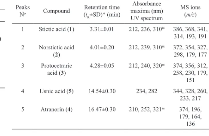
was 1.0 mL/min. The standards used were obtained from the following sources: norstictic acid (2, tR=4.01±0.20 min) was isolated from lichen Ramalina furinacea, stictic acid (1, tR=3.31±0.01 min) from lichen Xanthoparmelia conspersa, protocetraric acid (3, tR=4.28±0.05 min) from lichen Cetraria islandica, usnic acid (5, tR=14.54±0.30 min) and atranorin (6, tR=16.47±0.30 min) from lichen
Evernia prunastri. The standard samples were isolated in our laboratory and their structures were conirmed by spectral data. The retention times and UV spectra of these standards are shown in Table 2 and Figure 1.
Reference solutions of decreasing concentrations were obtained by dilution with eluent of the requisite standard solution. These solutions were analyzed and the corresponding peak areas plotted against the concentration of acid injected. The concentrations of the components in the analyzed samples (were calculated from the chromatogram peak areas using the normalization method. The identiication of the different compounds was achieved by comparison of both tR and the absorption spectra obtained for each diluted peak with those obtained for the standards. The data obtained were statistically processed by using a standard non-parametric variance analysis method (ANOVA) for determining signiicant intergroup
differences. Data are presented as the mean±standard deviation (SD) of three separate experiments performed on different samples.
LC-MS/MS analysis was carried out using an Agilent 1100 LC system consisting of degasser, binary pump, auto sampler, and column heater. The column outlet was coupled to an Agilent MSD Ion Trap XCT mass spectrometer equipped with an ESI ion source. For the chromatographic separation, a Zorbax 300Ĺ Extend-C-18 Column (2.1×150 mm, 1.8 μm ) was used. The mobile phase was pumped at 1 mLmin-1 low rate and consisted
of methanol-water-formic acid (80:20:0.9, v/v/v).
Determination of the total phenolics
The total phenolics content was determined using the Folin-Ciocalteau method (Singleton et al.,1999). Extract was diluted to the concentration of 1 mg/mL, and aliquots of 0.5 mL were mixed with 2.5 mL of FC reagent (previously diluted 10-fold with distilled water) and 2 mL of NaHCO3 (7.5%). After 15 min of staying at the 45 °C the absorbance was measured at 765 nm on spectrophotometer versus blank sample. Total phenols were determined as gallic acid equivalents (mg GA/g extract), and the values are presented as means of triplicate analyses.
Determination of total antioxidant capacity
The total antioxidant activity of the
Toninia candida extract was evaluated by the phosphomolybdenum method (Prieto et al., 1999). The assay is based on the reduction of Mo (VI)-Mo (V) by the antioxidant compounds and subsequent formation of a green phosphate/Mo (V) complex at acid pH. 0.3 mL of sample extract was combined with 3 mL of reagent solution (0.6 M sulfuric acid, 28 mM sodium phosphate and 4 mM ammonium molybdate). The tubes containing the reaction solution were incubated at 95 °C for 90 min. Then the absorbance of the solution was measured at 695 nm using spectrophotometer against blank after cooling to room temperature. Methanol (0.3 mL) in the place of extract was used as the blank. Ascorbic acid (AA) was used as standard and the total antioxidant capacity is expressed as milligrams of ascorbic acid per gram of the dry extract.
Determination of DPPH free radical scavenging activity
(2 mL) and allowed to stand for 30 min for any reaction to occur, and the absorbance was measured at 517 nm. Ascorbic acid (AA), gallic acid (GA) and butylated hydroxytoluene (BHT) were used as reference standards and dissolved in methanol to make the stock solution with the same concentration (1 mg/mL). Control sample was prepared containing the same volume without test compounds or reference antioxidants. Methanol 95% was used as blank. The DPPH free radical scavenging activity (%) was calculated using the following equation:
% inhibition =
x 100
The IC50 value, which is the concentration of the test material that reduces 50% of the free radical concentration, was calculated as µg/mL through sigmoidal dose-response curve.
Determination of the inhibitory activity toward lipid peroxidation
The antioxidant activity was determined by the thiocyanate method (Hsu et al., 2008). Serial dilutions were carried out with the stock solution (1 mg/mL) of the extracts, and 0.5 mL of each solution was added to linoleic acid emulsion (2.5 mL, 40 mM, pH 7.0). The linoleic acid emulsion was prepared by mixing 0,2804 g linoleic acid, 0.2804 g Tween-20 as emulsiier in 50 mL 40 mM phosphate buffer and the mixture was then homogenized. The inal volume was adjusted to 5 mL with 40 mM phosphate buffer, pH 7.0. After incubation at 37 °C in the dark for 72 h, a 0.1 mL aliquot of the reaction solution was mixed with 4.7 mL of ethanol (75%), 0.1 mL FeCl2 (20 mM) and 0.1 mL ammonium thiocyanate (30%). The absorbance of this mixture was measured at 500 nm, after it was stirred for 3 min. Ascorbic acid, gallic acid, α-tocopherol and BHT were used as a reference compounds. To eliminate the solvent effect, the control sample, which contained the same amount of solvent added to the linoleic acid emulsion in the test sample and reference compound, was used. Inhibition percent of linoleic acid peroxidation was calculated using following formula:
% inhibition = x 100
Measurement of ferrous ion chelating ability
The ferrous ion chelating ability was measured by the decrease in absorbance at 562 nm of the iron (II)-ferrozine complex (Carter, 1971; Yan et al., 2006). One milliliter of 0.125 mM FeSO4 was added to 1.0 mL sample (with different dilutions), followed by 1.0 mL of 0.3125 mM ferrozine. The mixture was allowed to equilibrate for
10 min before measuring the absorbance. The ability of the sample to chelate ferrous ion was calculated relative to the control (consisting of iron and ferrozine only) using the formula:
Chelating effect (%) = x 100
Determination of hydroxyl radical scavenging activity
The ability of Toninia candida to inhibit non site-speciic hydroxyl radical-mediated peroxidation was carried out according method described by Hinneburg et al. (2006). The reaction mixture contained 100 µL of extract dissolved in water, 500 µL of 5.6 mM 2-deoxy-D-ribose in KH2PO4-NaOH buffer (50 mM, pH 7.4), 200 µL of premixed 100 µM FeCl3 and 104 mM EDTA (1:1 v/v) solution, 100µL of 1.0 mM H2O2 and 100 µL of 1.0 mM aqueous ascorbic acid. Tubes were vortexed and incubated at 50 °C for 30 min. Thereafter, 1 mL of 2.8% TCA and 1 mL of 1.0% TBA were added to each tube. The samples were vortexed and heated in a water bath at 50 °C for 30 min. The extent of oxidation of 2-deoxyribose was estimated from the absorbance of the solution at 532 nm. The percentage inhibition values were calculated from the absorbance of the control (Ac) and of the sample (As), where the controls contained all the reaction reagents except the extract or positive control substance. The values are presented as the means of triplicate analyses.
Statistical analysis
All the results are presented as mean±standard deviations of three determinations. Statistical analyses were performed using Student´s t-test and one way analysis of variance. Multiple comparisons of means were done by LSD (least signiicant difference) test. A probability value of 0.05 was considered signiicant. All computations were made by employing the statistical software (SPSS, version 11.0). IC50 values were calculated determined by nonlinear regression analysis from the sigmoidal dose-response inhibition curve.
Results and Discussion
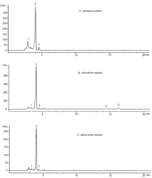
A representative chromatograms for standards and Toninia candida methanol, chloroform and petrol ether extracts eluted by HPLC are represented in Figure 1 and Figure 2. As it is evidenced in the chromatograms, there were the presence of depsidones as the most abundant substance class in the extracts examined. As the most abundant depsidone, norstictic acid (NOR) (tR=4.01±0.2 min, 2) was identiied. This compound was previously reported from the family Umbilicariaceae (Narui et al., Ac - As
Ac
Ac - As
Ac
Ac - As
1996) and had been reported to demonstrate antimicrobial effect on Bacillus subtilis, Listeria monocytogenes, Proteus vulgaris, Staphylococcus aureus, Streptococcus faecalis, Aeromonas hydrophila, Candida albicans and
Candida glabrat (Tay et al., 2004).
As can be seen in chromatograms, beside norstictic acid (2), stictic acid (tR=3.31±0.01 min, 1), protocetraric acid (tR=4.28±0.05 min, 3), usnic acid (tR=14.54±0.30 min, 5) and atranorin (tR=16.47±0.30 min, 4) were also identiied. Four detected compounds belonging to the depsidones. Atranorin is depside and usnic acid is antibiotic with dibenzofurane structure. The UV spectra of depsidones have three absorption maxima and are dissimilar from those of depsides and monocyclic compounds. The UV spectra of protocetraric acid are very similar to those of norstictic acid. Absorbance
maxima at 234 and 282 nm are characteristic for usnic acid. Except norstictic acid, other compounds were found in the extracts in small amounts. Identiication of these compounds was achieved by comparison of their tR values with the standard substances previously isolated from lichens. The UV absorbance spectral data (200-400 nm) also corresponded with those of standards and found in Refs. (Yoshimura at al., 1994; Huneck & Yoshimura, 1996). LC-MS analysis was also used to conirm the structures of detected molecules. Values of molecular and fragmentation ions were consistent with those published in literature (Huneck & Yoshimura, 1996). Table 2. shows the retention time of the detected lichen substances, their absorbance maxima (nm) and fragmentation ions. All compounds are identiied for the irst time in this lichen.
Figure 1. Chromatogram of the standards used for identiication of the compounds present in the Toninia candida. In detel can be observed the UV spectra of these compounds (200-400 nm).
Table 1. The amounts of presented compounds in the lichen
Toninia candida.
Compound Substance class
μg/g DW
MET CHL PET
Stictic acid (1) Depsidone 2.0±0.2 5.2±0.5 4.7±0.2 Norstictic acid (2) Depsidone 51.5±1.0 53.6±0.5 64.3±1.0 Protocetraric
acid (3) Depsidone 0.6±0.4 2.5±0.2 2.1±0.4 Usnic acid (5) Dibenzofurane - 0.6±0.1
-Atranorin (4) Depside - 0.8±0.1
-Table 2. Retention time of the examined lichen substances and their absorbance maxima (nm) and fragmentation ions.
Peaks
No Compound
Retention time (tR±SD)* (min)
Absorbance maxima (nm) UV spectrum
MS ions (m/z)
1 Stictic acid (1) 3.31±0.01 212, 236, 310m 386, 368, 341,
314, 193, 191 2 Norstictic acid
(2)
4.01±0.20 212, 239, 310m 372, 354, 327,
298, 179, 177 3 Protocetraric
acid (3)
4.28±0.05 212, 240, 320m 374, 356, 312,
258, 230, 179, 151 4 Usnic acid (5) 14.54±0.30 234, 282 344, 328, 260,
233, 217 5 Atranorin (4) 16.47±0.30 210, 252, 321m 374, 196,
Figure 2. HPLC hromatograms acquired at 256 nm of the methanol (A), chloroform (B) and petrol etar (C) extracts of Toninia candida. Chromatographic peaks identities are reported in Table 2.
Phenolic compounds have been reported to be associated with antioxidative action in biological systems, mainly due to their red-ox properties, which can play an important role in absorbing and neutralizing free radicals, quenching singlet and triplet oxygen, or decomposing peroxides (Saha et al., 2008). The results of determination of total phenolic and antioxidant capacity are given in Table 3. Total phenolic contents were determined and amounted to 76.26±0.32 mg GA/g, 45.25±0.72 mg GA/g and 42.98±0.15 mg GA/g, for methanol, chloroform and petrol ether extracts, respectively. The results showed that the methanolic, chloroform and petroleum ether extracts
possess antioxidant activity, with total antioxidant capacity of 78.45±0.58 μg AA/g, 56.67±0.30 μg AA/g and 51.45±0.31 μg AA/g, respectively.
that all tested extracts exhibited signiicant inhibitory activity towards lipid peroxidation (from 21.45±1.55 μg/ mL to 46.46±1.68 μg/mL). The results of metal chelating activity are also shown in the Table 4 and these values were very similar for all tested extracts.
The results of determination of hydroxyl radical scavenging activity (Table 4) showed that IC50 values were 67,11±0.23, 53.23±0.51 and 50.57±0.75 μg/mL for metanolic, chloroform and petrol ether extracts, respectively. These results revealed that the methanol, chloroform and petrol ethar extracts of T. candida organs were free radical scavengers, acting possibly as primary antioxidants. The strong antioxidant activity of T. candida
assessed by the different systems could be attributed to their high total polyphenolic contents. Moreover, the high yield of the different phenolic compounds (depsidones) found in T. candida thallus (Table 1) might contribute to the potent antioxidant activity of the tested extracts, since a positive correlation between phenolic composition and antioxidant activity was proved (Katalinic et al., 2006). Thus, antioxidant property of the lichen could be attributed to the signiicant amount of depsidones, especially norstictic acid, present in our study with the high amounts of 51.5±2.0, 53.6±1.0 and 64.3±1.5 μg/g
DW, respectively in the methanol, chloroform and petrol ether extracts. Other minor phenolic compounds should not be neglected, since synergy of the different chemicals with each other should be taken into consideration for the biological activity. The presence of the phenolic groups in the lichen metabolites is considered to be key element for the antioxidative eficiency (Markovic & Manojlovic, 2010). Norstictic acid posesses two phenolic groups in the molecule which probably play an important role in expression of their antioxidant activity.
In conclusion, this is the irst study focused on the chemical composition and biological activities of
T. candida. The methanol, chloroform and petrol ether extracts of the lichen showed signiicant antioxidant activity. Four depsidones, one depside and usnic acid were identiied, and norstictic acid was the dominant phenolic compound in the lichen. The present study provides data for supporting the use of T. candida
extracts as natural antioxidant agents, and conirms that these extracts represent a signiicant source of phenolic compounds. Future investigation will be focused on isolation of phenolic compounds and determination of their biological activities in vitro and in vivo.
O O O
O CHO H3CO
CH3
CH3
OH
HO O
1
O O O
O CHO HO
CH3
CH3
OH
HO O
2
O O O
CHO HO
CH3
OH
3
OH
H3C CO2H
O
O OH OHC
HO CH3
CH3
OH
CH3
CO2CH3
4
O H3C OH
HO
H3C O
OH CH3 O
CH3 O
5
Table 3. Total phenolic and total antioxidant capacity of the examined Toninia candida extracts
Total phenolic content (mg GA/g) Total antioxidant capacity (μg AA/g)
methanol extract chloroform extract petrol ether extract methanol extract chloroform extract petrol ether extract
Acknowledgements
This work was supported by the Ministry of Science and Environment of Serbia, projects No 172015.
References
Carter P 1971. Spectrophotometric determination of serum iron at the submicrogram level with a new reagent-ferrozine.
Anal Biochem 40: 450-458.
Correché E, Carrasco M, Giannini F, Piovano M, Garbarino
J, Daniel E 2002. Cytotoxic screening activity of secondary lichen metabolites. Acta Farm Bonaer 21: 273-278.
Gulluce M, Aslan A, Sokmen M, Sahin F, Adiguzel A, Agar G, Sokmen A 2006. Screening the antioxidant and
antimicrobial properties of the lichens Parmelia saxatilis, Platismatia glauca, Ramalina pollinaria, Ramalina polymorpha and Umbilicaria nylanderiana.
Phytomedicine 13: 515-521.
Hidalgo ME, Fernández E, Quilhot W, Lissi E 1994. Antioxidant activity of depsides and depsidones. Phytochemistry 37: 1585-1587.
Hinneburg I, Dorman HJD, Hiltunen R 2006. Antioxidant activities of extracts from selected culinary herbs and spices. Food Chem 97: 122-129.
Hsu CK, Chiang BH, Chen YS, Yang JH, Liu CL 2008. Improving the antioxidant activity of buckwheat
(Fagopyrum tataricm Gaertn) sprout with trace element
water. Food Chem 108: 633-641.
Huneck S 1999. The significance of lichens and their
metabolites. Naturwissenschaften 86: 559-570.
Huneck S, Yoshimura I 1996. Identiication of lichen substances. Berlin: Springer, p. 168-183.
Katalinic V, Milos M, Kulisic T, Jukic M 2006. Screening of
70 medicinal plant extracts for antioxidant capacity and
total phenols. Food Chem 94: 550-557.
Kumarasamy Y, Byres M, Cox PJ, Jaspars M, Nahar L, Sarker
SD 2007. Screening seeds of some Scottish plants for free-radical scavenging activity. Phytother Res 21: 615-621.
Manojlovic NT, Solujic S, Sukdolak S 2002. Antimicrobial
activity of extract and anthraquinones from Caloplaca schaereri. Lichenologist 34: 83-85.
Manojlović NT, Vasiljević P, Jusković M, Najman S, Janković S, Milenković-Andjelković A 2010a. HPLC analysis
and cytotoxic potential of extracts from the lichen,
Thamnolia vermicularis var. subuliformis. J Med Plants Res 4: 817-823.
Manojlovic NT, Vasiljevic PJ, Gritsanapan W, Supabphol R, Manojlovic I 2010b. Phytochemical and antioxidant
studies of Laurera benguelensis growing in Thailand.
Biol Res 43:169-176.
Manojlovic NT, Vasiljevic PJ, Markovic ZS 2010c.
Antimicrobial activity of extracts and various fractions of chloroform extract from the lichen Laurera benguelensis. J Biol Res-Thessalon 13: 27-34.
Markovic ZS, Manojlovic NT 2010. Analytical characterization
of lichexanthone in lichen: HPLC, UV spectroscopic, and DFT analysis of lichexanthone extracted from
Laurera benguelensis (Mull. Arg.) Zahlbr. Monatsh Chem 141: 945-952.
Narui T, Culberson CF, Culberson WL, Johnson A, Shibata S 1996. A contribution to the chemistry of the lichen family Umbilicariaceae (Ascomycotina). The Bryologist 99: 199-211.
Prieto P, Pineda M, Aguilar M 1999. Spectrophotometric quantitation of antioxidant capacity through the
formation of a phosphomolybdenum complex: speciic
application to the determination of vitamin E. Anal Biochem 269: 337-341.
Saha M R, Hasan SMR, Akter R, Hossain MM, Alam MS,
Alam MA, Mazumder MEH 2008. In vitro free radical
Table 4. The antioxidant activity of the examined extracts.
Lichen extracts/ standards
aIC50 (µg/mL)
DPPH scavenging activity Inhibitory activity toward
lipid peroxidation Metal chelating activity
Hydroxyl radical scavenging activity
Toninia candida
methanol extract 51.45±1.78 46.46±1.68 41.91±0.88 67,11±0.23
Toninia candida
48.98±1.45 41.68±1.09 40.45±0.45 53.23±0.51
chloroform extract
Toninia candida
50.10±0.95 21.45±1.55 39.50±0,53 50.57±0.75
petrol ether extract
Gallic acid 3.79±0.69 255.43±11.68 - 59.14±1.10
Ascorbic acid 6.05±0.34 > 1000 - 160.55±2.31
BHT 15.61±1.26 1.00±0.23 - 33.92±0.79
α-Tocopherol - 0.48±0.05 -
scavenging activity of methanol extract of the leaves of
Mimusops elengi Linn. Bangl J Vet Med 6: 197-202.
Shukla V, Joshi GP, Rawat MSM 2010. Lichens as a potential
natural source of bioactive compounds. Phytochem Rev 9: 303-314.
Singleton V, Orthofer R, Lamuela-Raventos RM 1999. Analysis of total phenols and other oxidation substrates and antioxidants by means of Folin-Ciocalteu reagent.
Method Enzymol 299: 152-175.
Takao T, Watanabe N, Yagi I, Sakata K 1994. A simple screening
method for antioxidants and isolation of several
antioxidants produced by marine bacteria from ish and shellish. Biosci Biotechnol Biochem 58: 1780-1783.
Tay T, Özdemir Türk A, Yılmaz M, Türk H, Kıvanc M 2004.
Evaluation of the antimicrobial activity of the acetone extract of the lichen Ramalina farinacea and its (+)-usnic acid, norstictic acid, and protocetraric acid
constituents. Z Naturforsch 59: 384-388.
Timdal E 1991. A monograph of the genus Toninia (Lecideaceae, Ascomycetes). Opera Bot 110: 1-137.
Yan LY, Teng LT, Jhi TJ 2006. Antioxidant properties of Guava
fruits: comparison with some local fruits. Sunway Academic Journal 3: 9-20.
Yoshimura I, Kurokawa T, Kinoshita Y, Yamamoto Y, Miyawaki
H 1994. Lichen substances in cultured lichens. J Hatt Bot Lab 76: 249-261.
*Correspondence
Nedeljko T. Manojlovic
Department of Pharmacy, Medical Faculty, University of
Kragujevac