190
EFFECTS OF HIGH FAT DIETS WITH BARU EXTRACT
AND CHOCOLATE ON ADIPOCYTE AREA OF RATS
SUBJECTED TO PHYSICAL EXERCISE
Fabricio Cesar de Paula Ravagnani1,2,3
Christianne de Faria Coelho Ravagnani2
José Antônio Braga Neto1
Fabricio Azevedo Voltarelli2
Arturo Alejandro Zavala Zavala2
Carlos Alexandre Habitante2
Celso Massaschi Inouye1
1. Post-Graduation Program in Health and Development in the Central- West Region – UFMS, Campo Grande, MS, Brazil. 2. Physical Aptitude, Metabolism and Health Group - NAFIMES/ UFMT, Cuiabá, MT, Brazil.
3. Physical Education Degree of the Federal Institute of Roraima – IFRR - Boa Vista - RR, Brazil.
Mailing address:
Rua Garcia Neto, 395, casa 14, Ed. Porto Seguro, Jardim Kennedy 78065-050 – Cuiabá, MT, Brasil. E-mail: [email protected]
ABSTRACT
Introduction: Fat tissue accumulation provokes several metabolic disorders, which may be attenuated by dietetic modulation and physical exercise. Objective: The effects of hypercaloric/hyperlipidic diets with additional baru extract associated to aerobic exercise on adipocytes from different regions as well as on hepatic triglycerides (TGLhep)of Wistar rats were evaluated. Methods: The animals, except for
the control ones (Nuvilab® diet: 3.48kcal/g), were fed with a chocolate-based diet (4.17 kcal/g) during
2 months in order to induce obesity. The animals were then distributed into 6 groups, according to the introduction of baru extract diet as well as to swimming training: Sedentary Control (SC); Trained Control (TC); Sedentary Baru (SB); Trained Baru (TB); Sedentary Chocolate (SCho) and Trained Chocolate (TCho). The trained animals were subjected to swimming exercise supporting overload equivalent to 2% of body weight, during 8 weeks, 5x/week, and 1h/day. At the end, the animals were killed and the TGLhep content was determined. The retroperitoneal (RET), inguinal (IN), and omental (OM) fat tissues were excised, weighted, and submitted to adipocyte area evaluation. Results:The hypercaloric diet increased both body weight and cell areas of RET if compared to the control diet (P<0.05). The physical training decreased the OM (TCho: 6370.91 ± 7776.13 < SCho: 7341.28 ± 2.24 µm2) and IN (TCho: 5147.49 ± 5712.71 < SCho: 7083.11±7682.40 µm2) cell areas of chocolate group as well as IN cell areas of TC (TE: 2212.87 ± 1920.34 < SC: 3386.11 ± 3973.09µm2). The TGL
hep contents of sedentary animals
were higher in comparison to the trained rats fed with hypercaloric diets (SB: 1.36 ± 0.50 > TB: 0.88 ± 0.43mg.100mg-1; SCho: 1.77 ± 0.64 > TCho: 0.86 ± 0.41mg.100mg-1). Conclusion: In the conditions of
the present study, the exercise training protocol seemed more effective than the diet protocol in attenuating lipid tissue accumulation in rats.
Keywords: physical training, baru, high fat diet, adipocyte area.
INTRODUCTION
Prevention or reduction of obesity and the metabolic syn-drome (MS) in the population is a goal of many health policies in developed and developing countries, which necessarily include physical activity and healthy diet in their course of actions1-4.
Inclusion of food with functional properties in the diet is a trend which has caused increase in the commercialization of these products and been source of curiosity from the pharma-ceutical industry. Especially in the developing countries, the therapeutic use of native plants and fruit is common, despite little known from the scientific point of view5.
Baru (Dipteryx alata Vog.) is a kind of fruit with abundant presence in the Brazilian tropical savanna (cerrado) regions. Isolate reports based on homemade remedies suggest that the oil extracted from the nut presents anti-rheumatic activity and sweating, tonic, and menstrua-tion regulating properties6. These characteristics of the fruit transfer
to it great potential of use in the food and pharmaceutical industries. Its nut is rich in unsaturated fatty acids (78.5% of the to-tal lipids, 44.5% of oleic acid and 31.7% of linoleic acid) and α-tocopherol (5mg/100g)7. This chemical composition enables
the clinical research, since the use of mono and polyunsaturated fats, fibers and antioxidant nutrients is part of the nutritional
conduct recommended by the metabolic syndrome guideline3.
Concerning physical exercises, it is known that moderate in-tensities promote muscular mass preservation, reduction of the cardiometabolic risk factors associated with obesity, increase in the energetic cost and body fat loss8.
However, it is not clear yet the way the skeletal muscle contractility regularly acts in each compartment of the adipose tissue (e.g. visceral abdominal and subcutaneous fat). The information about these effects is important, since the distribution of body fat, and not only obesity per se, is crucial to the metabolic profile9.
Studies which demonstrate the effects of regular physical exer-cise on the adipocytes area of different fat tissues, as well as which compromise possible functional allegations of the baru extract are scarce in the literature. For obvious reasons, a significant number of studies involving exercise and nutrition in laboratory animals, especially rats, as well as the application of different kinds of food with the aim to verify the association between them and physical exercise have been conducted.
Due to the aspects previously mentioned, the present re-search has the aim to study the effects of lipid-rich diets (choco-late and baru) and aerobic exercise on the adipocytes area of the different fat deposits of rats.
EXERCISE AND SPORTS SCIENCES
191 METHODOLOGY
Animals
42 Wistar rats (30 days old in the beginning of the experiment), which have been obtained from the Animal facility of the Federal University of Mato Grosso do Sul, Campo Grande, were used. The animals were kept in collective cages (five rats /cage), at steady temperature of 28°C and under 12-hour light/dark cycle. The present investigation was approved by the Ethics Committee in Animal Use, which is included in the Federal University of Mato Grosso do Sul (UFMS) (protocol # 183/2008).
Diet
All animals were fed with a chocolate-based diet, which was composed of: 25% of toasted peanuts, 25% of milk chocolate and 12.5% of cornstarch cookies, 18% of protein, 4.5% of cellulose, 5% of vitamins and minerals and 10% of fibers. These ingredients were grind, mixed and offered as pellets during two consecu-tive months, without physical exercise performance, and had the aim to induce obesity in the animals. After the end of this period, the animals were distributed in six groups, according to performance or not of the physical training and with introduction of a new diet based on baru extract. This diet was composed through the lipid substitution of the chocolate diet for the baru ethereal extract. The control group remained with standard diet (Nuvilab®
) during the entire experiment. The composition of the diets is found in Table 1.
Thus, six experimental groups were sorted: Trained baru (TB) (n = 6): animals fed with baru extract=based diet and trained; sedentary baru (SB) (n = 9): sedentary animals fed with baru extract-based diet; trained chocolate (TCho) (n = 9): animals fed with chocolate-based diet and trained; sedentary chocolate (SCho) (n = 10): animals fed with chocolate-based diet and kept sedentary; sedentary control (SC) (n = 4): sedentary animals fed with standard diet; trained control (TC) (n = 4): animals fed with standard diet and trained.
The physical exercise protocol was performed in a swimming pool with lanes (50cm-high by 15cm-wide). The animals performed the training with frequency of five days per week (one hour/day), supporting load equivalent to 2% of their body weight attached to their torsos, during eight weeks, with beginning at 90 days old. The 2% of their body weight intensity used in the present study may be considered light to moderate or subliminary, since it is already well-established that the load equivalent to the aerobic/anaerobic metabolic transition (anaerobic threshold) is obtained in average at 5% of body weight from the application of different tests adapted to the rat conditions during the swimming exercise10.
Evaluations
All animals were weighed and had eating intake recorded once a week during the entire experiment. Eating intake was de-fined as the difference between the quantity of food offered and the leftovers, divided by the number of animals per cage. After the eight weeks of physical training and 48 hours post-perfor-mance of the last exercise session have passed, the animals were killed by CO2 inhaling. The retroperitoneal, inguinal and omental
adipose tissues were immediately removed and weighed on an analytical scale. Choice of the adipose deposits was based on the body cavity and their localization. The retroperitoneal adipose tissue represents the subcutaneous abdominal adipose tissue, the omental represents the visceral abdominal and the inguinal, the peripheral subcutaneous tissue11.
Adipocytes area
Approximately 100mg of each adipose tissue was removed and placed in saline solution, stained in a 0.2M colidine buffer, containing 2% of osmium tetroxide, in greenhouse at 37oC, for an
approximate 48 to 72 hour period. After that step, the cells were washed and suspended in saline solution, immediately removed and spread on slides for subsequent reading/measurement of the adipocyte areas. This method was previously described by Hirsch and Gallian12 and standardized by Dâmaso13. The adipocytes area
was measured in approximately 50 cells of the same tissue with a Nikon Eclipse E200®
microscope and UTHSCSA image software, image Tool 3.0®
.
Statistical analysis
The data were analyzed using the variance analysis (ANOVA) to detect differences between the trained and sedentary groups fed with the same diet and among the three different diets, followed by the Tukey post-hoc test. The Kolmogorov-Smirnov test was used for the normality test and to convert not normal data to normal, the Box-Cox was used. The comparison between eating intake of the animals (kcal/day) was done with the Student’s t test for non-paired samples. All values were expressed as mean ± standard deviation and the dif-ferences set at p < 0.05.
RESULTS
Table 2 evidences that the energetic intake of the animals in the trained group was similar in the three diet models. The same fact occurred concerning the sedentary animals. Only the animals in the trained control group consumed more calories in comparison to their sedentary pairs (p < 0.05).
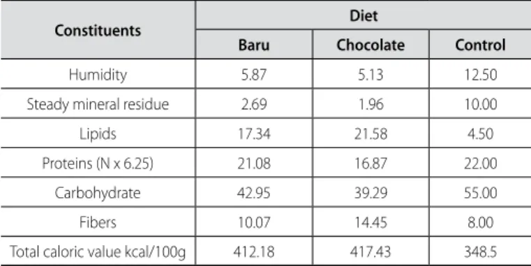
Table 1. Centesimal composition of the hyperlipidic experimental diets designed for rats of the chocolate group, soybean group, and baru group, expressed in humid basis, in g/100g.
Constituents Diet
Baru Chocolate Control
Humidity 5.87 5.13 12.50
Steady mineral residue 2.69 1.96 10.00
Lipids 17.34 21.58 4.50
Proteins (N x 6.25) 21.08 16.87 22.00
Carbohydrate 42.95 39.29 55.00
Fibers 10.07 14.45 8.00
Total caloric value kcal/100g 412.18 417.43 348.5
Adaptation to the water medium and physical training protocol
Adaptation consisted in keeping the animals in contact with shallow water at temperature of 32 ± 2°C, while they supported led loads inserted in cloth “backpacks” fastened with Velcro®
192
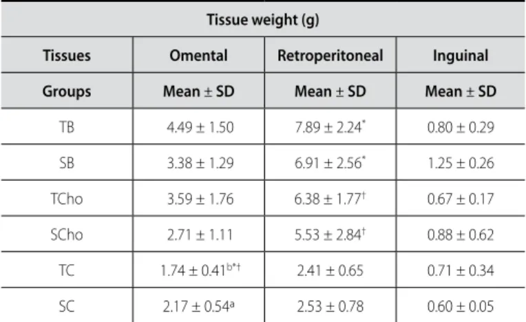
The data presented in tables 3 and 4 indicate that the hyper-caloric/hyperlipidic diets increased weight and area of the cells of the retroperitoneal adipose tissue compared to the control diet. Exercise was efficient in reducing the area of the cells of the omental and inguinal adipose tissues of the chocolate group and inguinal tissue in the control group. The trained and fed with baru diet group presented wider adipocytes area concern-ing the retroperitoneal and concern-inguinal tissues compared to their sedentary pairs.
Table 5 evidences that the content of triglycerides in the liver of the sedentary animals was higher than the trained and fed with hypercaloric/hyperlipidic diets animals (p < 0.05), and the values of the trained groups with these diets were not different from the ones presented by the animals with control diet.
Table 2. Energetic intake (kcal/day) of the sedentary (S) and trained (T) fed with baru diet (B), chocolate diet (CHO) and control diet (C) animals.
Energetic intake (kcal/day)
Groups Mean± SD
TB 77.16 ± 22.50*
SB 73.94 ± 21.96
TCho 62.28 ± 13.73
SCho 60.19 ± 10.01
TC 70.72 ± 13.10a
SC 61.04 ± 9.58b*
Values expressed in mean ± standard deviation. Diferent underlined letters reveal statistical diference between trained and sedentary animals from the same group (p < 0.05). * p < 0.05 diference between baru diet and control diet.
Table 3. Weight of the (g) omental, retroperitoneal and inguinal tissues of the sedentary (S) and trained (T) animals fed with baru (B), chocolate (CHO) and control diets (C).
Tissue weight (g)
Tissues Omental Retroperitoneal Inguinal Groups Mean ± SD Mean ± SD Mean ± SD
TB 4.49 ± 1.50 7.89 ± 2.24* 0.80 ± 0.29
SB 3.38 ± 1.29 6.91 ± 2.56* 1.25 ± 0.26
TCho 3.59 ± 1.76 6.38 ± 1.77† 0.67 ± 0.17
SCho 2.71 ± 1.11 5.53 ± 2.84† 0.88 ± 0.62
TC 1.74 ± 0.41b*† 2.41 ± 0.65 0.71 ± 0.34
SC 2.17 ± 0.54ª 2.53 ± 0.78 0.60 ± 0.05
Values expressed in mean ± standard deviation. Diferent underlined letters reveal statistical diference between trained and sedentary animals from the same group (p < 0.05).*p < 0.05 diference between baru hypercaloric diet and control diet; † p < 0.05 diference between chocolate and control diets.
Table 4. Adipocytes area(µm2) of the omental, retroperitoneal and inguinal
tissues of the sedentary (S) and trained animals (T) fed with baru diet (B), chocolate diet (CHO) and control diet (C).
Adipocytes area
Tissue Omental (µm2) Retroperitoneal (µm2) Inguinal (µm2)
Groups Mean ± SD Mean± SD Mean ± SD
TB 6.471.04 ± 3.412.29* 18.286.56 ± 12.344.85ª* 4.480.08 ± 3.442.84a*
SB 7.482.62 ± 5.836.58* 11.378.45 ± 12.695.21b* 3.259.32 ± 3.273.79b
Tcho 6.370.91 ± 7.776.13b 8.715.90 ± 7.300.99† 5.147.49 ± 5.712.71a†
SCho 7.341.28 ± 5.862.24ª† 8.549.01 ± 10.394.3† 7.083.11 ± 7.682.40b†
TC 4.300.62 ± 3.603.44 6.031.03 ± 6.133.87 2.212.87 ± 1.920.34a
SC 5.099.05 ± 4.642.03 6.835.89 ± 6.328.56 3.386.11 ± 3.973.09b
Values expressed in mean ± standard deviation. Diferent underlined letters reveal statistical diference between trained and sedentary animals from the same group (p < 0.05).*p < 0.05 diference between baru hypercaloric diet and control diet; † p < 0.05 diference between chocolate diet and control diet.
Table 5. Triglycerides in the liver of the sedentary (S) and trained (T) animals fed with baru diet (B), chocolate diet (CHO) and control diet (C) expressed in mg/100mg.
Triglycerides in the liver
Groups Mean ± SD
TB 0.88 ± 0.43a
SB 1.36 ± 0.50b*
TCho 0.86 ± 0.41a
Scho 1.77 ±0.64b†
TC 0.55 ± 0.08
SC 0.50 ± 0.25
Values expressed in mean ± standard deviation. Diferent underlined letters reveal statistical diference between trained and sedentary animals from the same group (p < 0.05).*p < 0.05 diference between baru hypercaloric diet and control diet; † p < 0.05 diference between chocolate and control diets.
DISCUSSION
In the present study, the administration of hyperlipidic diet with baru extract, both for the sedentary animals and trained animals, caused increase in adipose cells of the subcutaneous abdominal (retroperitoneal) and visceral abdominal (omental) tissues when compared to the control diet. The animals from the chocolate sedentary group presented adipocytes area wider than the controls in all the assessed tissues. Such results cor-roborate the findings by Chen and Farese14, which observed that
mice fed with lipid-rich diet (16%), during 16 weeks, presented adipocytes area of the epididimal region 2.5 times bigger than the control group.
The results obtained by us, which denoted the accumulation of adipose tissue predominantly in the abdomen region after administration of hypercaloric and hyperlipidic diets, are in agreement with other studies of this nature. The lipoprotein lipase activity, key-enzyme in the fat deposition in many adipose compartments, as well as the RNAm levels of the LPL, are bigger in the abdominal cells than in the gluteal ones of male individuals15.
Moreover, these tissues present higher expression of the fatty acids transporter proteins (FATPs)16.
According to Oller do Nascimento et al.17, in rats,
hyperlipi-dic diets reduce the activity of the lipogenic enzymes and the lipogenesis in the inguinal deposit and icnrease these enzymes in the visceral tissue. This fact would partly explain the lack of differences for the weight of the inguinal tissues between the animal groups with hyperlipidic or normolipidic diets.
Additionally, it is highlighted that obesity in the animals with hypercaloric/hyperlipidic diet could be observed even in the absence of hyperfasia, since there was no ingestion difference between rats of the chocolate and baru diets compared to those with control diet submitted to the same treatment.
In many animal models, the addition of lipids in the diet resulted in reduction of ingestion and weight increase18, while
physical activity promoted higher food intake (23%)11. In our
193 presented tendency to lower values in the adipocytes area in all
tissues, with remarkable significance to the subcutaneous ingui-nal adipose tissue. Furthermore, the total weight of the omental adipose tissue was lower (p < 0.05) in the trained control animals.
On the other hand, this same behavior was not reproduced in the retroperitoneal tissue or in the baru group tissues, raising the hypothesis that the ethereal baru extract contains dietetical factor which contribute to greater lipids deposition and/or may inhibit the exercise effects on the body adipose storage.
The results observed in the chocolate group agree with the ones found by Gollisch et al. (2009)19, who observed smaller
adi-pocytes area due to physical exercise in both deposits, inguinal and parametrial of rats fed with hyperlipidic diet.
These findings are also in agreement with another study which presents it is easier to move visceral abdominal fat than subctaneous abdominal fat with physical exercise20. The visceral
adipose tissue seems to be less responsive than the subcutane-ous abdominal to the antilipolytic and re-esterification stimulus of de fatty acids produced by insulin19. Thus, it can be suggested
that in the weight gain condition, the adipose accumulation would be predominant in the subcutaneous abdominal tissue and in the weight loss; the effect would be more remarkable in the visceral tissue.
In fact this result was observed in the present study, since the retroperitoneal tissue of the groups with hypercaloric diet was higher than the one of the control animals. In the condi-tions which there was effect of physical exercise in fat loss, it was perceived in the visceral tissue (omental).
According to Thomas et al.20, the interaction between the
different sources of lipid supply during exercise has not been totally elucidated yet, while its functioning remains obscure and disperse in distinct experimental work. The apparent differences concerning the exercise effects on the fat deposits may be due to many factors, including methodology for adipocytes analysis, diet, characteristics of the sample (e.g. obese and/or eutrophic subjects) and exercise protocols adopted, which vary according to the frequency, intensity and duration of the physical exertion. In a study conducted by Guerra et al.21, it was observed that
exercise performed at moderate intensity and chronic manner (five days/week) significantly decreased relative weight of central adiposity (retroperitoneal fat) and visceral adiposity (epididimal fat) after administration of two diets (lipid-rich and standard) when trained animals were compared to their respective sed-entary and fed with the same diets.
In another study22, it was observed that the cafeteria diet
induced abdominal obesity in rats. On the other hand, physi-cal training (swimming, 5d/week, 30min/session) minimized the negative effects attributed to this diet; that is to say, it was ef-fective in reducing both central adiposity and visceral adiposity of the animals.
It is important to highlight that in the two studies previously mentioned, the exercise load used during physical exertion was of 5% of body mass of the animals; that is, higher than the one adopted in our study (2%).
Fiorese23 observed that rats fed with hyperlipidic diet as well
as submitted to physical exercise (90min/day, 5x/week, during eight weeks) did not present alterations in the epididimal and
retroperitoneal white adipose tissues. In this study, the exercise load supported by the animals was 3% of the weight; that is, similar to the one used in our study.
Thus, the results of the present study suggest that the exer-cise intensity supported by the animals fed with different kinds of diet was not sufficient to avoid the increase of the adipocytes area located in the abdominal region, specifically, the retroperi-toneal portion.
In the present study, it can be observed that the values con-cerning the weight of the retroperitoneal tissue of the animals fed with hyperlipidic/hypercaloric diet were higher when compared with the ones obtained in the control animals with normocaloric diet However, there was no difference between the trained and sedentary groups fed with the same diet, a fact which would possibly confirm our hypothesis previously stated.
Increase of fat mass raises the question about how far this expo-nential factor is associated only with the excessive energy ingested or to sedentarism11. In this study, it seems that diet was the main
element for the increase of the cells area and weight of the adipose tissues analyzed by us. Physical exercise preferably acted on the fat storage in the omental region of the rats.
This finding was also observed by Laye et al.11, which
evi-denced that voluntary physical activity (on a wheel) prevented hypertrophy and hyperplasia of adipose cells of the omental region of obese rats.
The reduction of the cells area of omental tissue of the trained chocolate group is highly relevant from the clinical point of view, since the studies confirm that the adipocytes area of the visceral tissue is positively associated with several metabolic alterations in both sexes, such as insulin resistance and systemic inflamma-tion of low level24.
The contribution of the visceral fat to the onset of insulin re-sistance seems to be more related to the adipokines release than to the flow of free fatty acids in the muscular tissue. The visceral tissue secretes higher quantities of PCR, IL-6 and PAI if compared to the subcutaneous one15, and a large part of the fatty acids which
compose the visceral tissue is directly released in the portal vein, with consequent alteration of metabolism and accumulation of tri-glycerides in the liver25.
In the present study, it was observed that the animals in the sedentary and fed with hyperlipidic/hypercaloric diets groups pre-sented higher weight of the omental tissue as higher area of cells of the same tissue, besides high content of hepatic triglyceride. On the other hand, the values of the same variables, found in the groups in which the hypercaloric/hyperlipidic diet was associated with physical training, were different when compared with their sedentary pairs. Furthermore, such results are close to the values obtained by the groups with normocaloric diet, showing that the exercise performed at light to moderate intensity may be a protective factor in the fight to fat accumulation in the liver, even in the persistence of diets rich in calories and lipids.
These findings corroborate the ones in other studies, which have observed significant decrease in the triglycerides in the liver of the animals trained at light to moderate intensity23 or in
those voluntarily active26.
194
fatty acids in the liver. The study by Rector et al.26 evidences that
physi-cal training improves hepatic mitochondrial function, specifiphysi-cally the electron coupling of the beta oxidation, and boosts the functioning of the Krebs cycle and of the oxidative phosphorylation, which results in a more complete degradation of hepatic fatty acids of the obese rats. The present investigation has the goal to devise important perspectives concerning public health, since the non-alcoholic hepatic steatosis is more frequent in individuals with metabolic syndrome27, and they are the ones who present greater difficulty
in performing exercises at higher intensities.
Although this study of experimental nature presents some positive effects derived from the practice of aerobic exercises at light to moderate intensity, future studies are necessary. The fact the hyperlipidic diet based on baru extract has produced contrary effects to the expectation; it raises the need for more accurate analyses concerning the confirmation of its supposed functional characteristic. Some substances present in the extract, besides the unsaturated fatty acids, can somehow have interfered in the results obtained.
The addition of purified baru oil to an isocaloric diet, rather than the hyperlipidic/hypercaloric diet used by us, could produce distinct results and hypothetically, positive to health, as the case of many oils extracted from nuts.
Another limitation of the study refers to the absence of re-search which used the omental and inguinal adipose tissues, a fact which may have compromised our comparisons. Additionally and importantly, there is not a gold-standard technique described in
the literature for measurement of the adipocytes area, while the osmium tetroxide, used by us for this purpose seems to overesti-mate the results14.
CONCLUSION
The effects of aerobic exercise performed at light to moderate intensity, were significant and more remarkable in the reduc-tion of visceral and inguinal adipose deposits of the animals fed with control and chocolate diets. This same behavior was not reproduced with the diet based on baru extract. Moreover, the trained animals presented lower amount of hepatic triglyceride, even in the persistence of the hyperlipidic/hypercaloric diet with chocolate or baru extract.
ACKNOWLEDGEMENTS
To the Post-Graduation Program in Health and Development in the Central-West Region of the UFMS for the support; to the Education, Science and Technology Support and Development Foundation of Mato Grosso do Sul State (file 23/200.200/2008); to the Research Support Foundation of Mato Grosso State (file 23108.009511/09-1), for the financial support to the research; and to the physical education professors Brunno Elias Ferreira and Erika Alves Morel for the technical contribution.
All authors have declared there is not any potential conflict of interests concerning this article.
REFERENCES
1. Brasil. Ministério da Saúde. Secretaria de Vigilância em Saúde. Secretaria de Atenção à Saúde. Política nacional de promoção da saúde / Ministério da Saúde, Secretaria de Vigilância em Saúde, Secretaria de Atenção à Saúde. Brasília: Ministério da Saúde, 2006.
2. U.S. Department of Health and Human Services. 2001. The Surgeon General’s Call to Action to Prevent and Decrease Overweight and Obesity. Washington, DC: U.S. Government Printing Office. Disponível em http://www.surgeongeneral.gov/topics/obesity/calltoaction/CalltoAction.pdf
3. I Diretriz Brasileira de Diagnóstico e Tratamento da Síndrome Metabólica da ‘Sociedade Brasileira de Hipertensão’. Arq Bras Cardiol 2005; 84:3-28. Disponível em: http://www.scielo.br/pdf/abc/v84s1/ a01v84s1.pdf. Acesso em: 28 jan. 2010.
4. Brasil. Ministério da Saúde. Análise da estratégia global para alimentação saudável, atividade física e saúde. 2004 [atualizada 2007 acesso em 10 janeiro de 2010]. Disponível em: http://dtr2004.saude. gov.br/nutricao/documentos/doc_eg_final_submetido.pdf
5. Ikeda AA. Moraes A, Mesquita G. Considerações sobre tendências e oportunidades dos alimentos funcionais. Revista P&D em Engenharia de Produção 2010;8:40-56. Disponível em http://www. revista-ped.unifei.edu.br/documentos/V08N02/v8n2_artigo_03.pdf
6. Sano SM, Ribeiro JF, Brito MA. Baru: biologia e uso. Planaltina, DF: Embrapa Cerrados, 2004. [acesso em 02 abril 2007]. Disponível em: http// bbeletronica.cpac.embrapa.br/2004/doc/doc_116.pdf 7. Takemoto E, Okada IA, Garbelotti ML, Tavares M, Aued-Pimentel S, Composição química da semente
e do óleo de baru (Dipteryx alata Vog.) nativo do Município de Pirenópolis. Revista Instituto Adolfo Lutz 2001;60:113-7.
8. Hainer, V, Toplak H, Mitrakou A. Treatment modalities of obesity. What fits whom? Diabetes Care 2008;31:S269-77.
9. Weiss R. Fat distribution and storage: how much, where, and how? Eur J Endocrinol 2007;157:39-45. 10. Contarteze RVL, Manchado FB, Gobatto CA, Mello MAR. Biomarcadores de estresse em ratos exerci-tados por natação em intensidades igual e superior à máxima fase estável de lactato. Rev Bras Med Esporte 2007;13:169-74.
11. Laye MJ, Thyfault JP, Stump CS, Booth FW. Inactivity induces increases in abdominal fat. J Appl Physiol 2007;102:1341-7.
12. Hirsch J, Gallian E. Methods for determination of adipose cell size in man and animals. J Lipid Res1968;9:110-9. 13. Dâmaso AR. Efeitos do exercício agudo e crônico sobre o metabolismo lipídico e a celularidade adiposa de ratas no período de lactação e após o desmame. [Tese]. São Paulo: Escola Paulista de Medicina, Universidade Federal de São Paulo; 1996:120p.
14. Chen HC, Farese Jr RV. Determination of adipocyte size by computer image analysis. J Lipid Res 2002;43:986-9.
15. Wajchenberg BL. Subcutaneous and visceral adipose tissue: their relation to the metabolic syndrome. Endocr Rev 2000;21:697-738.
16. Shadid S, Koutsari C, Jensen M D. Direct Free Fatty Acid Uptake Into Human Adipocytes In Vivo. Relation to Body Fat Distribution. Diabetes 2007;56:1369-75.
17. Oller do Nascimento, CM, Ribeiro EB, Oyama L. Metabolism and secretory function of white adipose tissue: effect of dietary fat. An Acad Bras Cienc 2009;81:453-66.
18. Alaniz MHF, Takada J, Vale MICA, Lima FB. Adipose tissue as an endocrine organ: from theory to practice. J Pediatr 2007;83:192-203.
19. Gollisch KSC, Brandauer J, Jessen N, Toyoda T, Nayer A, Hirshman MF, et al. Effects of exercise train-ing on subcutaneous and visceral adipose tissue in normal- and high-fat diet-fed rats. Am J Physiol Endocrinol Metab 2009;297:495-504.
20. Thomas EL, Brynes AE, McCarthy J, Goldstone AP, Hajnal JV, Saeed N, et al. Preferential Loss of Visceral Fat Following Aerobic Exercise, Measured by Magnetic Resonance Imaging. Lipids 2000;35:769-76. 21. Guerra RLF, Prado WL, Cheik NC, Viana FP, Botero JP, Vendramini RC, et al. Effects of 2 or 5 consecutive
ex-ercise days on adipocyte area and lipid parameters in Wistar rats. Lipids in Health and Disease 2007;6:1-8. 22. Eguchi R, Cheik NC, Oyama LM, Nascimento CMO, Mello MT, Tufik S, et al. Efeitos do exercício crônico sobre a concentração circulante da leptina e grelina em ratos com obesidade induzida por dieta. Rev Bras Med Esporte 2008;14:182-7.
23. Fiorese MS. Efeitos do exercício acumulado sobre o controle daadiposidade visceral, leptinemia e gordura hepática em ratos alimentados com dieta hiperlipídica. [Tese]. São Carlos: Universidade Federal de São Carlos; 2008; 90 p.
24. Tchoukalova YD, Koutsari C, Karpyak MV, Votruba SB, Wendland E, Jensen MD. Subcutaneous adipocyte size and body fat distribution. Am J Clin Nutr 2008;87:56-63.
25. Frayn KN. Visceral fat and insulin resistance – causative or correlative? Br J Nutr 2000;83:71-7. 26. Rector RS, Thyfault JP, Morris RT, Laye MJ, Borengasser SJ, Booth FW, et al. Daily exercise increases
hepatic fatty acid oxidation and prevents steatosis in otsuka long-evans tokushima fatty rats. Am J Physiol Gastrointest Liver Physiol 2008;294:619-26.