Article
J. Braz. Chem. Soc., Vol. 25, No. 6, 1116-1123, 2014. Printed in Brazil - ©2014 Sociedade Brasileira de Química 0103 - 5053 $6.00+0.00
A
*e-mail: [email protected]
Vanadium-Binding Protein in Marine Plankton from Tropical South Atlantic Ocean
Vinicius T. Kütter,*,a Maria Montes-Bayón,b Silvia M. Sella,c Alfredo Sanz-Medelb and
Emmanoel V. Silva-Filhoa
aGeochemistry Department, Federal Fluminense University, Outeiro São João Batista s/n, 24020-141 Niterói-RJ, Brazil
bPhysical and Analytical Chemistry Department, Faculty of Chemistry, University of Oviedo, Julian Clavería 8, 33006 Oviedo, Spain
cAnalytical Chemistry Department, Federal Fluminense University, Outeiro São João Batista s/n, 24020-141 Niterói-RJ, Brazil
Nós investigamos o papel do plâncton no ciclo do vanádio (V) em Cabo Frio, Rio de Janeiro, Brasil, uma região com alta concentração de V nas partículas atmosféricas, devido ao aerossol marinho. As concentrações de V no plâncton variaram de 0,08-20,9 µg g-1 (zooplâncton),
0,1-28,4 µg g-1 (fitoplâncton > 64 µm) e < 0,0005-49,0 µg g1 (pequeno fitoplâncton > 20 µm). A
especiação de V associado a biomoléculas foi realizada por duas estratégias: (i) o acoplamento de cromatografia de exclusão de tamanho (SEC) para o fracionamento de espécies com espectrometria de massas com plasma indutivamente acoplado (ICP-MS) e (ii) cromatografia de exclusão de tamanho por troca aniônica com detector UV-Vis acoplado a espectrometria de massas com plasma indutivamente acoplado (SEC-AE-UV-Vis-ICP-MS). Os resultados mostraram uma única fração contendo V associada a uma biomolécula na faixa de 8 a 16 kDa, com ponto isoelétrico acima de 8. Resultados preliminares usando MALDI-TOF não permitem identificar a biomolécula, considerando a larga faixa de massa molecular obtida.
We investigated the role of plankton in the vanadium (V) cycle at Cabo Frio, Rio de Janeiro state, Brazil, a region with high V concentration in the atmospheric particles due to marine aerosol. The concentrations of V in plankton vary from 0.08 to 20.9 µg g-1 (zooplankton), 0.1 to 28.4 µg g-1
(phytoplankton > 64 µm) and < 0.0005 to 49.0 µg g-1 (small phytoplankton > 20 µm). The V
speciation in biomolecules was performed by the use of two strategies: (i) coupling of size exclusion chromatography (SEC) for the fractionation of species with inductively coupled plasma mass spectrometry (ICP-MS) and (ii) with size exclusion anionic exchange chromatography with UV-Vis detector coupled to inductively coupled plasma mass spectrometry (SEC-AE-UV-Vis-ICP-MS). The results showed a single fraction containing V associated with a biomolecule in the range of 8 to 16 kDa, with isoeletric points above 8. The preliminary analyses using matrix-assisted laser desorption-ionization-time of flight (MALDI-TOF) do not permit to identify such biomolecule, considering the broader size range of the proteins obtained.
Keywords: bioaccumulation, biogeochemistry, chemical ecology, HPLC-ICP-MS, metallothionein
Introduction
Vanadium (V) is the 21st most abundant element in
Earth’s crust, being more common in the environment than many other elements, such as zinc and copper.1 In aquatic
environment the concentration levels are highly variable, typically ranging from 2 to 300 µg L-1 in freshwater, while
the concentration found in the ocean varies from 1 to 3 µg L-1.
The oxidation states of this element naturally found in the environment are +3, +4 and +5. However, it can exist in the oxidative states of –2 to +5.2
Concerning its biological role, vanadium has no well-defined function in higher life forms. However, the best evidence for its biological role comes from bacteria (so-called alternative nitrogenases in which vanadium replaces molybdenum in the FeMo-cofactor of some
Azotobacter species) and plants (vanadium-dependent haloperoxidases found in some algae, lichens and fungi). The substitution of vanadium for nitrogenases occurs in situations of molybdenum deficiency. Moreover, the enzyme containing vanadium is more effective that the one containing molybdenum when the temperature is significantly reduced, which may be an important evolutionary mechanism for organisms living in the poles or at high altitudes.3,4
On the other hand, laboratory experiments have shown that in animals the deficiency of vanadium increases abortion rate, reduces milk production during lactation and causes thyroid disorders.4 From the medical point of
view, this element has been indicated as a promising drug for treating diabetes by mimicking insulin as well as in the treatment and prevention of certain types of cancer.5
Since Martin Henze discovered in 1911 that ascidians accumulate high vanadium concentrations (up to 105 to
106 times higher than the values found in sea water), the
interest in investigating the function of this metal in hyper-accumulator organisms has been increased.6 However,
no-one has been able to determine the function of this element in these organisms. After the Henze discovery some other vanadium hyper-accumulator species of marine organisms were found including holothurians.7
Recently, some polychaeta species were discovered as vanadium hyper-accumulators8,9 and the accumulation
mechanism was similar to that found in ascidians despite phylogenetic distance between these organisms.10 This fact
has brought attention to the discussion about the role of this metal. Many data are found in the literature about the function of this metal in hyper-accumulator organisms (as an oxygen carrier, energy source, anti-microbial defense and anti-predation), but none of these hypotheses have been confirmed.11
Regarding the mechanism of vanadium speciation in biomolecules of marine organisms, the only known protein was isolated from ascidians and named vanabin.12,13
The vanabin family consists of at least five small proteins: vanabin 1 through 4 and vanabin P, which are composed of approximately 90 amino acids including 18 cysteine residues. Recombination of vanabin 1, P and 2 were found to bind up to 20 vanadium ions in the +4 oxidation state (V+4) with dissociation constant of ca.
2 × 10⁻⁵ mol L-1.14 This biomolecule group has a molecular
weight ranging from 10.46 to 28.03 Da.13,15-18
Michibata et al.19 elucidated the incorporating
process of V+5 present in seawater by ascidians. The V+5
is incorporated into vanadocytes, where V is bound to vanabin and reduced to +4 oxidative state with NADPH produced by the pentose-phosphate route. The V+4 bound
with vanabin is transferred to an unknown protein to the vacuolar membrane, where it is trapped with metal-binding domains of a metal ATPase on the vacuolar membrane surface and is stored into vanadocytes vacuoles having both high levels of H+ and SO
4
2–. The V+4 is further reduced
to V+3 by an unknown reducing agent, whose oxidative
state predominates in vanadocytes in a proportion of 97.6 (V+3):2.4(V+4).
This complex vanadium accumulation process by tunicates aroused great interest, considering that there is an abundance and diversity of these organisms in the region of Cabo Frio: more than 17 species of ascidians and large amounts of planktonic tunicates (Doliolum sp and
Thalia democratica). The latter is the one of the largest groups of zooplankton occurring in the region, especially during coastal upwelling events.20-23
In addition, the high vanadium concentrations found in atmospheric particulate matter from Cabo Frio suggest biogenic origin associated with planktonic tunicates during upwelling events.24
The aim of the present work was to investigate the vanadium-binding proteins in different plankton samples from the Cabo Frio region in the southeast of Brazil.
Experimental
Study area
The Cabo Frio region is located at the southeast coast of Brazil (23º01’ S, 42º00’ W) where the main upwelling events in Brazil occur. In this area the South Atlantic Central Water (SACW), which is rich in nutrients and has cold temperature, arises to the surface mainly due to the strong winds of E-NE quadrant and also by topographic and hydrological characteristics, which are peculiar in this region.25 However, the upwelling events are intermittent,
differently from other regions of the planet. Even though this region is located in the tropics there are many occurrences of planktonic species characteristic of cold regions, due to the low seawater temperature of South Atlantic Central Water during upwelling events.
Sampling and storage
(mean 12 m deep) and usually receives cold water plume from South Atlantic Central Water (SACW) during the upwelling events that occur outside the bay. The bay has a surface area of approximately 7 km2, and is embedded
between the Cabo Frio promontory and Cabo Frio Island (see Supplementary Information). It has access to the sea via two openings. The 150 m wide and 18 to 38 m deep Boqueirão Channel, set at its southern premises between the promontory and Cabo Frio Island, and the 1.5 km wide and 9 to 15 m deep opening in the north, between Cabo Frio and Porcos islands.
A total of 25 plankton samples were collected monthly along two different periods, utilizing horizontal hauls with several mesh nets: (i) > 64 µm to < 100 µm (phytoplankton) and > 100 µm (zooplankton) during the 2003-2005 period and (ii) > 20 µm to < 64 µm (small phytoplankton + inorganic particles), > 64 µm to < 150 µm (phytoplankton) and > 150 µm (zooplankton) during the 2008-2009 period.
Although the fractionation using net mesh size has drawbacks such as a mixture of phytoplankton/zooplankton/ inorganic particulate matter in different proportions, it is justified because the predominant phytoplankton in this region are diatoms < 65 µm.26 Furthermore, due to small
size and mass of individuals the separation of species for metal and vanadium-binding protein determination is impracticable.
The samples were transported to the laboratory and immediately lyophilized (24 h) for preservation and subsequent analysis.
Determination of total vanadium in plankton samples
One aliquot (0.3 g) of the lyophilized sample was digested with an acid mixture of HNO3:HCl (both
suprapur® Merck) in the proportion of 7:2 (v/v) in a closed system utilizing a microwave oven (Anton PAAR MW3000). The program was set according to the manufacturer. The extracts obtained (n = 3) were used for total vanadium determination using an inductively coupled plasma mass spectrometer (ICP-MS, Thermo Fisher Xseries II). The quality control analysis was verified through blank samples, internal standard (Rh) and certified reference material (BCR 414) analysis.
Determination of vanadium-binding protein employing multidimensional techniques
In order to investigate the vanadium-binding protein a sample of phytoplankton (> 64 µm) with total vanadium content of 13.0 ± 1.7 µg g-1 was selected. The biomolecule
extraction efficiency was tested with 0.2 g of lyophilized plankton sample using ammonium acetate (0.050 mol L-1)
at different pH values (5.0, 7.0 and 8.8), as well as different times (2, 2:30, 12 and 24 h) with mechanical mix only or associated with ultrasonic bath. Every procedure was executed at room temperature (25 ºC).
After the extraction procedure was established, the analysis employing multidimensional techniques was performed, as follows:
Size exclusion chromatography with UV-Vis detector coupled to inductively coupled plasma mass spectrometry (SEC-UV-Vis-ICP-MS)
The extracts obtained (0.2 g of plankton sample in 0.050 mol L-1 ammonium acetate, pH 5.0, mechanically mixed
for 12 h) were immediately submitted to chromatographic analysis using size exclusion chromatography (SEC). A SuperdexTM 75 5/150 GL column, UV-Vis detector for
macromolecules monitoring and ICP-MS detector for
51V monitoring were used. The column was previously
calibrated with standard solutions (Sigma-Aldrich®)
in the concentration of 1 mg mL-1 prepared from
apo-transferrin (79 kDa), superoxide dismutase (SOD, 32 kDa),
α-lactalbumin (14 kDa), insulin (5 kDa) and vitamin B12
(1.4 kDa). The elution times of the standards were used to estimate the molecular mass. The mobile phase used was 0.050 mol L-1 ammonium acetate (pH 7.0) with flow
rate of 0.3 mL min-1 and sample injection volume of
10 µL. The samples were analysed with high performance liquid chromatography (HPLC) with UV-Vis detector (Agilent 1100) and inductively coupled plasma mass spectrometry (Thermo Fisher Xseries II).
Anionic exchange chromatography with UV-Vis detector coupled to inductively coupled plasma mass spectrometry (AE-UV-Vis-ICP-MS )
The extracts obtained (0.2 g of plankton sample in 0.050 mol L-1 ammonium acetate, pH 5.0, mechanically
mixed for 12 h) were immediately submitted to chromatographic analysis using anionic exchange chromatography (AE, Mono Q 4.6/100 PE), a gradient of 0.010-0.250 mol L-1 (pH 9.0) of ammonium acetate, flow
rate of 1 mL min-1 and sample injection volume of 50 µL.
The samples were analysed by HPLC-UV-Vis and ICP-MS. In order to identify the molecular mass of protein that binds to vanadium, the extracts from AE were evaluated using matrix-assisted laser desorption-ionization-time of flight (MALDI-TOF), previously calibrated with
trifluoroacetic acid (TFA, 0.1%), which was mixed with a sample in a proportion of 1:1 (v/v).
Results and Discussion
Total vanadium concentration in plankton
In order to evaluate the method accuracy, including sample digestion and vanadium determination by ICP-MS, the certified reference material (BCR 414, Institute for Reference Materials and Measurements, IRMM) was analyzed. Excellent recovery was obtained (96.0%, n = 4) with similar repeatability to that recommended by IRMM. The vanadium concentrations in the phytoplankton of Cabo Frio Bay are of the same order of magnitude compared to data from literature, in contrast to zooplankton that showed vanadium concentration values more than 5 times below other sites (Table 1).
The differences found in the zooplankton from Cabo Frio Bay in relation to other works can be related to the taxon composition of sample that is governed by the seawater characteristics. Cabo Frio Bay has a peculiar characteristic in comparison to other sites shown in Table 1, it is located in a tropical area and the seawater temperature varies (sometimes warm and cold during upwelling events). Due to these characteristics the plankton taxon composition in Cabo Frio Bay oscillates (sometimes the tropical species are more abundant, other times cold species predominate, and even a mixing of the two species can
be found) in response to environmental conditions. The vanadium concentration in the plankton shown in these different sites can be related to evolution and adaptation of organisms to live in this site. It is known that species from cold weather accumulate more vanadium than species from warm weather.3,4
The total vanadium concentration found in the small phytoplankton/inorganic particles can be related to different taxon composition in comparison to the phytoplankton (> 64 µm). Furthermore, the higher vanadium values in the phytoplankton/inorganic particles (> 20 µm) can be attributed to the presence of vanadium in inorganic matter. Accord to Kütter et al.31 these samples showed less than
5% of carbon. Pyle and Tieh32 demonstrated that the shell
of dead pteropods, a zooplanktonic gastropod, accumulates high vanadium concentrations (50 to 290 µg g-1). However
it is not clear if the pteropod shell accumulates vanadium when the organism is alive or dead.
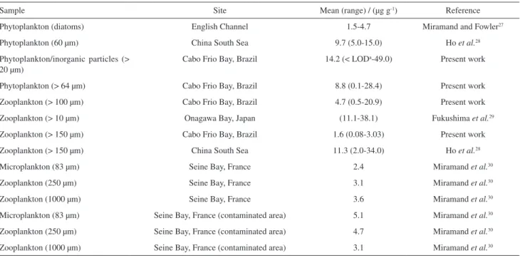
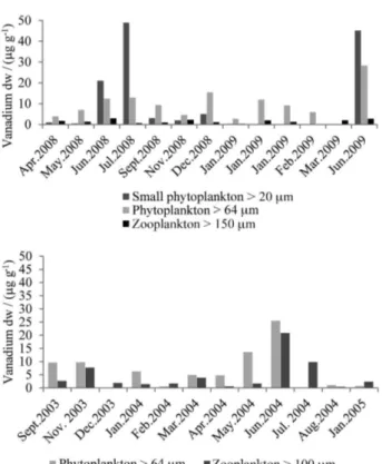
Figure 1 shows data from total vanadium concentration in the plankton samples during the studied period. It is observed that the maximum vanadium concentration occurred in the small phytoplankton in July 2008 and June 2009. This element showed a marked seasonal distribution like other metals: aluminum, chromium, iron and nickel,33
where the highest average concentrations of each group of plankton were recorded during winter season. This result is probably related to the increase of coastal plumes in this season. During the winter the occurrence of cold fronts promotes wave intensification leading to an increase in
Table 1. Vanadium concentration) in plankton from Cabo Frio region compared to data from literature
Sample Site Mean (range) / (µg g-1) Reference
Phytoplankton (diatoms) English Channel 1.5-4.7 Miramand and Fowler27
Phytoplankton (60 µm) China South Sea 9.7 (5.0-15.0) Ho et al.28
Phytoplankton/inorganic particles (> 20 µm)
Cabo Frio Bay, Brazil 14.2 (< LODa-49.0) Present work
Phytoplankton (> 64 µm) Cabo Frio Bay, Brazil 8.8 (0.1-28.4) Present work
Zooplankton (> 100 µm) Cabo Frio Bay, Brazil 4.7 (0.5-20.9) Present work
Zooplankton (> 10 µm) Onagawa Bay, Japan (11.1-38.1) Fukushima et al.29
Zooplankton (> 150 µm) Cabo Frio Bay, Brazil 1.6 (0.08-3.03) Present work
Zooplankton (> 150 µm) China South Sea 11.3 (2.0-34.0) Ho et al.28
Microplankton (83 µm) Seine Bay, France 2.4 Miramand et al.30
Zooplankton (250 µm) Seine Bay, France 3.1 Miramand et al.30
Zooplankton (1000 µm) Seine Bay, France 3.6 Miramand et al.30
Microplankton (83 µm) Seine Bay, France (contaminated area) 5.1 Miramand et al.30
Zooplankton (250 µm) Seine Bay, France (contaminated area) 4.7 Miramand et al.30
sediment re-suspension and consequent major availability of metals for incorporation by plankton.34
Test for vanadium-binding protein extraction in plankton
The parameters optimized for sample extraction, utilizing mechanical mixture for 12 h, are shown in Table 2. Despite the low recovery of vanadium, we opted for extraction with ammonium acetate buffer because it is the method that has less interference in the determination of vanadium by ICP-MS. The major interference is due to chloride that corresponds to 75% (16O + 35Cl) and 24.5%
(14N + 37Cl). The use of ammonium acetate buffer as
extractor of metal associated with biomolecules reduces
interference and has been shown to be efficient for elements such as arsenic (As).35
Determination of vanadium-binding protein employing multidimensional techniques
SEC-UV-Vis-ICP-MS
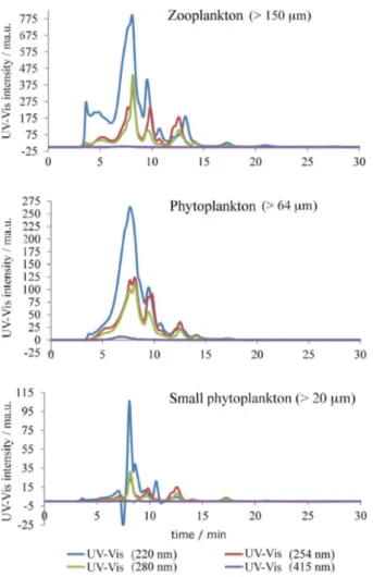
Data from the literature reports different wavelengths for monitoring the vanadium-binding proteins in ascidians. Some authors employ reverse phase column using a wavelength of 220 nm36,37 while Michibata et al.12 uses
SEC employing various wavelengths, which demonstrates that each species of Ascidia has a maximum absorbance at a specific wavelength. An example is Ascidia gemmate
that showed maximum absorbance in the visible range (756 nm). The data from our study showed the best results for the wavelengths of 220 and 254 nm (Figure 2).
In the SEC-ICP-MS chromatograms (Figure 3), obtained for different groups of plankton, the presence of vanadium and sulfur (S) peaks are observed between 5-10 min, which seems to indicate that vanadium is associated with the SH group, whose biomolecule has an estimated molecular mass of 8-16 kDa, which is characteristic of vanabins. These results corroborate the ones obtained by Michibata et al.19 that described three
types of vanadium-binding proteins with molecular masses of 12.5, 15 and 16 kDa.
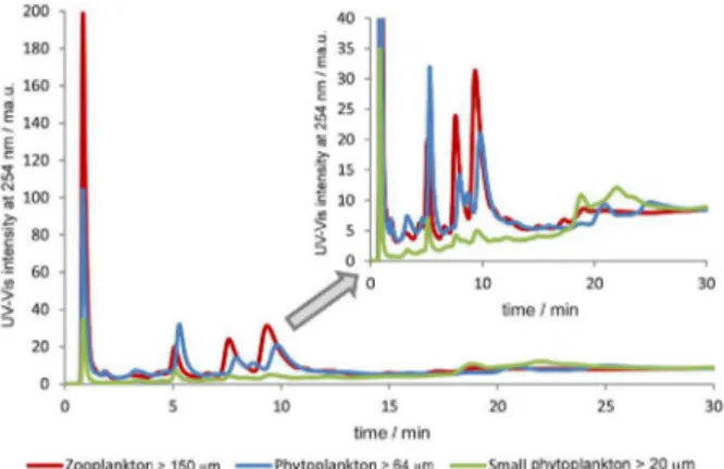
AE-UV-Vis-ICP-MS
The column used in these experiments was a Mono Q (strong anion exchanger) with a mobile phase having a gradient with increasing concentration of ammonium acetate (0.010-0.25 mol L-1) buffering the mobile phases
at different pH values (Figure 4). It is possible to observe that at pH 7.0 most of the species elute at the void volume, showing no interaction with the chromatographic column. When increasing the pH to 8.0 it is possible to observe that the species are retained in the column, meaning that they have slightly negative charge. This is even more noticeable
Table 2. Mean ± standard deviation (n = 3) optimized parameters for extraction efficiency of vanadium-binding protein in plankton samples containing 13.0 ± 1.7 µg g-1 of vanadium
Ammonium acetate / (0.05 mol L-1)
Mechanical mix Mechanical mix + ultrasonic bath
2 h 12 h 24 h 2 h + 30 min
V / (µg g-1) Recovered / % V / (µg g-1) Recovered / % V / (µg g-1) Recovered / % V / (µg g-1) Recovered / %
pH 5 2.9 ± 0.5 22.3 ± 0.1 4.0 ± 1.0 30.7 ± 0.1 4.0 ± 1.1 30.4 ± 0.3 2.7 ± 1.0 20.8 ± 0.1
pH 7 2.3 ± 0.8 18.1 ± 0.1 3.3 ± 0.3 25.1 ± 0.4 2.8 ± 0.2 21.6 ± 0.1 2.0 ± 0.4 15.7 ± 0.3
pH 8.8 2.5 ± 1.0 19.2 ± 0.2 3.3 ± 0.4 25.5 ± 0.1 3.8 ± 0.7 29.1 ± 0.1 2.1 ± 0.8 16.3 ± 0.5
when the mobile phase is adjusted to pH 9.0. In this latter case, the analytes show higher interaction (longer retention times) and better resolution. Therefore, according to the retention characteristics their isoelectric point should be 8 or above. These results would support the possibility for this proteinaceous species to be vanabins.
The chromatographic profile at this pH seems to be identical for all plankton groups (Figure 5), despite differences on absorbance values.
Figure 5 also shows the presence of the same proteinaceous material in all the analyzed fractions (small phytoplankton > 20 µm, phytoplankton > 64 µm and zooplankton > 150 µm) exhibiting slightly different distribution among them. However, the separation process via anion exchange column (strong exchanger) seems to lead to the disruption of the interaction between the proteins and the V ion (no V was detected in the separated peaks by ICP-MS). This disruption was confirmed by the incubation of the plankton sample with V+4 (25 nmol L-1), which demonstrated a single peak of
vanadium in the void volume of the chromatographic
run (data not shown). These results are consistent with those described by Kanda et al.38 that verified the loss of
up to 90% of vanadium associated with molecule during the process of sample preparation. Nevertheless, in this study it was possible to separate the proteins associated with vanadium using an anion exchange column with a gradient of 0-0.4 mol L-1 NaCl at pH 7.4.
Even with V loss during AE fractionation, the extracts were used to identify the possible biomolecules present in the extracts. Figure 6 shows the plot from UV-Vis and MALDI-TOF from phytoplantkton samples. From the three fractions collected (A, B and C) the only one that showed a detectable MALDI mass spectrometry signal corresponded to fraction B. The molecular mass obtained from this peak was 8.2 kDa, which indicates that the biomolecule associated with vanadium could be a type of vanabin.
Figure 2. SEC-HPLC-UV-Vis chromatogram of plankton from Cabo Frio Bay.
Conclusions
Among the three plankton groups studied, small phytoplankton showed the higher total vanadium concentration, followed by phytoplankton and zooplankton.
The results of the use of multidimensional strategies (SEC and AE) and UV-Vis (SEC and AE)-ICP-MS for vanadium speciation, showed only one fraction of vanadium associated with the biomolecule in the range of 8 to 16 kDa. This fraction contains isoelectric points above 8.
The preliminary results obtained by MALDI-TOF do not permit to identify such biomolecule, considering the broader size range of the proteins obtained. Furthermore, the results showed that plankton may play an important role in the local V cycle, since the high V levels in atmospheric particulate matter in this region is a result of ocean-atmospheric interaction, due to the formation of marine aerosols as suggested by Sella et al..24
Supplementary Information
Supplementary information is available free of charge at http://jbcs.sbq.org.br as PDF file.
Acknowledgements
This study was supported by CAPES (PDEE 455209-1), Instituto Nacional de Ciência e Tecnologia (INCTTMCOcean 573601/2008-9), Instituto de Pesquisa da Marinha Almirante Paulo Moreira (IEAPM), Geochemistry Network from PETROBRAS/Cenpes, the National Petroleum Agency (ANP), Brazil (grant 0050.004388.08.9) within the scope of the Upwelling Project (Projeto Ressurgência). E. V. S.-F. is a senior researcher of the National Council for Research and Development (CNPq, Brazil) and Rio de Janeiro Research Foundation (FAPERJ, Brazil).
Figure 4. Anionic exchange chromatogram with UV-Vis detector (phytoplankton 64 µm) at different pH with a gradient of 0.010-0.25 mmol L-1 of ammonium acetate (undiluted sample).
Figure 5. Anionic exchange chromatogram with UV-Vis detector from different plankton groups with a gradient of 0.010-0.25 mol L-1 of
ammonium acetate at pH 9.0 (undiluted sample).
References
1. Nriagu, J. O.; Vanadium in the Environment: Part I, Chemistry and Biochemistry, 1st ed.; John Wiley & Sons: Hoboken, 1998.
2. Crans, D.; Amin, S. S.; Keramidas, A. D. In Vanadium in the Environment: Part I, Chemistry and Biochemistry, 1st ed.;
Nriagu, J. O., ed.; John Wiley & Sons: Hoboken, 1998. 3. Tracey, A. S.; Willsky, R. G.; Takeuchi, E. S.; Vanadium:
Chemistry, Biochemistry, Pharmacology, and Practical
Applications, 1st ed.; Taylor and Francis: Boca Raton, 2007.
4. Baran, E. J.; Chem. Biodiversity2008, 5, 1475.
5. Rehder, D.; Bioinorganic Vanadium Chemistry, 1st ed.; John
Wiley & Sons: Chichester, 2008.
6. Henze, M.; Hoppe-Seyler’s Z. Physiol. Chem.1911, 72, 494. 7. Ciereszko, L. S.; Ciereszko, E. M.; Harris, E. R.; Lane, C. A.;
Comp. Biochem. Physiol. 1962, 7, 127.
8. Ishii, T.; Kakai, I.; Numako, C.; Okoshi, K.; Otake, T.;
Naturwissenschaften1993, 80, 268.
9. Fattorini, D.; Notti, A.; Nigro, M.; Regoli, F.; Environ. Sci. Pollut. Res. 2010, 17, 220.
10. Uyama, T.; Nose, Y.; Wuchiyama, J.; Moriyama, Y.; Michibata, H.; Zool. Sci.1997, 14, 43.
11. Michibata, H.; Ueki, T.; Biomol. Concepts 2010, 1, 97. 12. Michibata, H.; Hirose, H.; Sugiyama, K.; Ookubo, Y.; Kanamori,
K.; Biol. Bull.1990, 179, 140.
13. Ueki, T.; Adachi, T.; Kawano, S.; Aoshima, M.; Yamaguchi, N.; Kanamori, K.; Michibata, H.; Biochim. Biophys. Acta 2003,
1626, 43.
14. Kawakami, N.; Ueki, T.; Amata, Y.; Kanamori, K.; Matsuo, K.; Gekko, K.; Michibata, H.; Biochim. Biophys. Acta2009, 1794, 674.
15. Trivedi, S.; Ueki, T.; Yamaguchi, N.; Michibata, H.; Biochim. Biophys. Acta2003, 1630, 64.
16. Yamaguchi, N.; Kamino, K.; Ueki, T.; Michibata, H.; Mar. Biotechnol. 2004, 6, 165.
17. Yoshihara, M.; Ueki, T.; Watanabe, T.; Yamaguchi, N.; Kamino, K.; Biochim. Biophys. Acta 2005, 1730, 206. 18. Hamada, T.; Asanuma, M.; Ueki, T.; Hayashi, F.; Kobayashi, N.;
Yokoyama, S.; Michibata, H.; Hirota, H.; J. Am. Chem. Soc.
2005, 127, 4216.
19. Michibata, H.; Yamaguchi, N.; Uyama, T.; Ueki, T.; Coord. Chem. Rev. 2003, 237, 41.
20. Valentin, J. L.; Macedo, F.; Monteiro, W.; Mureb, A.; Publ. Inst. Pesqui. Mar.1975, 86, 1.
21. Valentin, J. L. In Coastal Marine Ecosystems of Latin America, 1st ed.; Seeliger, U.; Kjerfve, H., eds.; Springer-Verlag: Berlin,
2001.
22. Nogueira, C. R.; Santos, L. H. S.; Bonecker, S. L. C.; Dias, C. O.; Reis, J. M. L.; Oecologia Brasiliensis1999, 7, 73. 23. Rocha, R. M.; Costa, L. V. G.; Iheringia, Ser. Zool. 2005, 95,
57.
24. Sella, S. M.; Neves, A. F.; Moreira, J. C.; Silva-Filho, E. V.;
Atmos. Environ. 2006, 40, 6181.
25. Carvalho, W. F.; Gonzalez-Rodriguez, E.; Braz. J. Oceanogr.
2004, 52, 35.
26. Guenther, M.; Gonzalez-Rodriguez, E.; Carvalho, W. F.; Rezende, C. E.; Mugrabe, G.; Valentin, J. L.; Mar. Ecol.: Prog. Ser.2008, 363, 109.
27. Miramand, P.; Fowler, S. W. In Vanadium in the Environment: Part I, Chemistry and Biochemistry, 1st ed.; Nriagu, J. O., ed.;
John Wiley & Sons: Hoboken, 1998.
28. Ho, T. Y.; Wen, L. S.; You, C. F.; Lee, D. C.; Limnol. Oceanogr.
2007, 52, 1776.
29. Fukushima, M.; Suzuki, H.; Saito, K.; Chatt, A.; J. Radioanal. Nucl. Chem.2009, 282, 85.
30. Miramand, P.; Bentley, D.; Guary, J.; Brylinski, M.; Oceanol. Acta1993, 16, 125.
31. Kütter, V. T.; Wallner-Kersanach, M.; Sella, S. M.; Albuquerque, A. L. S.; Knoppers, B. A.; Silva-Filho, E. V.; Environ. Monit. Assess. 2014, 186, 559.
32. Pyle, T. E.; Tieh, T. T.; Limnol. Oceanogr.1970, 15, 153. 33. Kütter, V. T.; personal communication.
34. Belém, A.; personal communication.
35. Montes-Bayón, M.; Meija, J.; Leduc, D. L.; Terry, N.; Caruso, J. A.; Sanz-Medel, A.; J. Anal. At. Spectrom.2004, 19, 153. 36. Ueki, T.; Satake, M.; Kamino, K.; Michibata, H.; Biochim.
Biophys. Acta2008, 1780, 1010.
37. Yoshihara, M.; Ueki, T.; Yamaguchi, N.; Kamino, K.; Michibata, H.; Biochim. Biophys. Acta2008, 1780, 256. 38. Kanda, T.; Nose, Y.; Wuchiyama, J.; Uyama, T.; Moriyama, Y.;
Michibata, H.; Zool. Sci. 1997, 14, 37.
Submitted: January 26, 2014