Quim. Nova, Vol. 37, No. 2, 260-267, 2014
Artigo
http://dx.doi.org/10.5935/0100-4042.20140045
*e-mail: [email protected]
BIOMOLECULES PRODUCED IN LIQUID-STATE FERMENTATION BY A MARINE-DERIVED FUNGUS, Penicillium roqueforti
Roberto Miosoa,*, Francisco J. T. Marantea, Irma H. Bravo de Lagunab, Juan E. G. Gonzálezc and Juan J. S. Rodríguezc aDepartamento de Química, Universidad de Las Palmas de Gran Canaria, Gran Canaria 35017, Spain
bDepartamento de Biología, Universidad de Las Palmas de Gran Canaria, Gran Canaria 35017, Spain
cDepartamento de Ingeniería de Procesos, Universidad de Las Palmas de Gran Canaria, Gran Canaria 35017, Spain
Recebido em 04/06/2013; aceito em 20/09/2013; publicado na web em 01/11/2013
Screening of biomass of a new marine-derived strain of Penicillium roqueforti, as produced by liquid-state fermentation, led to the
identification of several volatile organic compounds active in the fatty acid pathway as well as fragments produced by their catabolism, terpenoids, and metabolites from the shikimic acid pathway. In addition, five non-volatile organic compounds, triolein, ergosterol peroxide, 9(11)-dehydroergosterol peroxide, 4-hydroxybenzaldehyde, and D-mannitol, were isolated and identified by spectroscopy. The results showed that this fungal strain did not produce any mycotoxin in the culture conditions applied, and thus is useful for industrial applications, where high value-added biomolecules are generated.
Keywords: Penicillium roqueforti; chemical characterization; high value-added biomolecules.
INTRODUCTION
The filamentous fungus Penicillium roqueforti is well-known for its use in biotechnological applications and has been extensively used in the dairy industry to add flavor and veining to internally mould--ripened blue cheeses.1 It is a common contaminant mould found in silages, food, and feed, and its ability to produce a large number of biologically active extrolites, including various toxins, has attracted the interest of many researchers.2
Although it is described as a terrestrial fungus, some studies have shown that P. roqueforti strains have high salt tolerance,3 with spore germination inhibited only at sodium chloride concentrations of over 100 g L−1.4 This extraordinary tolerance to salt, along with its tolerance to high osmotic pressure, indicates that this hyphomycete adapts to life as a marine facultative fungus.5 Note that the average seawater salinity is 35 g L−1.
Moreover, under certain culture conditions, this fungus has sho-wn its pronounced capacity to biosynthesize secondary metabolites,6 some of which have antiparasitic7 and bacteriostatic properties.8 Its proteolytic enzymes allow it to be applied in various processes, such as removal of scales of some fish, in the fishing industry9 and in biotechnology for the production of high-quality fat from food waste.10
With regard to the P. roqueforti chemical composition, it is worth noting the extensive research performed on its fatty acid profile and lipid metabolism.11 Chromatographic studies on the total lipid fraction of P. roqueforti have shown the presence of palmitic, stearic, oleic, and linoleic acids sterified in the form of acyl-glycerides and free fatty acids, whereas the more polar lipids are composed of phospholipids and glycolipids,12 and the free steroids have an ergosterol skeleton.13 Further data are available on the biogenesis of unsaturated fatty acids depending on the phases of growth.14
However, the applicability of this fungi in foodstuffs became limited after the discovery that these organisms are also capable of producing dangerous secondary metabolites.15,16 These toxic strains can be identified using GC–MS in line with the chemical tracers proposed by Demyttenaere et al.,17 Jelen,18 and Calvert et al.19
P. roqueforti produces several mycotoxins, such as PR-toxin, roquefortine C, mycophenolic acid, patulin, and penicillic acid,20 some of which are known to be generally unstable or incapable of causing serious damage at low concentrations.21 Although they can occur under natural conditions in feed, both roquefortine C and mycophenolic acid are considered to be low-toxicity myco-toxins with less significance.20-22 PR-toxin is the most significant because it is reported to cause damage to the liver and kidney in rats16 and is also potentially carcinogenic.23 However, it should be noted that fungal metabolite production depends on several variables such as the isolate,24 growth medium, and environmental factors.25
Thus, the present study examined the metabolites biosynthesized by the fungus P. roqueforti that was isolated from marine biota and grown in marine culture media. Detailed screening of mycelia was performed and volatile and non-volatile organic compounds were identified using GC–MS and NMR spectroscopy. It is anticipated that the chemo-specific information may offer crucial information for improving its biotechnological applications.
EXPERIMENTAL
Isolation and identification of the fungus
Biomolecules produced in liquid-state fermentation by a marine-derived fungus, Penicillium roqueforti 261
Vol. 37, No. 2
Liquid-state fermentation
After the purification of colonies, the mycelial spores were scraped and transferred to Erlenmeyer flasks (2 L capacity) con-taining the sterile modified KMV broth, which was autoclaved at 121 °C for 20 min. The modified KMV broth contained 1 g yeast extract, 1 g hydrolyzed gelatin, 1 g peptone, and 5 g glucose in 1 L filtered seawater.
Fungal biomass production was performed in liquid-state fer-mentation in static polypropylene boxes (83 × 46 × 18 cm) that were previously sterilized with sodium hypochlorite and steamed for 5 min. Once the steam was condensed, water was drained out of the boxes. The spore solution was then homogenized for 10 min in a magnetic mixer using a bar stir and transferred directly to each box (20 units with 1.2 L of culture broth/unit). After 10–12 days of incubation at room temperature (22–25 °C), the supernatant mycelia were separated by filtration and dried by IR radiation.
Apparatus and analytical methods
Normal-phase chromatography was performed on silica gel (Scharlau) using a 0.06-0.2 mm particle sized adsorbent and a 0.04-0.06 mm particle sized stationary phase. Chromatography was performed at medium (Büchi Chromatography System) or low pres-sure using Fluid Metering Inc. motors connected in series with an Ace Glass Inc. column. Reverse-phase chromatography was performed on a LiChroprep RP-18 (40-63 µm particle size; Merck) column connected to a low-pressure chromatography system also based on the Fluid Metering Inc. apparatus.
Size-exclusion chromatography was performed on lipophilic Sephadex® LH-20 (Sigma). The column was eluted first with an-hydrous methanol (2 h) and then with a mixture of CH2Cl2/CH3OH (50:50, 2 h). The extracts were applied at the top of the column and eluted with CH2Cl2/CH3OH (50:50) at a rate of 1.0 mL min−1.
Normal-phase TLC was performed on silica gel plates (0.25 mm diameter; Tracer Analitica) using a combination of n-hexane, ethyl acetate, chloroform, and methanol as the eluent, in the proportions specified for each. Reverse-phase TLC was performed on RP-18F254 plates (0.25 mm; Merck) using CH3CN/CH3OH/H2O (80:18:2) as the other mobile phase. In all cases, spots were revealed by spraying with oleum [sulfuric acid (4%) + acetic acid (80%) + water (16%)] and heating at 120 °C for 20 min.
Normal-phase semipreparative HPLC was performed using an Alltech Econosphere silica column (10 µm particle size, 250 × 4.6 mm, and 100 Å pore size), and reverse-phase semipreparative HPLC was performed on a Waters ODS column (10 µm particle size, 250 × 4.6 mm, and 100 Å pore size). Both these processes were conducted using a semipreparative HPLC apparatus coupled with a Spectra-physics P100 isocratic pump and in line with a Hewlett Packard 1050 UV-vis variable wavelength detector working at room temperature (26 °C). Analytical chromatography was performed using a Shimadzu HPLC system with an LC-9A pump connected to a UV SPD-6AV detector (254 nm). The conditions used for the normal-phase column were combinations of n-hexane and ethyl acetate as the eluent; in case of the size-exclusion chromatography column (Shodex OH Pak SB 806 HQ), a mixture of water and 0.05% sodium azide was used as the eluent. An eluent flow rate of 1.0 mL min−1 was used for all analyses.
Infrared spectra were recorded on a Shimadzu FTIR-8400S spectrophotometer, with chloroform (Merck) as a solvent for spec-troscopy. The samples were sandwiched between two sodium chloride plates, and the spectrum was calibrated against the 1603 cm−1 band of polystyrene.
1H, 13C, and 2D-NMR experiments were recorded at 250 or 300 MHz on AC or AMX Bruker apparatus, respectively. A Varian UNITY INOVA 400 MHz NMR spectrometer was used for high-resolution analysis. Tetramethylsilane was used as an internal standard for 1H, and deuterated chloroform (δ 77.00) or deuterated methanol (δ 49.00) was used for the calibration of 13-carbon NMR spectra.
Electrospray ionization mass spectrometry was performed either at low or high resolution with a common electron impact mass spec-trometer (IE) or by fast atom bombardment (FAB). Positive mode was performed on a FAB-MS at 70 eV with a FISONS VG Micromass Autospec apparatus, with NBA (3-nitrobenzyl alcohol) as the matrix. Melting points were established using a Gallenkamp apparatus and were left uncorrected.
Gas chromatography–mass spectrometry (GC–MS) was per-formed on a chromatograph model Varian CP3800 with an ion-trap mass spectrometer model Saturn 2000 and under the following conditions: CP-Sil 8 low bleed/MS capillary column. The injector temperature was kept isothermal at 270 °C, initial split conditions on, and 0.01 min off and 5 min on,with a split ratio of 1:50; the oven was set at 50 °C for 5 min and then ramped at 15 °C min−1 to 250 °C and held for 10 min (for a total run time of 28.33 min for each sample) with a flux of 1 mL min−1, using the mass detector in the EI mode (20–400 m/z).
Compounds lacking reference standard were quantified using the response factor for alkanes (Dr. Ehrenstorfer GmbH Alkanes-Mix 10), fatty acid methyl esters (SupelcoTM 37 Component FAME Mix), 1-alkenes (Fluka Chemika), and 1-alkanols (Fluka Chemika). The remaining compounds were assigned by structural analogy to the above. Thus, tetradecanoic acid 1-(hydroxymethyl)-1,2-ethanediyl ester was assigned to the factor obtained experimentally for hexa-decanoic acid methyl ester (764.117 × 10⁻¹² mg K counts−1). This same factor was used for other FAME such as tetra-unsaturated 6,9,12,15-docosatetraenoic acid methyl ester.
RESULTS AND DISCUSSION Chemical analysis
Each box of culture broth (1.2 L) yielded 38.71–49.67 g of fresh mycelia, corresponding to 3.75–5.42 g of dry matter. The total wet mass of mycelia resulting from the sum of yields was 888.34 g, which produced 89.9 g of dry matter after IR desiccation. The crude extract was obtained by maceration in CH2Cl2 (×3, 24 h) and CH3OH (×3, 24 h) at room temperature. After filtration, evaporation, and vacuum desiccation, 14.8 g of brown oil was obtained. The whole extract was fractionated by polarity in a liquid–liquid extraction system, accord-ing to a modified version of the Kupchan method.27 The process flow diagram can be found in supplementary material (Figure 2S). Each fraction was screened using chromatography (column chromatogra-phy, size-exclusion chromatograchromatogra-phy, and thin-layer chromatography) and analyzed using GC–MS (for volatile compounds) or spectroscopy (NMR, MS, and IR), allowing the identification of the following categories of compounds:
Volatile organic compounds
Mioso et al.
262 Quim. Nova
Table 1. GC–MS of volatile organic compounds produced by P. roqueforti mycelia
N° Rt (min) (mean ± SD) Compound (structure1); concentration (mg × kg−1)
1 12.075 ± n.d. Dodecane (1, n = 9); 0.004 2 12.437 ± n.d. 1-Dodecanol (3, n = 10); 0.002 3 12.524 ± 0.007 2-Butyl-1-octanol (4, n = 3, m = 3); 4.3 4 13.235 ± n.d. Tetradecane (1, n = 11); 0.07
5 13.252 ± n.d. 2-Hexyl-1-octanol (4, n = 3, m = 5); 0.6 6 13.454 ± n.d. Benzaldehyde, 4-hydroxy (26); 1.1
7 13.684 ± 0.004 Non-anionic acid, 9-oxo-, methyl ester (21); 39.7 8 13.972 ± n.d. 1-Pentadecene (2, n = 12); 1.1
9 14.005 ± n.d. Geranyl isovalerate (23); 114.4 10 14.471 ± n.d. 1-Dodecanol, 3,7,11-trimethyl (24); 3.78
11 14.751 ± n.d. Tetradecanoic acid, 1-(hydroxymethyl)-1,2-ethanediyl ester (16); 0.6 12 14.771 ± 0.005 Tetradecanoic acid (5; n = 12); 0.01
13 14.843 ± 0.002 1-Hexadecene (2; n = 13); 10.7 14 14.857 ± 0.010 1-Tridecanol (3; n = 11); 3.2 15 15.642 ± 0.000 Heptadecane (1, n = 14); 0.3
16 15.655 ± 0.015 2-Hexyl-1-decanol (4, n = 5; m = 5); 4.1 17 15.890 ± 0.008 Tetradecanoic acid, methyl ester (6, n = 12); 1.8 18 15.906 ± n.d. Pentadecanoic acid (5, n = 13); 2.5
19 16.338 ± 0.010 1-Hexadecanol (3, n = 14); 3.6 20 16.348 ± 0.022 1-Eicosanol (3, n = 18); 20.0 21 16.418 ± n.d. Octadecane (1, n = 15); 2.2
22 16.501 ± n.d. 9-Hexadecenoic acid, tetradecyl ester, (Z)- (20, n = 5); 1.6 23 16.538 ± n.d. 9-Hexadecenoic acid(7, n = 5, m = 7); 11.2
24 16.604 ± 0.020 Pentadecanoic acid, methyl ester (6, n = 13); 32.2 25 16.711 ± n.d. 2-Pentadecanone, 6,10,14-trimethyl- (25); 15.9
26 17.160 ± 0.015 9-Hexadecenoic acid, methyl ester, (Z)- (8, n = 5, m = 7); 382.7 27 17.273 ± 0.006 Pentadecanoic acid, 14-methyl-, methyl ester (9, n = 11); 166.9 28 17.289 ± 0.019 Hexadecanoic acid, methyl ester (6, n = 14); 1884.1
29 17.928 ± 0.020 Heptadecanoic acid, methyl ester (6, n = 15); 9.7 30 18.352 ± n.d. Tridecanoic acid, 13-formyl-, ethyl ester (22); 1.1 31 18.360 ± 0.008 9-Octadecenamide (10); 9.6
32 18.385 ± 0.004 11-Octadecenoic acid, methyl ester (8, n = 5, m =9); 291.2 33 18.389 ± n.d. Triolein (15); 2.9
34 18.394 ± 0.009 8,11-Octadecadienoic acid, methyl ester (11, n = 5, m = 6); 100.8 35 18.550 ± 0.028 Octadecanoic acid, methyl ester (6, n = 16); 60.7
36 18.558 ± n.d. Heptadecanoic acid, 16-methyl-, methyl ester (9, n = 13); 11.6
37 18.811 ± n.d. 9,12-Octadecadienoic acid (Z,Z)-, 2-hydroxy-1-(hydroxymethyl) ethyl ester (17); 0.7 38 18.962 ± n.d. Eicosanoic acid (5, n = 18); 30.9
39 19.542 ± n.d. 7,10,13-Eicosatrienoic acid, methyl ester (13); 140.2 40 19.748 ± 0.005 Tricosane (1, n = 20); 5.9
41 19.793 ± 0.024 9-Octadecenoic acid (Z)-, 9-octadecenyl ester, (Z)- (19); 11.1 42 19.814 ± 0.004 13-Docosenoic (erucic) acid, (Z)- (8, n = 7, m = 11); 10.3
43 19.817 ± 0.001 9-Octadecenoic (oleic) acid (Z)-, tetradecyl ester (20, n = 7); 65.0 44 20.205 ± n.d. 6,9,12,15-Docosatetraenoic acid, methyl ester(14); 86.0 45 21.454 ± n.d. 9,12-Octadecadienoic (linoleic) acid (Z,Z)-, ethyl ester (12); 68.7 46 21.820 ± n.d. Pentacosane (1, n =22); 31.8
47 22.143 ± n.d. 9,12-Octadecadienoic (linoleic) acid (Z,Z)-, 2,3-dihydroxypropyl ester (18); 48.9
48 22.244 ± n.d. Docosanoic acid, methyl ester (6, n = 20); 5.1 49 22.486 ± n.d. 1-Docosanol (3, n = 20); 25.4
50 25.823 ± n.d. Tetracosanoic acid, methyl ester (6, n = 22); 12.7
1“Structure” refers to the compound number in figures; R
Biomolecules produced in liquid-state fermentation by a marine-derived fungus, Penicillium roqueforti 263
Vol. 37, No. 2
monoglycerides (17); wax esters (19 and 20); lipid catabolites (21 and 22); mono-, sesqui-, and straight-chain terpenes (23-25); and an aromatic hydrocarbon from the shikimic acid route (26).
Occurrence of methyl-branched fatty acids
Methyl-branched fatty acids are present as minor lipid compo-nents in various living organisms and as major compocompo-nents of lipids in various bacteria.28 It has been ascertained that they are formed by the selective incorporation of methylmalonyl-CoA, catalyzed by the fatty acid synthetase enzyme.29
This biogenetic pathway is a characteristic of bacteria that pro-duce relatively high concentrations of these iso-methyl-branched fatty acids, which are therefore accepted as molecular markers of organic matter produced by these organisms.30 Therefore, identifi-cation of methyl ester in 14-methyl-pentadecanoic acid (9, n = 11) and 16-methyl-heptadecanoic acid (9, n = 13), the two iso -methyl--branched fatty acids derivatives, is an indirect evidence of the pre-sence of bacteria associated with this fungus. Apart from the GC–MS fingerprint, the iso-methyl-substitution proposed in 9 (n = 11 and 13) was confirmed by the relatively intense fragment ion peak at M+-43 (m/z = 227 and 255, respectively) observed using GC–MS, together with a decrease in the intensity of the M+-29 (m/z = 241 and 269, respectively) fragment.31
Figure 1. Volatile lipid compounds identified in Penicillium roqueforti
Figure 2. Volatile lipid compounds identified in Penicillium roqueforti
Figure 3. Volatile terpenoid/shikimate compounds identified in Penicillium
roqueforti
Non-volatile organic compounds
Mioso et al.
264 Quim. Nova
of the 1H-NMR spectrum and were classified as high-molecular weight alkanes (1.29%), waxes (0.37%), unsaturated steroidal waxes (1.11%), other waxes (2.73%), unsaturated triglycerides [triolein (15) and other, 2.56%], ergosterol peroxide (27, 12.63%), 9(11)-dehydro-ergosterol peroxide (DHEP; 28, 2.15%), 4-hydroxybenzaldehyde (26, 0.92%), unidentified phospho- and glycolipids (0.31%), mannitol (29, 5.0%), and unidentified polyhydroxy compounds (70.93%). Note that some of these described compounds were also detected as volatile components in the previous section.
The major substances, triolein, ergosterol peroxide (EP), 9(11)- DHEP, and mannitol, were the only compounds to be purified and characterized in these non-volatile lipid compounds (Figure 4). Among monocyclic aromatic compounds from the shikimic acid route, 4-hydroxybenzaldehyde could be purified and characterized (Figure 3).
Triolein (15) was obtained from one of the less polar fractions of the mycelia extract as yellowish oil and was identified using IR, 1H-NMR, 13C-NMR, 1H-1H-(COSY, TOCSY, NOESY), DEPT_135, HSQC, HMBC, and mass spectroscopic data.
Although several factors may affect either the fatty acid com-position or the percentage of total lipids found in fungi,32 fatty acid analysis of the cultured P. roqueforti indicates a limited capacity of lipid accumulation in this strain. However, the characteristic oleic acid as a major constituent of the unsaturated fatty acid fraction (0.4% DW of mycelia) offers functional benefits of oxidative stability and nutritional attributes to this fungus.
Another biomolecule that was isolated in its purest form was 5α, 8α-epidioxyergosta-6,22-dien-3β-ol (27) usually denominated ergosterol peroxide (EP). The product, which crystallizes from metha-nol, has a melting point of 178–180 °C and was identified using IR, 1H-NMR, 13C-NMR, 1H-1H-(COSY, TOCSY, NOESY), DEPT_135, HSQC, HMBC, and mass spectroscopic data (Table 2).
Previous studies have offered contradictory explanations for the origin of sterol peroxides that may be formed by the photo-oxygenation of sterols with double conjugated bonds at C-5/C-6 carbons and at C-7/C-8 carbons.33 Thus, EP in extracts from fungi was regarded as an artifact rather than as a natural product, which was produced by the photo-oxygenation of ergosterol during the ex-perimental procedures or was possibly sensitized by fungal pigments during mycelia growth.34
Although there are no references to the existence of sterol per-oxides as biological membrane constituents, enzymatic and photo-oxidative conversions of ergosterol into EP were observed in vivo in Penicillium sp. and Gibberella sp.22 Similarly, Sheikh and Djerassi35
working with a sterol mixture from the marine sponge Tethya aurantia suggested that EP was formed by biological processes.
EP is also a major antitumor sterol produced by edible or me-dicinal mushrooms.36-38 This compound can be either extracted from another filamentous fungal species such as Paecilomyces variotii,39 P. tenuipes,40 and P. herquei41 or synthesized from ergosterol by photosensitized oxygenation with eosin.42
The 9(11)-dehydro derivative (28) was not isolated in its purest form, and it was found with the aforementioned EP (27) in the form of a white solid, with a melting point (Mp) of 171 °C–176 °C [α]D20 = −9.7 (CHCl3, c 1.2), and was identified using IR, 1H-NMR, 13C-NMR, 1H-1H-(COSY, TOCSY, NOESY), DEPT_135, HSQC, HMBC, and mass spectroscopic data. The 1H-NMR spectrum showed two super-imposed AB systems centered at δ 6.41 and 6.48, respectively, with J = 8.7 Hz for both. After consulting the literature, this material was recognized as a mixture of EP and its 9(11)-dehydro derivative.43 The quantitative ratio of these products in the mixture was obtained by integrating the olefinic protons in the AB system NMR spectrum (400 MHz), resulting in a mix of EP (79%) and 9(11)-DHEP (21%), structures 27 and 28, respectively.
This quantitative relation was confirmed using the optical rotation method. A graph was then drawn using the experimental values of optical rotation of the mixture versus the percentage of EP. The graph of the optical rotation data provided by Fisch et al.42 and Mediavilla43 (Table 1S in the supplementary material) showed a linear relationship between the value of [α]D and the percentage of EP, where
% EP = 70 − 0.91 × [α]D
Figure 4. Non-volatile compounds identified in Penicillium roqueforti
Table 2. Ergosterol peroxide (27) spectra (13C-NMR and 1H-NMR, CDCl 3,
400 MHz)
C δ (13C) δ (1H)
1 34.683 1.0-2.2 (20 H, m)
2 30.106
3 66.465 3.93 (m, 1H)
4 28.630
5 82.135
6 135.390 6.20 (1H, d, J = 8.40 Hz), syst. AB 7 130.733 6.46 (1H, d, J = 8.40 Hz), syst. AB
8 79.406 9 51.081 10 36.956 11 20.866 12 39.335 13 44.550 14 51.671 15 23.389 16 29.686 17 56.195
18 12.859 0.771 (3H, s)
19 18.163 0.839 (3H, s)
20 39.712
21 19.624 0.954 (3H, d, J = 6.4 Hz)
22 135.183 5.08 (1H, m)
23 132.295 5.12 (1H, m)
24 42.760
25 33.053
Biomolecules produced in liquid-state fermentation by a marine-derived fungus, Penicillium roqueforti 265
Vol. 37, No. 2
By introducing the value [α]D20 = −9.7 into this equation, the optical rotation obtained for this material was 78.8% for EP, which was consistent with the 79.0% optical rotation obtained by integrating the 1H-NMR spectrum.
Finally, it should be noted that in the 13C-NMR spectrum of this mixture, six small signs of olefinic carbons can be detected from the DHEP, with the signals assigned relevant for C-9 carbon at δ 130.970 (δ 51.081 in EP) and C-11 carbon at δ 125.00 (δ 20.866 in EP). Moreover, the 1H-NMR spectrum of this mixture gives a signal at δ 5.48 (0.21 H, m) that has been assigned to vinyl proton on C-11 carbon.
Another non-volatile compound, 4-hydroxybenzaldehyde (26), was obtained pure by crystallization of methanol (Mp = 121–122 °C). Its structure was elucidated from its spectroscopic data, mainly NMR spectroscopy (Table 3).
Although it is the first time that this has been described in fungi, this substance is structurally related to other previously known me-tabolites in the said organisms that are involved in the shikimic acid pathway, such as oxime-2-(4-hydroxyphenyl)-2-oxo acetaldehyde, a metabolite previously isolated from P. olsonii.44
Finally, D-mannitol, the last non-volatile compound, was ob-tained from the most polar fraction, and was characterized based on the spectroscopic data of its hexa-acetate derivative. The product was crystallized in the shape of transparent hexagonal crystals. The melting point of the mix (with authentic mannitol acetate) was 123 °C–124 °C and the optical rotation was [α]D20 = +23.68 (CHCl
3, c 1.93), which are consistent with the literature.45
Polyhydroxy alcohols or sugar alcohols are biomolecules pro-duced by many organisms, including plants, bacteria, and fungi.46 In fungi, mannitol is the most common polyol found in large quantities in spores, fruiting bodies, sclerotia, and mycelia.47 In Aspergilus niger conidiophores, for example, this compound may make up 10%–15% of the dry weight.48 Thus, the use of raw materials derived from renewable sources remains an excellent choice for the development of new high value-added substances.
Mannitol is produced commercially by catalytic hydrogenation of fructose syrups or by inverting sugar with the co-production of another sugar alcohol sorbitol. Typically, hydrogenation of a 50/50 fructose/glucose mixture results in a 30/70 mixture of mannitol and sorbitol.49 For better yield, some alternative processes based on the use of microbes have been suggested in the literature. For example, yeast, fungi, and lactic acid bacteria in particular are known to produce mannitol effectively without the co-formation of sorbitol.50
Thus, the production of mannitol by fermentation from alternative sources such as the filamentous fungi P. roqueforti could have inter-esting applications because mannitol has widespread use in clinical medicine,51 with applications in the food and cosmetics industries.52
Absence of mycotoxins
Strain development of filamentous fungi has focused both on
productivity and safety, and the latter is widely exploited as factories of cells in the food and beverage industry worldwide.53 In some cases, related strains may produce toxins because of which Penicillium mycotoxins have been well documented.54 However, because of the use of the present methodology for biomass production, it was not possible to detect potential bioactive toxic compounds in the myce-lia of P. roqueforti nor in the intermediary metabolites involved in a biogenetic route that could produce these substances. Consistent with this observation, no non-volatile organic compound was found that could bind biogenetically with the mycotoxins such as patulin, PR toxin, and mycophenolic acid54,55 that have been previously described in the literature for Penicillium sp. Moreover, there are no indications that the studied strain may produce botryodiplodin, a mycotoxin described by Moreau et al.56 in a P. roqueforti strain that did not produce the PR-toxin. However, such volatile compounds were found in the NIST and Wiley mass spectral database using the Varian Saturn GC–MS equipment. This suggests the potential use of this strain in food (both animal and human).
CONCLUSIONS
In all, 50 volatile compounds were identified in the P. roqueforti mycelia, which included n-alkanes; 1-alkenes; 1-alkanols; 2-alkyl-1--alkanols; saturated and unsaturated free fatty acids; fatty acid amides; saturated and unsaturated fatty acid methyl and ethyl esters; unsatura-ted triglycerides and diglycerides; unsaturaunsatura-ted monoglycerides; wax esters; lipid catabolites; mono-, sesqui-, and straight-chain terpenes; and an aromatic hydrocarbon from the shikimic acid route.
Five non-volatile compounds were identified, which have been described in P. roqueforti for the first time, namely, triolein, ergosterol peroxide, 9(11)-DEPH, 4-hydroxybenzaldehyde, and D-mannitol.
In summary, an unusual strain of P. roqueforti that did not produce any toxins was found. This study allowed identification of compounds that were already reported in the literature, but it also detected new compounds for this fungus. The cultured strain of P. roqueforti does not contain any component of the volatile chemical components involved in the biogenesis of PR-toxin, which is consistent with the chemical tracers proposed by Demyttenaere et al.,17 Jelen,18 and Calvert et al..19 Moreover, there are no indications the studied strain may produce botryodiplodin, the mycotoxin described by Moreau et al.56 in a P. roqueforti strain that does not produce the PR-toxin.
The results of this study support the idea that the metabolic profiles of the mycelia of this P. roqueforti strain can be potentially used as therapeutic agents and natural sources for the production of nutraceuticals and functional foods. Although relatively closely studied, there is no doubt that these halotolerant filamentous fungi still represent an intriguing area of research for the production of new, high-value biomolecules.
SUPPLEMENTARY MATERIAL
A detailed descriptive mycelial study together with the 1H and MS spectra of the compounds 15, 26, 27, 28; 13C and TOCSY spectra of the compound 26; HSQC and HMBC spectra of the compounds 27 and 28; and GC–MS fingerprint of the compounds 5 (n = 12, 13, and 18) and 26 can be seen at http://quimicanova.sbq.org.br, in PDF file, with free access. The GC–MS spectra of additional compounds and further spectroscopy data are available on request.
ACKNOWLEDGMENTS
The authors would like to thank the CAPES agency (Coordenação de Aperfeiçoamento de Pessoal de Nível Superior, Brazil) for the Table 3. 13C and 1H-NMR spectra from 4-hydroxybenzaldehyde (CD
3OD,
300 MHz)
C δ (13C), CDCl
3 δ (1H), CDCl3
1 128.299
2 and 6 132.195 7.812 (2H, d, J = 8.541 Hz) 3 and 5 115.556 6.949 (2H, d, J = 8.541 Hz)
4 163.226
Mioso et al.
266 Quim. Nova
PhD fellowship and the European Commission for a Marie Curie Training Site Fellowship, both granted to Roberto Mioso. We grate-fully acknowledge the financial support given to the project SI-697 (ULPAPD-08/01-5) by the Canary government (Agencia Canaria de Investigación, Innovación y Sociedad de la Información, ACIISI) and the ICIC (Instituto Canario de Investigación del Cáncer).The authors also express their thanks to Margaret Hart for revising the English of this manuscript.
REFERENCES
1. Bourdichon, F.; Casaregola, S.; Farrokh C.; Frisvad, J. C.; Gerds, M. L.; Hammes, W. P.; Harnett, J.; Huys, G.; Laulund, S.; Ouwehand, A.; Powell, I. B.; Prajapati, J. B.; Seto, Y.; Schure, E. T.; Boven, A. V.; Vankerckhoven, V.; Zgoda, A.; Tuijtelaars, S.; Hansen, E. B; Int. J. Food Microbiol. 2012, 154, 87.
2. Frisvad, J. C.; Andersen, B.; Thrane, U.; Mycol. Res. 2008, 112, 231.
Rasmussen, R. R.; Rasmussen, P. H.; Larsen, T. O.; Bladt, T. T.; Bind-erup, M. L.; Food Chem. Toxicol. 2011, 49, 31.
3. Kubeczka, K. H.; Archiv. Mikrobid. 1968, 60, 139. Van den Tempel, T.; Nielsen, M. S.; Int. J. Food Microbiol. 2000, 57, 193.
4. Godinho, M; Fox, P. F.; Milchwissenschaft 1981, 36, 205.
5. Vansteelandt, M.; Kerzaon, I.; Elodie Blanchet, E.; FossiTankoua, O.; Robiou du Pont, T.; Joubert, Y.; Monteau, F.; Le Bizec, B.; Frisvad, J. C.; Pouchus, Y. F.; Grovel, O.; Fungal Biol., in press.
6. Engel, G.; Spicher, G.; Getreide, Mehl und Brot 1985, 39, 123.; Engel, G.; Teuber, M.; Milchwissenschaft 1983, 38, 513. Peberdy, J. F.; In Biol-ogy of Industrial Microorganisms; Demain, A. L.; Solomon, N. A., eds.; The Benjamin/ Cummings Pub. Co., Inc.: London, UK, 1985.; Sharpell, F. H. Jr.; In Comprehensive Biotechnology; Pergamon Press: New York, 1985. Szoltysek, K; Roczniki PZH 1982, 33, 39.
7. Lee, B. H.; Clothier, M. F.; Dutton, F. E.; Nelson, S. J.; Johnson, S. S.; Thompson, D. P.; Geary, T. G.; Whaley, H. D.; Haber, C. L.; Marshall, V. P.; Gabe, I.; McNally, P. L.; Ciadella, J. I.; Martin, D. G.; Bowman, J. W.; Baker, C. A.; Coscarelli, E. M.; Alexander-Bowman, S. J.; Davis, J. P.; Zinser, E. W.; Wiley, V.; Lipton, M. F.; Mauragis, M. A; Curr. Top.Med. Chem. 2001, 2, 779.; Mrozik, H.; US pat.88-233785 1989.
Ruddock, J.; UK pat. GB 90-17922 1992.
8. Kopp-Holtwiesche, B.; Rehm, H. J.; J. Environ. Pathol. Toxicol. Oncol.
1990, 10, 41.
9. Woloszyn, J.; Szoltysek, K.; Ziobrowski, J.; Pol. Przemysl Spozywczy
1988, 42, 297.; Venugopal, V.; Lakshmanan, R.; Doke, S. N.; Bongirwar, D. R.; Food Biotechnol. 2000, 14, 21.
10. Hassanien, F. R.; Ragab, M.; El-Makhzangy, A; Fette, Seifen, Anstrich-mittel 1986, 88, 33.; Ragab, M.; Hassanien, F. R.; El-Makhzangy, A.; Fette, Seifen, Anstrichmittel 1986, 88, 72.
11. Lawrence, L.C.; J. Gen. Microbiol. 1967, 46, 65; Salvadori, P.;
Salva-dori, B. B.; Le Lait 1967, 47, 605; Lawrence, R. C.; Hawke, J. C.; J. Gen. Microbiol. 1968, 51, 289; Fan, T. Y.; Kinsella, J. E.; J. Sci. Food Agric. 1976, 27, 745.
12. Lomascolo, A.; Dubreucq, E.; Perrier, V.; Galzy, P.; Grimaud, J.; J. Dairy Sci. 1994, 77, 2160.; Shimp, J. L.; Kinsella, J. E.; J. Food Sci. 1977, 42, 681.
13. Kaufmann, H. P.; Ahmad, A. K. S.; Radwan, S. S.; Fette, Seifen, An-strichmittel 1966, 68, 498.
14. Lawrence, L. C.; J. Gen. Microbiol. 1967, 46, 65; Salvadori, P.; Salva-dori, B. B.; Le Lait 1967, 47, 605; Lawrence, R. C.; Hawke, J. C.; J. Gen. Microbiol. 1968, 51, 289; Fan, T. Y.; Kinsella, J. E.; J. Sci. Food Agric. 1976, 27, 745.
15. Kanota, K.; Eisei Shikensho Hokoku 1969, 87, 31.; Kanota, K.; Proceed-ings of the U.S.-Japan First Conf. Toxic Micro-Organisms, Honolulu,
Hawaii, 1968; Kolousek, J.; Michalik, S.; Sbornik Ceskoslov. Akad. Zemedel. Ved. 1954, 27, 281.
16. Scott, P. M.; Kennedy, B. P. C.; Harwig, J.; Blanchfield, B. J.; Appl. Environ. Microbiol. 1977, 33, 249.
17. Demyttenaere, J. C. R.; Adams, A.; Van Belleghem, K.; De Kimpe, N.; Konig, W. A.; Tkachev, A. V.; Phytochemistry 2002, 59, 597. 18. Jelen, H. H.; J. Agric. Food Chem. 2002, 50, 6569.
19. Calvert, M. J.; Ashton, P. R.; Allemann, R. K.; J. Am. Chem. Soc. 2002,
124, 11636.
20. Pitt, J. I.; In The Genus Penicillium, Academic Press: London, UK, 1999. 21. Frisvad, J. C.; Smedsgaard, J.; Larsen, T. O.; Samson, R. A.; Stud.
Mycol. 2004, 49, 201; Smith, J. E.; Lewis, C. W.; Anderson, J. G.; Solo-mons, G. L.; In Mycotoxins in human nutrition and health, Eur. Comm.
Dir. Gen., Brussels, rep. EUR 16048EN, 1994.
22. Pitt, J. I.; Leistner, L.; In Mycotoxins and Animal Foods; J. E. Smith J.
E.; Henderson, R. S., eds.; CRC Press: Boca Raton, USA, 1991. 23. Chang, S. C.; Wei, Y. H.; Wei, D. L.; Chen, Y. Y.; Jong, S. C.; Appl.
Environ. Microbiol. 1991, 57, 2581.
24. O’Brien, M.; Nielsen, K. F.; O’Kiely, P.; Forristal, P. D.; Fuller, H. T.; Frisvad, J. C.; J. Agric. Food Chem. 2006, 54, 9268; Edel, V.; Poly-merase Chain Reaction in Mycology - an Overview. In Application of PCR in Mycology; Bridge, P. D.; Arora, D. K.; Reddy, C. A.; Elander, R. P., eds.; CAB International: Wallingford, 1998. Bugni, T. S.; Ireland, C. M.; Nat. Prod.Rep. 2004, 21, 143.
25. Masuma, R.; Yamaguchi, Y.;Noumi, M.; Omura, S.; Michio, N.;
Mycoscience 2001, 42, 455; Furtado, N. A. J. C.; Said, S.; Ito, I. Y.; Bastos, J.K.; Microbiol. Res. 2002, 157, 207.
26. Seimour, R. L.; Fuller, M. S.;Collection and isolation of water molds(Saprolegniaceae)from water and soil. In Zoosporic fungi in teaching and research; Fuller, M. S.; Jaworowskia, A., eds.; Southestern Publishing: Athens, 1987.
27. Kupchan, S. M.; Briton, R. W.; Ziegler, M. F.; Siegel, C. W.; J. Org. Chem. 1973, 38, 178.
28. Carballeira, N. M.; Miranda, C.; Lozano, C. M.; Nechev, J. T.; Ivanova, A.; Ilieva, M.; Tzvetkova, I.;Stefanov, K.; J. Nat. Prod. 2001, 64, 256;
Duncan, W. R. H.; Lough, A. K.; Garton, G. A.; Brooks, P.; Lipids 1974,
9, 669; Rezanka, T.; Nedbalova, L., Elster, J.; Cajthaml, T.; Sigler, K.; Phytochemistry 2009, 70, 655; Rezanka, T.; Sigler, K.; Prog. Lipid Res.
2009, 48, 206.
29. Buckner, J. S.; Kolattukudy, P. E.; Biochemistry 1975, 14, 1774; Seyama, Y.; Oaci, K.; Imamura, T.; Kasama, T.; Otsuka, H.; J. Biochem. 1983, 94, 1231.
30. Cooper, W. J.; Blumer, M.; Deep Sea Res. 1968, 15, 535; Cranwell, P.
A.; Chem. Geol. 1973, 11, 307; Cranwell, P. A.; Chem. Geol. 1974, 14, 1; Grimalt, J. O.; Albaigés, J.; Mar. Geol. 1990, 95, 207; Kaneda, T.; Microbiol. Rev. 1991, 55, 288; Leo, R. G.; Parker, P. L.; Science 1966,
152, 649.
31. Anderson, B. A.; Progress in the Chemistry of Fats and other Lipids
1978, 16, 279.
32. Dyal, S.; Narine, S. S.; Food Res. Int. 2005, 38, 445.
33. González, A. G.; Barrera, J. B.; Marante, F. J. T.; Phytochemistry 1983, 22, 1049.
34. Windaus, A.; Brunken, J.; Liebigs Ann. Chem. 1928, 460, 225; Bowen,
E. J.; In Advances in Photochemistry; Noyes, W. A.; Hammond, G. S.; Pitts, J. N., eds.; Wiley Interscience: New York, USA, 1963.
35. Sheikh, Y.; Djerassi, C.; Tetrahedron 1974, 30, 4095.
36. Kim, S. W.; Park, S. S.; Min, T. J.; Yu, K. H.; Bull. Korean Chem. Soc.
1999, 20, 819.
37. Duarte, N.; Ferreira, M. J.; Martins, M.; Viveiros, M.; Amaral, L.;
Phytotherapy Research 2007, 21, 601.
38. Bok, J. W.; Lermer, L.; Chilton, J.; Klingeman, H. G.; Towers, G. H.; Phytochemistry 1999, 51, 891; Yaoita, Y.; Ebina, K.; Kakuda, R.; Machida, K.; Kikuchi, M.; Nat. Med. 2002, 56, 117; Takei, T.; Yoshida,
Biomolecules produced in liquid-state fermentation by a marine-derived fungus, Penicillium roqueforti 267
Vol. 37, No. 2
39. Marante, F. J. T.; Mioso, R.; Barrera, J. B.; González, J. E. G.; Rodríguez, J. J. S.; Bravo de Laguna, I. H.; Ann. Microbiol. 2012, 62, 1601.
40. Nam, K. S., Jo, Y. S., Kim, Y. H., Hyun, J. W.; Kim, H. W.; Life Sci.
2001, 69, 229.
41. Marinho, A. M. R.; Marinho, P. S. B.; Rodrigues Filho, E.; Quim. Nova
2009, 32, 1710.
42. Fisch, M. H.; Ernst, R.; Flick, B. H.; Arditti, J.; Barton, D. H. R.; Mag-nus, P. D.; Menzies, I. D.; J. Chem. Soc. Chem. Commun. 1973, 23, 530.
43. Mediavilla, M. J.; PhD Thesis, Universidad de La Laguna, Spain, 1984. 44. Amade, P.; Mallea, M.; Bouaiacha, N; J. Antibiot.1994, 47, 201.
45. Peat, S., Whelau, W. J., Lawdey, H. G.; J. Chem. Soc. 1958, 729. 46. Wiseelink, H. W.; Weusthuis, R. A.; Eggink, G.; Hugenholtz, J.;
Grob-ben, G. J.; Int. Dairy J. 2002, 112, 151. 47. Lewis, D.; Smith, D.; New Phytol. 1967, 66, 143.
48. Stoop, J. M. H.; Mooibroek, H.; Appl. Environ. Microbiol. 1998, 64, 4689;Ruijter, G. J.; Bax, M.; Patel, H.; Flitter, S. J.; van de Vondervoort, P. J.; de Vries, R. P.; vankuyk, P. A.; Visser, J.; Eukaryotic Cell 2003, 2, 690.
49. Soetaert, W.; Vanhooren, P. T.; Vandamme, E. J.; Methods in Biotechnol-ogy 1999, 10, 261; Makkee, M.; Kieboom, A. P. G.; Van Bekkum, H.;
Starch 1985, 37, 136.
50. Itoh, Y.; Tanaka, A.; Araya, H.; Ogasawara. K.; Inabi, H.; Sakamoto, Y.; et al.; EU pat. 486 024 1992.
51. Oliveira, P. S. M.; Ferreira, V. F.; de Souza, M. V. N.; Quim. Nova 2009,
32, 441.
52. Von Weymarn, N.; Hujanen, M.; Leisola, M.; Process Biochemistry
2002, 37, 1207.
53. Archer, D. B.; Current Opinion in Biotechnology 2000, 11, 478. 54. Abbas, A.; Dobson, A. D. W.; In Encyclopedia of Dairy Sciences, 2nd ed.,
2011; Fujimoto, H.; In Encyclopedia of Dairy Sciences, 2nd ed., 2011.
55. Frisvad, J. C.; Filtenborg, O.; Mycologia 1989, 81, 836; Paterson, R. R.
M.; Venâncio, A.; Lima, N.; Revista Iberoamericana de Micología 2006,
23, 155.
Quim. Nova, Vol. 37, No. 2, S1-S15, 2014
Supplementary Material
*e-mail: [email protected]
BIOMOLECULES PRODUCED IN LIQUID-STATE FERMENTATION BY A MARINE-DERIVED FUNGUS, Penicillium roqueforti
Roberto Miosoa,*, Francisco J. T. Marantea, Irma H. Bravo de Lagunab, Juan E. G. Gonzálezc and Juan J. S. Rodríguezc aDepartamento de Química, Universidad de Las Palmas de Gran Canaria, Gran Canaria 35017, Spain
bDepartamento de Biología, Universidad de Las Palmas de Gran Canaria, Gran Canaria 35017, Spain
cDepartamento de Ingeniería de Procesos, Universidad de Las Palmas de Gran Canaria, Gran Canaria 35017, Spain
Table 1S. Optical rotation described to Ergosterol Peroxide (27) and 9(11)-dehydroergosterol peroxide (28)
Compounds mixture (EP: DHPE, %) [α]D 20
0:100 (DHEP pure) +80,0
Experimental P. roqueforti mixture -9,7
81,5: 18,5 (Mediavilla)48 -12,5
84:16 (Fisch et al.)47 -14,2
100:0 (EP pure) -32,9
Mioso et al.
S2 Quim. Nova
Figure 2S. Solvent-solvent processing scheme used for partitioning of
Penicil-lium roqueforti mycelia, adapted from Kupchan et al. (1973)
Descriptive mycelial study
89.9 g of dried mycelium was obtained by maceration extraction in CH2Cl2 (x3, 24 h each one, at room temperature). After the filtration, evaporation and drying (vacuum), this gave 14.804 g of raw extract. The 1H-NMR spectrum of this mixture of substances revealed the presence of aromatic protons at δ 8.4-7.2, olefinic protons at δ 6.6-5.0, geminal to heteroatom protons at δ 4.0-3.2, and typical hydrocarbon chain protons at δ 3.0-0.8.
This raw extract was subjected to the partitioning scheme de-scribed in Figure 2S. Thus, it was dissolved in 200 ml of CH2Cl2 and was partitioned in a separatory decantation funnel with an ad-ditional 200 mL of H2O. The organic phase was separated and the aqueous phase re-extracted with CH2Cl2 (x3). The organic phases were combined to yield 1.637 g of the crude liposoluble fraction (Prm-L-0).
The 1H-NMR spectrum of this mixture revealed the presence of olefinic protons at δ 5.8-5.0, geminal to heteroatom protons at δ 4.0-3.0 and typical hydrocarbon chain protons at δ 3.0-0.8.
Finally, from the liposoluble fraction, three different sub-fractions were obtained: Prm-L-1, Prm-L-2 and Prm-L-3. The aqueous phase was re-extracted with 2-butanol to give the “hidrosoluble (Prm-H-0) fraction”.
Study of the liposoluble-1 fraction (Prm-L-1)
540 mg of a colourless oil was produced that, on TLC analysis; was seen to be a mixture of five major substances. The following volatile substances were identified by GC-MS:
2-Hexyl-1-decanol (4; n= 5, m= 5; Rt= 15.670; 0.083 mg) Pentadecanoic acid, methyl ester (6; n= 13; Rt= 16.622; 0.61 mg) 9-Hexadecenoic acid, methyl ester, (Z)- (8; n= 5, m= 7; Rt= 17.175; 0.316 mg)
Hexadecanoic acid, methyl ester (6; n= 14; Rt= 17.302; 12.16 mg) Heptadecanoic acid, methyl ester (6; n= 15; Rt= 17.942; 0.44 mg) 8,11-Octadecadienoic acid, methyl ester (11; n= 5, m= 6; Rt= 18.403; 8.060 mg)
Octadecanoic acid, methyl ester (6; n= 16; Rt= 18.568; 1.93 mg) The product weighed a total 409.24 mg that corresponded to non-volatile material formed by components that did not volatilize at the injector temperature used, or were outside the predetermined scanning time in the method. The 1H-NMR spectrum detected an AB system at δ 6.8-6.0, olefinic protons (δ 5.6-5.0), geminal to heteroatom protons (δ 4.1-3.0) and typical protons of hydrocarbon chains (δ 3.0-0.5). The 13C-NMR spectrum showed carbonyl carbons (δ 178-173), olefinic carbons (δ 142-116), geminal to heteroatom carbons (δ 83-60) and saturated carbons typical of hydrocarbon chains (δ 56-11). This suggests the presence of ergosterol peroxide and unsaturated triglycerides.
It was filtered through lipophilic Sephadex LH-20 eluting with CH2Cl2: MeOH (1: 1), which gave three homogeneous fractions by TLC: Prm-L-1-a (290 mg); Prm-L-1-b (105 mg); and Prm-L-1-c (110 mg).
The Prm-L-1-a mixture was fractionated by semi-preparative HPLC (normal phase, Hex: EtOAc, 80: 20) to give three sub-fractions: Prm-L-1-a-1, Prm-L-1-a-2 and Prm-L-1-a-3. All of these were analyzed by NMR and GC-MS as described below.
Prm-L-1-a-1
This gave 45 mg of an oil in which the following volatile sub-stances were identified by GC-MS:
Octadecane (1; n= 15; Rt= 16.418; 0.2 mg)
Hexadecanoic acid, methyl ester (6; n= 14; Rt= 17.297; 1.891 mg) Heptadecanoic acid, methyl ester (6; n= 15; Rt= 17.933; 0.12 mg) 8,11-Octadecadienoic acid, methyl ester (11; n= 5, m= 6; Rt= 18.391; 3.62 mg)
Heptadecanoic acid, 16-methyl-, methyl ester (9; n= 13; Rt= 18.558; 1.04 mg)
9-Octadecenoic (oleic) acid (Z)-, tetradecyl ester (20; n= 7; Rt= 19.817; 0.47 mg)
This gave a total of 36.039 mg of non-volatile material, presum-ably formed by components that did not volatilize at the injector temperature used, or that were outside the pre-determined scanning time in the method. This material was identified, by integrating the 1H-NMR spectrum, as a mixture (1.0: 1.6) of ergosterol peroxide (27) {δ 6.55-6.14 (AB system characteristic)} and unsaturated triglyceride triolein type (15) {δ 5.28 (m); δ 4.34-4.01 (two characteristic dd); δ 2.79 (t); δ 2.31 (t); δ 2.04 (m); δ 1.63 (m); δ 1.29 (m); δ 0.92 (t)}.
Prm-L-1-a-2
This gave 187 mg of an oil in which the following volatile sub-stances were identified by GC-MS:
Nonanoic acid, 9-oxo-, methyl ester (21; Rt= 13.687; 3.42 mg) Tetradecanoic acid, methyl ester (6; n= 12; Rt= 15.898; 0.07 mg) Pentadecanoic acid, methyl ester (6; n= 13; Rt= 16.598; 1.57 mg) Hexadecanoic acid, methyl ester (6; n= 14; Rt= 17.270; 154.73 mg) 11-Octadecenoic acid, methyl ester (8; n= 5, m= 9; Rt= 18.389; 25.38 mg)
Biomolecules produced in liquid-state fermentation by a marine-derived fungus, Penicillium roqueforti S3
Vol. 37, No. 2
Prm-L-1-a-3
This gave 53 mg of a yellow oil in which the following volatile substances were identified by GC-MS:
1-Dodecanol (3; n= 10; Rt= 12.437; 0.0002 mg) 1-Tridecanol (3; n= 11; Rt= 14.847; 0.2912 mg) 1-Hexadecanol (3; n= 14; Rt= 16.329; 0.3238 mg) Triolein (15; Rt= 18.389; 0.26 mg)
Eicosanoic acid (5; n= 18; Rt= 18.962; 2.78 mg) 1-Docosanol (3; n= 20; Rt= 22.482; 2.28 mg)
It gave 47.065 mg of non-volatile components that did not volatilize at the injector temperature used or that were outside the pre-determined scanning time in the method. Using 1H-NMR, this was identified as a mixture of unsaturated triglycerides similar to triolein (15) {δ 5.22 (m); δ 4.35-3.90 (two characteristic dd); δ 2.78 (t); δ 2.33 (t); δ 2.04 (m); δ 1.62 (m); δ 1.29 (m); δ 0.91 (m)}. The 13C-NMR spectrum also supports the carbonyl carbon allocation at δ 173.979-173.948; olefinics at δ 130.054-127.927; geminal to oxygens at the structural sub-unit of the glycerol (δ 72.124-62.001), and the same for the hidrocarbon aliphatic chains at δ 34.286-14.163.
Via re-chromatography over silica gel with hexane- ethyl acetate (98: 2), some 2.17 mg of a homogeneous oil was obtained by TLC, the spectroscopic data of which were consistent with the structure of triolein (15):
I.R (CHCl3) - 3029.48; 3006.99; 1738.68; 1653.08; 1232.81; 1168.45 cm-1
1H-NMR (CDCl
3) - δ 5.34 (6H, t, J= 5.6 Hz); 5.27 (1H, dd, J= 4.3 Hz; J= 5.9 Hz); 4.30 (2H, dd, J= 4.3 Hz; J= 11.9 Hz); 4.14 {2H, dd (J= 5.9 Hz; J= 11.9 Hz); 2.30 (6H, t, J= 7.5 Hz); 2.01 (12H, m); 1.61 (6H, m); 1.27 (60H, m); 0.89 (9H, t, J= 6.6 Hz)}.
MS, m/z (%) - 603.5567 (M+-C18H33O2; 49.16 %); 602.5514 (M+ -C18H34O2; 30.03 %); 265.2749 (C18H33O; 23.14 %); 55.0413 (100 %). 13C-NMR (300 MHz, CDCl
3) - δ 14.018; 22.606; 24.767; 27.087; 27.130; 29.022; 29.094; 29.248; 29.454; 29.623; 29.684; 31.835; 33.930; 34.092; 62.006; 68.810; 129.592; 129.892; 172.723; 173.132; 173.73.
The Prm-L-1-b fraction (105 mg) was a mixture of at least six substances, a fact that was revealed by analytical TLC. However, using HPLC, ten major substances were separated of which only the following volatile components were identified by GC-MS:
Hexadecanoic acid, methyl ester (6; n= 14; Rt= 17.288; 5.01 mg) Octadecanoic acid, methyl ester (6; n= 16; Rt= 18.554; 0.67 mg)
The fractionation was carried out by semi-preparative HPLC (nor-mal phase, Hex: EtOAc, 80: 20) to give the following sub-fractions: Prm-L-1-b-1 and Prm-L-1-b-2.
Prm-L-1-b-1
This gave 78 mg of a semi-solid material which GC-MS analysis allowed to identify as the following volatile substances:
1-Hexadecene (2; n= 13; Rt= 14.845; 0.96 mg)
Pentadecanoic acid, methyl ester (6; n= 13; Rt= 16.584; 0.67 mg) 2-Pentadecanone, 6,10,14-trimethyl-(25; Rt= 16.711; 1.43 mg) 9-Hexadecenoic acid, methyl ester, (Z)- (8; n= 5, m= 7; Rt= 17.145; 34.08 mg)
7,10,13-Eicosatrienoic acid, methyl ester (13; Rt= 19.542; 12.6 mg) 6,9,12,15-Docosatetraenoic acid, methyl ester (14; Rt= 20.205; 7.73 mg)
9,12-Eicosadienoic (linoleic) acid (Z,Z)-, ethyl ester (12; Rt= 21.454; 6.18 mg)
9,12-Octadecadienoic (linoleic) acid (Z,Z)-, 2,3-dihydroxypropyl ester (18; Rt= 22.143; 4.4 mg)
Docosanoic acid, methyl ester (6; n= 20; Rt= 22.244; 0.46 mg) Tetracosanoic acid, methyl ester (6; n= 22; Rt= 25.823; 1.14 mg)
The total weight was 8.35 mg of non-volatile material that, as before, did not volatilize at the injector temperature used, or were outside the pre-determined scanning time for the method. A study of the integral curve of the 1H-NMR spectrum of this fraction indi-cated that it was a mixture 2.3: 0.4 of ergosterol peroxide (27), and 9(11)-dehydroergosterol peroxide (28) {δ 6.55-6.20 (two character-istic AB systems slightly offset from one another); δ 5.43-5.10 (m, vinyl protons); δ 3.96 (m, characteristic of H geminal to hydroxyl at C-3)}; δ 2.14-0.60 (m, CH2; and CH3). These deductions are con-firmed by the 13C-NMR spectrum, wherein the following is observed: carbonyl carbons (δ 179.440), aromatics (δ 167.800), olefinics (δ 135.000-126.000); geminal to oxygen atoms (δ 82.000-65.000); and methine/ methylene /methyls (δ 55.000-12.500).
Prm-L-1-b-2
This gave 23 mg of a semi-solid compound with the following volatile substances identified by GC-MS:
Nonanoic acid, 9-oxo-, methyl ester (21; Rt= 13.680; 0.1500 mg) Tetradecanoic acid, 1-(hydroxymethyl)-1,2-ethanediyl ester (16; Rt= 14.751; 0.0501 mg)
1-Hexadecene (2; n= 13; Rt= 14.842; 0.2276 mg)
Tetradecanoic acid, methyl ester (6; n= 12; Rt= 15.882; 0.094 mg) Pentadecanoic acid, methyl ester (6; n= 13; Rt= 16.595; 0.436 mg) Pentadecanoic acid, 14-methyl-, methyl ester (9, n= 11; Rt= 17.267; 14.39 mg)
Heptadecanoic acid, methyl ester (6; n= 15; Rt= 17.908; 0.308 mg) 11-octadecenoic acid, methyl ester (8; n= 5, m= 9; Rt= 18.382; 0.7964 mg)
Octadecanoic acid, methyl ester (6; n= 16; Rt= 18.522; 1.46 mg) There were 5.0429 mg of non-volatile components that did not volatilize at the injector temperature used, or that were outside the pre-determined scanning time in the method. A study of the integral curve of the 1H-NMR spectrum of this fraction indicated that it was a mixture 6.0: 1.0 (86%: 14%) of ergosterol peroxide (27), and 9(11)-dehydroergosterol peroxide (28) {δ 6.65-6.21 (two characteris-tic AB systems, slightly offset); δ 5.45-5.02 (m, vinyl protons); δ 3.97 (m, characteristic of H geminal to hydroxyl at C-3}; δ 3.68 (hydroxyl proton); δ 2.42-0.72 (m, methylenes and methyls). This fraction was subjected to semi-preparative HPLC (normal phase, hexane-ethyl acetate, 80: 20) which yielded 3 mg of pure ergosterol peroxide (27): M.P.= 177- 179 ºC
IR (CHCl3) - 3618.18; 3024.99; 3011.06; 2957.69; 2933.7; 2871.04; 1603.24; 1461.19;
1377.92; 1221.18; 1208.93 and 973.53 (∆22-trans) cm-1.
MS, m/z (%): 428.329 (1.8 %); 410.042 (1.0 %); 396.113 (100 %); 363.099 (88.2 %);
303.052 (1.7 %); 271.062 (19.0 %); 253.076 (17.2 %); 217.061 (11.6 %).
1H-NMR (250 MHz, CDCl
3) - δ 6.58-6.11 (2H, characteristic AB system, CH=CH); δ 5.17-5.00 (2H, m, C=CH); δ 3.98 (1H, m, CH-OH); δ 2.15-1.19 (20H, m, CH and CH2); δ 1.08-0.74 (18H, CH3). 13C-NMR (250 MHz, CDCl
3) - δ 12.921; 17.595; 18.231; 19.691; 19.986; 20.669; 20.933; 23.433; 28.681; 29.721; 30.140; 33.090; 34.721; 36.987; 39.363; 39.767; 42.794; 44.580; 51.101; 51.707; 56.209; 66.504; 79.453; 82.046; 130.703; 132.305; 135.209; 135.411.
Fraction Prm-L-1-c
Mioso et al.
S4 Quim. Nova
identified/ quantified these volatile components: 2-Butyl-1-octanol (4; n= 3, m= 3; Rt= 12.528; 0.14 mg) 2-Hexyl-1-octanol (4; n= 3, m= 5; Rt= 13.252; 0.055 mg) 1-Dodecanol 3,7,11-trimethyl (24; Rt= 14.471; 0.34 mg) 2-Hexyl-1-decanol (4; n= 5, m= 5; Rt= 15.647; 0.131 mg) Hexadecanoic acid, methyl ester (6; n= 14; Rt= 17.290; 5.78 mg) Octadecanoic acid, methyl ester (6; n= 16; Rt= 18.552; 0.86 mg) 13-Docosenoic (erucic) acid, (Z)- (8; n= 7, m= 11; Rt= 19.815; 0.3 mg)
This gave 93.936 mg of non-volatile material that was fractionated by semi-preparative HPLC into two fractions: fraction Prm-L-1-c-1 and fraction Prm-L-1-c-2.
Prm-L-1-c-1
This consisted of 55 mg of a white solid substance which was identified again as ergosterol peroxide (27) on account of its physio-chemical constants and spectroscopic data:
M.P.= 178-180 ºC (crystallized from methanol) [α]D20ºC (CHCl
3, c 1.24 )= -25º
IR (CHCl3) - 3618.18; 3024.99; 1603.24; 1461.19; 1377.92; 1221.18; 1208.93; 973.53
(∆22-trans) cm-1.
MS, m/z (%): 428.917 (1.4 %); 410.921 (3.0 %); 395.069 (33.6 %); 362.991 (16.2 %);
336.997 (7.2 %); 252.956 (7.1 %); 151.972 (28.8 %); 80.981 (48.6 %); 68.991 (93.7 %); 28.105 (100 %).
HRMS, m/z (formula): 428.32890 (C28H44O3); 410.31140 (C28H42O2); 396.33612
(C28H44O); 303.19601 (C19H27O3). 1H-NMR (250 MHz, CDCl
3) - δ 6.58-6.19 (2H, characteristic AB system, CH=CH); δ 5.23 (2H, m, C=CH); δ 4.01 (1H, m, CH-OH); δ 3.65 (1H, OH); δ 2.20-1.18 (20H, m, CH and CH2); δ 1.08-0.75 (18H, CH3).
1H-NMR (400 MHz, CDCl
3) - See table 2. 13C-NMR (250 MHz, CDCl
3) - δ 12.890; 17.595; 18.200; 19.675; 19.986; 20.653; 20.902; 23.417; 28.681; 29.721; 30.140; 33.090; 34.705; 36.972; 39.363; 39.782; 42.794; 44.580; 51.086; 51.691; 56.209; 66.504; 79.453; 82.185; 130.784; 132.337; 135.240; 135.442. 13C-NMR (400 MHz, CDCl
3)- See table 2. Prm-L-1-c-2
This gave 50 mg of a semi-solid substance with some minor volatile components identified by GC-MS:
1-Pentadecene (2; n= 12; Rt= 13.972; 0.098 mg) 1-Eicosanol (3; n= 18; Rt= 16.327; 0.073 mg)
9-Octadecenoic acid (Z)-, 9-octadecenyl ester, (Z)- (19; Rt= 19.769; 0.533 mg)
This weighed a total 48.666 mg of non-volatile material that did not volatilize at the injector temperature used or that were outside the pre-determined scanning time in the method. A study of the 1H-NMR spectrum indicates that it was ergosterol peroxide again (27) {δ 6.57-6.17 (2H, characteristic AB system, CH=CH); δ 5.21 (2H, m, C=CH); δ 4.10 (1H, m, CH-OH); δ 2.43 (1H, OH); δ 2.20-1.16 (20H, m, CH and CH2); δ 1.09-0.72 (18H, CH3)}.
Study of the liposoluble fraction-2 (Prm-L-2)
710 mg were obtained for this material, semi-solid in appearance. By analysis with TLC, five major substances were identified. The 1H-NMR spectrum revealed signs of aromatic protons (δ 8.4-6.91), olefinic (δ 5.54-5.22), geminal to heteroatom (δ 4.51-3.49) and
hydrocarbon saturated chains (δ 2.84 to 0.75). The 13C-NMR spectrum gave carbonyl carbons (δ 173.529), aromatics (δ 146.419-142.103), olefinics (δ 132.305-127.880), geminal to heteroatom (δ 76.642-54.439) and typical aliphatic hydrocarbon chains (δ 42.825-14.117). These data indicate the presence of an aromatic hydrocarbon mixture, endoperoxides and polyunsaturated triglycerides. This fraction was filtered through Sephadex (lipophilic LH-20), resulting in the following subfractions: Prm-L-2-a, Prm-L-2-b, Prm-L-2-c and Prm-L-2-d.
Prm-L-2-a
This gave 35 mg of an oily material in which only one volatile component present at the trace level was identified by GC-MS: 13-Docosenoic (erucic) acid, (Z)- (8; n= 7, m= 11; Rt= 19.816; 0.034 mg)
It gave 34.918 mg of non-volatile material that did not volatilize at the injector temperature used or that was outside the pre-determined scanning time in the method. A study of the 1H-NMR spectrum (δ 1.92-0.73) indicated that it was a mixture of high molecular weight alkanes.
Prm-L-2-b
This gave 355 mg of an oil with the following volatile trace substances detected by GC-MS:
2-Butyl-1-octanol (4; n= 3, m= 3; Rt= 12.527; 0.0287 mg) Tridecanoic acid, 13-formyl-, ethyl ester (22; Rt= 18.352; 0.0988 mg) 9,12-Octadecadienoic acid (Z,Z)-, 2-hydroxy-1-(hydroxymethyl) ethyl ester (17; Rt= 18.811; 0.0637 mg)
By analytical HPLC (normal phase, Hexane: AcOEt, 1: 1) two major peaks were observed; they were separated by semi-preparative HPLC under the same conditions, resulting in the following fractions: Prm-L-2-b-1 and Prm-L-2-b-2.
Prm-L-2-b-1
This gave 270 mg of a white solid which crystallized from methanol to give crystals with m.p.= 171-176 °C and [α]D20= -9.7 (CHCl3, c 1.2). In its IR spectrum, hydroxyl groups are to be observed at 3400.00 cm-1. By the rest of the spectroscopic data (mainly the integral data of the 1H-NMR spectrum at 400 MHz), in the area of the two AB systems that are resolved in the range at δ 6.55-6.15, this was identified as a mixture 79: 21 of the ergosterol peroxide (27) and 9(11)-dehydroergosterol peroxide (28). This deduction is confirmed again by the 13C-NMR spectrum at 250 MHz which shows carbons in the major component (27) at δ 12.875; 17.579; 18.185; 19.660; 19.970; 20.638; 20.902; 23.402; 28.665; 29.686; 30.094; 33.075; 34.690; 36.972; 39.332; 39.767; 42.779; 44.564; 51.070; 51.691; 56.194; 66.488; 79.453; 82.185; 130.768; 132.321; 135.224; 135.426 and some of the minor at δ 125.000; 130.784; 130.970; 132.337; 135.240; 135.442; and the mass spectrum by high resolu-tion where peaks can be seen at m/z 428.32761 (4.7 %; calculated for C28H44O3 428.32905); 426.31196 (1.5 %; calculated for C28H42O3 426.31340); 410.3170 (10.7 %); 408.30139 (5.4 %); 396.3778 (32.2 %); 394.32213 (20.3 %); 303.19458 (5.9 %); 301.17893 (5.9 %).
Prm-L-2-b-2
This gave 30 mg of an oily material with the following substances identified by GC-MS analysis:
Tetradecane (1; n= 11; Rt= 13.235; 0.0058 mg) Heptadecane (1; n= 14; Rt= 15.642; 0.0240 mg)
Biomolecules produced in liquid-state fermentation by a marine-derived fungus, Penicillium roqueforti S5
Vol. 37, No. 2
It gave 29.94 mg of non-volatile material that did not volatilize at the injector temperature used, or that were outside the pre-determined scanning time in the method. By 1H-NMR sprectroscopy, this was revealed to be a mixture of steroidal unsaturated waxes {δ 5.56-5.03 (C=CH); 4.81-4.57 (COOCH); 2.84-2.64 (C=C-CH2-C=C); 2.15-1.40 (CH2COO, CH and CH2 of steroid skeletons); 1.38-1.11 (CH2, intense, long hydrocarbon chains); 1.04-0.72 (CH3, intense, angular methyl steroid skeletons)}. The attempts to separate these substances were unsuccessful.
Prm-L-2-c
This gave 109 mg of a semi-solid material with the following volatile substances as identified by GC-MS analysis:
1-Eicosanol (3; n= 18; Rt= 16.358; 0.0093 mg)
This means that this fraction contains 107.1924 mg of non-volatile components that did not volatilize at the temperature used for the injector or that exceeded the pre-determined scanning time in the method. Two subfractions were obtained by normal phase semi-preparative HPLC: Prm-L-2-c-1 and Prm-L-2-c-2.
Prm-L-2-c-1
This gave 10 mg of an oily material, the 1H-NMR spectrum of which revealed signals indicative of waxes structurally similar to those identified by GC-MS in other fractions {δ 5.57-5.26 (C=CH); 4.81-4.54 (COOCH); 2.83-2.69 (C=C-CH2-C=C); 2.21-1.48 (CH2CO, C=C-CH2); 1.43-1.19 (CH2, intense, long hydrocarbon chains); 1.01-0.74 (terminal CH3)}.
Prm-L-2-c-2
This gave 83 mg of a semi-solid material with only one volatile substance identified by GC-MS analysis:
1-Eicosanol (3; n= 18; Rt= 16.357; 1.66 mg)
This gave 82.961 mg of non-volatile material that did not volatilize at the injector temperature used or that were outside the pre-determined scanning time in the method. This component was identified by the 1H-NMR spectrum as ergosterol peroxide (27) {δ 6.57-6.17 (2H, characteristic AB system, CH=CH); 5.21 (2H, m, C=CH); 4.10 (1H, m, CH-OH); 2.17-1.10 (m, CH and CH2 of the steroid skeleton); 1.09-0.72 (angulars CH3)}.
Prm-L-2-d
This gave 105 mg of a viscous oil which revealed the following volatile components by GC-MS analysis:
Benzaldehyde, 4-hydroxy (26; Rt= 13.454; 0.1 mg) 1-Eicosanol (3; n= 18, Rt= 16.352; 0.062 mg)
Pentadecanoic acid, methyl ester (6; n= 13; Rt= 16.622; 0.036 mg) Hexadecanoic acid, methyl ester (6; n= 14; Rt= 17.290; 1.97 mg) 9-Octadecenamide (10; Rt= 18.366; 0.85 mg)
8,11-Octadecadienoic acid, methyl ester (11; n= 5, m= 6; Rt= 18.389; 1.0 mg)
Octadecanoic acid, methyl ester (6; n= 16; Rt= 18.556; 0.54 mg) Tricosane (1; n= 20; Rt= 19.753; 0.53 mg)
9-Octadecenoic (oleic) acid (Z)-, tetradecyl ester (20; n= 7; Rt= 19.817; 0.86 mg)
This revealed a total of 98.99 mg of non-volatile material,the components of which did not volatilize at the injector temperature used or that were outside the pre-determined scanning time in the method. By 1H-NMR spectrum analysis, a mixture made up of phenol aldehydes was determined {δ 12.29; 12.10; 9.88; 7.91-7.50; 7.15- 6.67} together with waxes. This was purified by low pressure
CC (silica gel, hexane- ethyl acetate, 90: 10) that yielded the fraction Prm-L-2-d-1 (25 mg), proved to be homogeneous by TLC. This was crystallized from methanol to give colorless prisms of 4-hydroxy-benzaldehyde (26):
M.P. - 118-119 ºC (Methanol).
I.R (CHCl3) - 3590.81; 3338.71; 3002.84; 1687.99; 1604.06; 1586.85; 1511.54; 1442.68; 1274.83; 1223.19; 1156.48; 1102.69; 859.3; 838.01 cm-1.
1H-NMR - See table 3. 13C-NMR - See table 3.
MS, m/z (%): 106.0794 (M+; 2.5 %); 105.0747 (M+-H; 16.7 %); 79.0599 (52.61 %);
78.0516 (M+ -CO; 25.43 %); 77.0444 (M+-H -CO; 72.4 %).
Study of the liposoluble fraction-2 (Prm-L-3)
This gave 380 mg of a viscous oil which 1H-NMR spectrum (DOCD3) showed to be one or more polyhydroxy compounds {
5.16 (1H, s wide; 4.05-3.35 (6H, m)}. This material was puriied by reverse-phase semi-preparative HPLC, resulting in the fraction
Prm-L-3-1 (310 mg). This was also analyzed by GC-MS, allowing us to identify the following volatile substance:
1-Eicosanol (3; n= 18; Rt= 16.357; 1.66 mg)
There were 305.45 mg of non-volatile material, the components of which did not volatilize at the injector temperature used or were outside the pre-determined scanning time in the method. By 1H-NMR
spectrum analysis this seemed to be made up of one or more mono-saccharides in the form of α-pyranose {δ 5.10 (1H, d, J= 3.6 Hz, CH anomeric); 3.83-3.21(9H, m, CH+CH2 geminals to OH groups)}; the
13C-NMR spectrum (DOCD
3) also conirmed that deduction (δ 94.948
for the anomeric carbon and 74.442-54.716 for the other carbons). Through an analysis of this fraction by HPLC with a Shodex OH Pak SB806 HQ column thermostated at 30 °C (water 0.05% NaN3
as eluent at a low of 1.0 ml/ min and a refractive index detector), and after iltration through a Sep-Pak C18 of Water cartridge, this
was found to be made up of two components, a minor component (30.63%) with a retention time (Rt= 11.481 min) matching that of the D-(-)mannitol {a mixed injection of the Prm-L-3-1 fraction with an authentic sample of D-(-)-mannitol had the same chromatogram with the peak of the minor substance (Rt= 11.481) increased in intensity (57.38%)}, and another major substance (69.11%) with a retention time (Rt= 12.711) which did not coincide with authentic samples of commercial monosaccharides as D-(+)-Glucose (Rt= 11.481) or D-(+)-mannose (Rt= 11.188). A more detailed study of this gluco-pyranose will be published shortly.
Study of the hidrosoluble fraction (Prm-H-0)
The fraction which was obtained directly from the resultant aqueous phase by separating the liposoluble element from the crude extract, was re-extracted (equal volume, x1) with 2-butanol. The evaporation of the solvent gave 1761 mg of a viscous oil (Prm-H-0) which was shown to be “highly polar” by thin layer chromatography in its normal phase. In the 1H-NMR spectrum of this crude fraction,
geminal to heteroatom protons were observed (δ 3.83-3.21) together with the typical unsaturated hydrocarbon chains (δ 5.32 and 2.31-0.79). The 13C-NMR spectrum shows oleinic carbons at δ
Mioso et al.
S6 Quim. Nova
Geranyl isovalerate (23; Rt= 14.005; 10.28 mg)
9-Octadecenoic (oleic) acid (Z)-, tetradecyl ester (20; n= 7; Rt= 19.818; 4.98 mg)
Pentacosane (1; n= 22; Rt= 21.820; 2.86 mg)
Two subfractions were separated by semi-preparative reverse phase HPLC: Prm-H-0-1 and Prm-H-0-2.
Prm-H-0-1
This gave 8.5 mg of a viscous oil. By GC-MS, the following volatile compounds were detected:
Dodecane (1; n= 9; Rt= 12.075; 0.00032 mg)
2-Butyl-1-octanol (4; n= 3, m= 3; Rt= 12.517; 0.00053 mg) Tetradecanoic acid (5; n= 12; Rt= 14.766; 0.00131 mg) 9-Octadecenamide (10; Rt= 18.352; 0.00475 mg)
9-Octadecenoic acid (Z)-, 9-octadecenyl ester, (Z)- (19; Rt= 19.796; 0.00401 mg)
This gave 8.49 mg of non-volatile material, the components of
which did not volatilize at the injector temperature used or that were outside the pre-determined scanning time for the method. By analysis of the 1H-NMR spectrum, this appeared to be made of phospho- and
glycolipids that remained unidentiied.
Prm-H-0-2
This gave 1670 mg of a semi-solid material with the following volatile components detected by GC-MS:
2-Butyl-1-octanol (4; n= 3, m= 3; Rt= 12.528; 0.22 mg) 2-Hexyl-1-decanol (4; n= 5, m= 5; Rt= 15.649; 0.16 mg) Pentadecanoic acid (5; n= 13; Rt= 15.906; 0.22 mg)
Pentadecanoic acid, 14-methyl-, methyl ester (9, n= 11; Rt= 17.279; 0.61 mg)
13-Docosenoic (erucic) acid, (Z)- (8; n= 7, m= 11; Rt= 19.810; 0.59 mg)
This gave a total of 1664.93 mg of non-volatile material that did not volatilize at the injector temperature used or that were outside the pre-determined scanning time for the method.
Biomolecules produced in liquid-state fermentation by a marine-derived fungus, Penicillium roqueforti S7 Vol. 37, No. 2
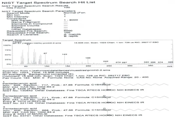
Figure 4S. GC-MS fingerprint of the pentadecanoic acid (5, n= 13)
Mioso et al.
S8 Quim. Nova
Figure 7S. 1H-NMR spectrum (CDCl
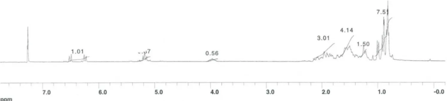
3, 300 MHz) of the Triolein (15)
Biomolecules produced in liquid-state fermentation by a marine-derived fungus, Penicillium roqueforti S9 Vol. 37, No. 2
Figure 8S. MS spectrum of the Triolein (15)
Figure 9S. 1H-NMR spectrum (CDCl
Mioso et al.
S10 Quim. Nova
Figure 10S. 1H-NMR spectrum (CDCl
3, 250 MHz) of the ergosterol peroxide (27) and its derivative, 9(11)-dehydroergosterol peroxide (28)
Biomolecules produced in liquid-state fermentation by a marine-derived fungus, Penicillium roqueforti S11 Vol. 37, No. 2
Figure 12S. 1H-NMR spectrum (CDCl
3, 400 MHz) of the ergosterol peroxide (27) and its derivative, 9(11)-dehydroergosterol peroxide (28)
Mioso et al.
S12 Quim. Nova
Figure 14S. 1H-NMR spectrum (CDCl
3, 250 MHz) of the ergosterol peroxide (27) and its derivative, 9(11)-dehydroergosterol peroxide (28)
Biomolecules produced in liquid-state fermentation by a marine-derived fungus, Penicillium roqueforti S13 Vol. 37, No. 2
Figure 16S. 13C-NMR spectrum (CD
3OD, 300 MHz) of the 4-hydroxybenzaldehyde (26)
Mioso et al.
S14 Quim. Nova
Figure 18S. Mass spectrum (MS) of the 4-hydroxybenzaldehyde (26)
Figure 19S. 1H-NMR spectrum (CD
Biomolecules produced in liquid-state fermentation by a marine-derived fungus, Penicillium roqueforti S15 Vol. 37, No. 2
Figure 20S. 1H-NMR spectrum (CD