1Neonatologist, Post-Graduate Student, Programa de Pos-Graduação em Ciências da Saúde, Universidade Federal do Rio Grande
do Norte, Natal RN, Brazil (UFRGN); 2P rofessor of Pediatric Neuro l o g y, Hospital de Pediatria and Maternidade Escola Januário Cicco,
Programa de Pos-Graduação em Ciências da Saúde, UFRGN.
Received 14 June 2005, received in final form 1 September 2005. Accepted 25 October 2005.
D r. Manoel Reginaldo Rocha de Holanda - Rua Gal. Felizardo Brito 2916 - 59078-410 Natal RN - Brazil. E-mail: re g h o l a n d a @ digizap.com.br
COMPARATIVE CLINICAL STUDY OF PRETERM
AND FULL-TERM NEWBORN NEONATAL SEIZURES
Manoel R.R. Holanda
1, Áurea N. de Melo
2ABSTRACT - Objective: To compare the characteristics of neonatal seizures between pre t e rm and full-term infants in intensive care unit. Method: A prospective study was developed with 104 high-risk newborn, 30 pre t e rm and 74 full-term infants, with clinical seizures. The dependent variable was gestational age. Statistical analyses: Fisher’s exact test, odds-ratio and Mann Witney U test. Results: T h e re were signifi-cant differences (p<0.05): i) premature neonates develop neonatal seizures later, probably related to the etiologies of the seizures; ii) etiologically, there is a predominance of peri-intraventricular hemorrhage in p re t e rm and of asphyxia in full term neonates; iii) clonic seizures are most frequent in pre t e rm and subtle s e i z u res in full term neonates. Conclusion: Although the study had a clinical basis, it was possible to iden-tify differences when the dependent variable was gestational age.
KEY WORDS: neonatal seizures, gestational age, comparative study, neonate, pre t e rm infant, full-term infant.
Estudo clínico comparativo das convulsões neonatais entre recém nascidos pretermo e termo
RESUMO - Objetivo: Comparar as características das convulsões neonatais entre recém nascidos prematu-ros e de termo internados em Unidades de Terapia Intensiva. Método: O estudo prospectivo neonatal avaliou 104 recém nascidos, 30 neonatos pre m a t u ros e 74 de termo, com crises convulsivas. As crises foram diagnosticadas pelas características clínicas. A variável dependente foi a idade gestacional. Na análise esta-tística utilizamos o teste exato de Fisher, odds ratio e o teste U de Mann Whitney. Resultados: Observa-mos diferenças estatísticas significantes (p<0,05): i) os neonatos prematuros manifestaram crises convulsi-vas mais tardiamente, provavelmente, relacionadas à etiologia; ii) observamos predomínio da hemorr a g i a peri-intraventricular nos neonatos prematuros e a asfixia nos recém nascidos de termo; iii) as crises clôni-cas foram mais freqüentes nos neonatos pre m a t u ros e as sutis nos neonatos de term o . Conclusão: E m b o r a o estudo tenha uma base clínica, foi possível identificar diferenças quando a variável dependente foi idade gestacional.
PA L AV R A S - C H AVE: convulsões neonatais, idade gestacional, estudo comparativo, neonatos pre t e rm o , neonatos de termo.
The occurrence of any manifestation of neonatal s e i z u res (NS) indicates a primary or secondary disfunc-tion of the central nervous system1 - 3. Thus, determ i n
a-tion of etiology is critical, because it gives the oppor-tunity to treat and to make a meaningful statement about the pro g n o s i s3 - 5. Nowadays, NS is defined by
v i d e o - e l e c t roencephalographic monitoring, by clin-ical observation associated to ictal or interictal elec-t roencephalogram (EEG), by elecelec-trographic discharg e without associated clinical manifestation or by neona-tal polysomnography4 , 6 - 1 2. However, in clinical
practi-ce at the pediatric or neonatal intensive care units (ICU), in developing countries where synchro n i z e d video-EEG monitoring is practically non-existent, clin-ical observation becomes the key to the diagnosis3.
On the other hand, we know that seizure pheno-menon in pre t e rm infant (PN) is less organized than it is in full-term infant (FT)3 , 1 3 , 1 4. So, when we
compa-re the clinical diagnosis of NS having gestational age as dependent variable, is the clinical profile diff e r-ent between the preterm and full-term infants?
NS diff e rences between PT and FT infants who were admitted to neonatal intensive care or pediatric ICU, based on clinical observations.
METHOD
We perf o rmed a prospective study from August 1s t, 2001
to July 31s t, 2002 in 9 ICU in Natal, Brazil. We selected al l
neonates that presented seizures during ICU stay. Pre p a r a-t o ry educaa-tional sessions were held in order a-to sa-tandard- standard-ize and improve the quality of observation and identifica-tion of NS. Semiology of seizures as well as stimulaidentifica-tion and restraint maneuvers were also discussed. These sessions included teams of neonatologists, intensive care pediatri-cians and nursing staff. A standard protocol of studying NS was developed jointly with the aforementioned teams. A c c o rd i n g l y, NS were defined as a paroxysmal phenome-non, re q u i red to have at least one of the following clinical manifestations: changes in behavior, stereotyped or peri-odic motor activities or autonomic dysfunctions3. We used
the classification proposed by Vo l p e1 5featuring four
essen-tial types: subtle, clonic, myoclonic, and tonic.
Gestational age was estimated based on the date of the last menstrual period, from obstetric ultrasonography or from a clinical estimates (Capurro ’s method or New Ballard Score). The newborn were determined as preterm when the gestational age was less than 37 weeks and full-term from 37-43 weeks.
The inclusion criteria w ere as follows: 1) detailed and unequivocal description of NS by the team; 2) seizures unre-sponsive to restraining maneuvers and unprovoked by stim-ulation and 3) occurrence of the first seizure up to 28 days old.
The exclusion criteria were: 1) uncertain clinical mani-festations, 2) adm ission to ICU with NS diagnosis but not p resenting any such episode during the stay, 3) those who had the first seizure after 28 days of life and 4) unidenti-fied gestational age.
The dependent variable was gestational age; the quan-titative independent variables were maternal age, number of gestations , maternal parity, Apgar at 1 and 5 minutes and the age at the first seizure. Independent category vari-ables were: sex, type of deliver y, depressed Apgar (0 to 6 s c o res), severe depressed Apgar (0 to 3 scores), seizure s type, seizure re c u rrence, presumed etiologies and mort a l-i t y. Recurrence were defl-ined when sel-izure repeats 24 hours after to get controlled. Deliveries w ere classified as vagi-nal and operative (caesarean section and forceps). The seizu-re ’s etiology was defined based on ICU’s guidelines. Neuro-ultrasonography scan were obtained, when possible, uti-lizing a GE Logiq Pro 400 machine.
The data were re c o rded in Epi-Info 6.0. The statistical analysis was perf o rmed by SPSS (Statistical Package for So-cial Science for Windows). Significance was obtained by Fi-s h e r’Fi-s exact teFi-st. OddFi-s ratio waFi-s analyzed when the inde-pendent variables were dichotomous. Mann Witney U test w e re perf o rmed for quantitative variables. The significance was considered when p value <0.05.
RESULTS
During the period of the study were admitted to the 9 ICU 1646 neonates, of these 1376 (83.3%) were in public hospitals, and 270 (16.4%) in private hospi-tals. We observed NS in 114 (6.9%) of the newborn infant, of which 103 (90.0%) were in public hospitals and 11 (10.0%) in private institutions. In accord a n c e with inclusion and exclusion criteria we selected 30 (28.8%) PN and 74 (71.2%) FT.
The average birth weight of PN was 1676.6g, stan-dard deviation 794.3g; median 1676.4g; mode 900g and range from 790 to 3650g. In FT the average was 3167.9g, standard deviation 559.2g; median 3155g; mode 3000g and range from 1605 to 4340g. The aver-age gestational aver-age of PN was 31.5 weeks; standard deviation 3.8 weeks; median 32 weeks; mode 31 weeks; and range from 23 to 36 weeks. The average of gestational age of FT was 39.6 weeks; standard deviation of 1.4 weeks; median 40 weeks; mode 39 weeks and range from 37 to 43 weeks.
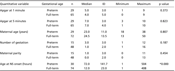
In Table 1 we demonstrated median, well as dis-persion and variability of the quantitative independ-ent variables. The Mann Whitney U test (Table 1) sho-wed a significant statistical diff e rence for the age of NS onset, later in PN (p=0.000).
Table 2 shows the data relative to the categorical variables. The male sex predominated in both full-t e rm and pre full-t e rm groups. In 1 FT ifull-t was nofull-t possible to determine the sex (ambiguous genitalia). General m o rtality was 21.15% (n=22). Comparing the two g roups, there was a mortality rate of 36.6% (n=11) in PN and 14.8% (n=11) in the FT, with statistical signi-ficance (p=0.014). Depressed Apgar at 1 minute was less common in PN and the diff e rence was statistical-ly significant (p=0.006).
Table 3 re p resents data re f e rent to NS classifica-tion. Subtle seizures predominated in FT (72.9%) and clonic seizures (50%) in PN. Analysis showed a signif-icant statistical diff e rence for subtle seizures being less frequent in PN (p=0.000) and for clonic seizures being more frequent in PN (p=0.000). In 28 (27%) n e w b o rn infants, 8 (26.6%) pre m a t u re and 20 ( 2 7 . 0 % ) f u l l - t e rm, we observed more than one type of NS. R e c u rrence of NS occurred in 20 (19.3%) of the neo-nates. In full-term neonates there was a re c u rre n c e in 16 (21.6%) and in the premature in 4 (13.3%).
N e u roultrasonography scan were perf o rmed on 58 (55.7%) of the neonates and 17 (29.0%) of these w e re found to be abnormal. The findings were ab-normal in 9 (30.0%) of PN and in 8 (10.8%) of FT.
Table 1. Median, dispersion and variability of quantitative variables in 30 pre t e rm and 74 full-term infants with neonatal seizures.
Quantitative variable Gestational age n Median ID Minimum Maximum p value
Apgar at 1 minute Preterm 29 5.0 3.0 1 9 0.373
Full-term 65 4.0 5.0 0 9
Apgar at 5 minutes Preterm 29 7.0 3.0 3 10 0.823
Full-term 65 7.0 4.0 1 10
Maternal age (years) Preterm 29 23.0 11.0 18 38 0.807
Full-term 72 24.5 13.5 13 50
Number of gestation Preterm 15 3.0 3.0 1 12 0.187
Full-term 48 1.0 2.0 1 16
Maternal parity Preterm 15 1.0 3.0 0 11 0.454
Full-term 48 0.0 2.0 0 13
Age at NS onset (hours) Preterm 30 72.0 141.7 1 504 *0.000
Full-term 74 12.0 23.0 1 408
N, neonatal seizures; ID, interquartile distance; p, value obtained with Mann Whitney U test; *p, significance.
Table 2. Category variables in 30 preterm and 74 full-term infants with neonatal seizures.
Gestational age
Category variables Preterm Full-term OR C.I. OR (95%) p value
n % n %
Male 17 56.6 41 56.1 1.01 0.55-1.86 0.963
Female 13 43.4 32 43.9 0.98 0.38-2.51 0.865
Operative delivery 12 40.0 42 56.7 0.61 0.33-1.14 0.121
Vaginal delivery 18 60.0 32 43.3 0.18 0.76-5.15 0.185
1 minute-SD Apgar 10 33.3 29 39.1 0.74 0.38-1.42 0.357
5 minutes-SD Apgar 3 10.0 9 12.1 0.72 0.12-3.21 0.459
1 minute-D Apgar 13 43.3 48 64.8 0.29 0.10-0.79 *0.006
5 minutes-D Apgar 9 30.0 28 37.8 0.59 0.21-1.63 0.269
NS Recurrence 4 13.3 16 21.6 0.64 0.25-1.64 0.331
Mortality 11 36.6 11 14.8 2.15 1.21-3.83 *0.014
NS, neonatal seizures; OR, odds ratio; CI, confidence interval; SD Apgar, severe depressed Apgar (0 to 3 scores); D Apgar, d e p ressed Apgar (0 to 6 scores); p, value obtained with Fisher’s exact test; *p, significance. 1, newborn full-term with ambiguous genitalia.
Table 3. Neonatal seizures types in 30 preterm and 74 full-term infants with neonatal seizures.
Gestational age
NS type PN FT OR CI OR (95%) p value
n % n %
Subtle 8 26.6 54 72.9 0.13 0.05-0.38 *0.000
Tonic 10 33.3 20 27.0 1.35 0.48-3.66 0.520
Clonic 15 50.0 12 16.2 5.17 1.81-14.78 *0.000
Myoclonic 3 10.0 4 5.4 1.94 0.27-12.25 0.320
FT (p=0.000) and more peri-intraventricular hemor-rhage (PVIH) (p=0.000). In 65 neonates (62.5%), of whom 12 (40.0%) were pre m a t u re and 53 (71.6%) w e re full-term, we noted a single probable etiology for NS. In 19 (18.2%) neonates, of whom 10 (33.3%) w e re PN and 9 (12.1%) FT, it was not possible to de-termine the etiology. In 15 (14.4%) of the neonates
we observed epileptic status, 12 (16.2%) in FT and 3 (10.0%) in PN.
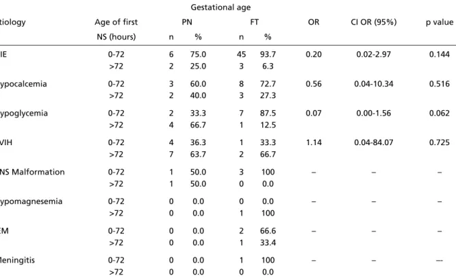
Table 5 shows the relation between the age at the onset of the seizures and etiology. In HIE, the onset of NS is early in both FT and PN, while in PVIH, which predominates in PN, seizure onset was slight-ly delayed.
Table 5. Relation between age of onset of neonatal seizures and probable etiologies in 30 pre t e rm and 74 full-term infants.
Gestational age
Etiology Age of first PN FT OR CI OR (95%) p value
NS (hours) n % n %
HIE 0-72 6 75.0 45 93.7 0.20 0.02-2.97 0.144
>72 2 25.0 3 6.3
Hypocalcemia 0-72 3 60.0 8 72.7 0.56 0.04-10.34 0.516
>72 2 40.0 3 27.3
Hypoglycemia 0-72 2 33.3 7 87.5 0.07 0.00-1.56 0.062
>72 4 66.7 1 12.5
PVIH 0-72 4 36.3 1 33.3 1.14 0.04-84.07 0.725
>72 7 63.7 2 66.7
CNS Malformation 0-72 1 50.0 3 100 – – –
>72 1 50.0 0 0.0
Hypomagnesemia 0-72 0 0.0 0 0.0 – – –
>72 0 0.0 1 100
IEM 0-72 0 0.0 2 66.6 – – –
>72 0 0.0 1 33.4
Meningitis 0-72 0 0.0 1 100 – –
–->72 0 0.0 0 0.0
NS, neonatal seizures; OR, odds ratio; PN, pre t e rm infant; FT, full-term infant; HIE, hipoxic-ischemic encephalopathy; PVIH, peri-intraventric-ular hemorrhage; CNS, central nervous system; IEM, inborn errors metabolism; CI, confidence interval; p, value obtained with Fisher’s exact test; (–), not observed.
Table 4. Probable etiologies in 30 preterm and 74 full-term infants with neonatal seizures.
Gestational age
Etiology PN FT OR CI OR (95%) p value
n % n %
HIE 8 26.6 48 64.8 0.20 0.07-0.55 *0.000
Hypocalcemia 5 16.6 11 14.8 1.15 0.28-4.04 0.817
Hypoglycemia 6 20.0 8 10.8 2.06 0.53-7.55 0.213
PVIH 11 36.6 3 4.0 13.7 3.10-81.39 *0.000
CNS malformation 2 6.6 3 4.0 1.69 0.13-15.51 0.572
Hypomagnesemia 0 0.0 3 4.0 – – –
IEM 0 0.0 1 1.3 – – –
Meningitis 0 0.0 1 1.3 – – –
DISCUSSION
It is well recognized that seizures are more com-mon in the neonatal period than in any other stage of life3. In the NS literature we find excellent
stud-ies, such as those by Mizrahi and Kellaway1 6, with
detailed discussions on various aspects, but not tak-ing into account the consequences of gestational age. Curre n t l y, with medical and technological advan-ces in ICU, 85% of PN surv i v e1 7, thus turning into a
c a t e g o ry with its own characteristics in relation to its i m m a t u re central nervous system and their neuro n a l lesion mechanisms18,19. In this way, in relation to NS,
it is important to bear in mind the diff e rences bet-ween PN and FT group.
Our study had three parameters which were fun-damental in attaining its objectives: 1) to be pro s p e c-tive in view of the neonatal period; 2) to use a pop-ulation that had been treated exclusively in ICU divid-ing it into two groups, accorddivid-ing to gestational age and rigorous clinical observation; and 3) using stim-ulation and restraint techniques to accurately define NS. Studies based on clinical data are uncommon in the literature, however we would like to mention the study of Brunquell et al.2 0and the similarity of
their approach to our emphasis on clinical observ a-tion. Saliba et al.2 1also emphasize gestational age as
a NS risk factor and discuss the diff e rence between term and preterm neonates.
The incidence of NS in our study was 6.9% this was slightly lower than that found by Sheth who re p o rted an incidence of 8.6% in 4165 newborn ad-mitted to ICU2 2. This finding can be partly explained
by the reduced number of public ICU in Natal and by the low socio-economic level of the study popula-tion, that have not access to private ICU. Our figure s exceed those of Brunquell et al.2 0. This diff e rence may
be related to the method of these authors, who per-formed a retrospective study, using data from med-ical re c o rds. In the electroclinmed-ical study by Sheth2 3,
the seizures correlated with EEG alterations in 63% of PN at 28 weeks and in 77% of FT. The lower neu-ronal organization may explain the lower clinical and electroclinical expressiveness of the condition in PN with a resultant lower seizure frequency with our study.
An important finding in this study was the statis-tically significant diff e rence for the time of NS onset, which occurred later in the pre m a t u re. Unlike of Sheth et al.2 2that re p o rted seizures manifested
ear-lier in infants <30 weeks and >36 weeks gestational c o m p a red with neonates 30 to 36 weeks. This diff e r-ence might have etiological reasons of the seizures:
HIE was the predominant etiology in the FT, causing early-onset NS in the first 48 hours of life2 4. This
find-ing is in agreement with the data re p o rted by Ahn et al.2 5who correlated time of NS onset and
under-lying neurological lesions. On the other hand, in PN peri-intraventricular hemorrhage was the most fre-quent etiology and its clinical picture intensifies after 72 hours of life, thus causing later seizures manifes-t a manifes-t i o n3 , 5 , 2 6. However, when HIE was the likely
etiolo-g y, NS had early-onset in both etiolo-groups. This findinetiolo-g a g ree with re p o rts stressing neonatal asphyxia as the most frequent cause of early-onset NS3 , 1 6. Where a s
Calciolari et al.2 4re p o rted neonatal asphyxia as the
p redominant etiology in both PN and FT. The NS are caused by hypoxic-ischemic encephalopathy in 50 to 60% of neonates, independent of gestational age, a c c o rding to Vo l p e3. However, determining the exact
etiology of the seizures is very difficult, since a larg e number of etiological factors may coexist part i c u l a r-ly: HIE, metabolic disorders and intracranial hemor-rhages. In this study, 62.5% of the neonates were presumed to have an etiology, while the number of babies with undetermined etiology (18.2%) was sub-stantial. This impossibility of pinning down likely eti-ologies was also due to the unavailability of certain diagnostic tests. For instance, access to cranial ultra-sounds examination was possible in only 55.7% of the neonates selected. Despite theses difficulties, our overall findings were not basically diff e rent from cur-rent literature.
The presence of severe depressed Apgar (0 to 3 s c o res) did not show significant statistical diff e re n c e between the groups. As to depressed Apgar (0 to 6 s c o res), there was a significant statistical diff e re n c e at 1 minute of life, with lower frequency in the PN. This diff e rence dissipated at 5 minutes probable due to reanimation techniques promptly initiated at birt h in the delivery room. PN have greater risk for peri-natal asphyxia and need for reanimation in the deliv-e ry room dudeliv-e to ddeliv-eficideliv-ency of thdeliv-e pulmonary surf a c-tant system, they are most susceptible to heat loss, have the greatest risk for intra-uterine infection and the most fragile cerebral capillaries2 7. Surprisingly,
our data of PN showed lower frequency of depre s s e d Apgar when compared with FT. These figures how-ever, could be deceptive due to our emphasis being placed on NS. According to Vo l p e3, the parameter of
asphyxia based on Apgar scores has greater pre d i c-tive value for the neurological outcome rather than for NS.
the newborn, similar to the data re p o rted by Calcio-lari et al.2 4and by Brunquell et al.2 0. Arpino et al.
re-ferred to generalized tonic seizures as the most fre-quent seizure type and in 71% of the neonates, an association of more than one clinical type of NS was observed28Ronan et al. reported that tonic seizures
p redominated in FT, and clonic seizures were equal-ly common in both gro u p s2 9. When we compared the
two groups, subtle seizures were most fre q u e n t l y o b s e rved in FT, in accordance with the literature3 , 1 6 , 2 4.
In PN, however, clonic seizures were the most fre-quent. This results were too observed by Scher et al.3 0
who used electro clinical data.
The general mortality rate of the sample was sim-ilar to that observed in the 1990s, which was aro u n d 2 0 %2 6and, in our material higher in PN (p=0.014).
The odds ratio indicates that the risk of death in PN is twice as high as that of FT. The historically gre a t e r m o rtality rate of PN is due to the wealth of serious pathologies of to prematurity and there f o re unre-lated to presence or absence of NS. In conclusion, we wish to point out that: 1) PN develop NS later, pro b-ably related to the etiologies of the seizures; 2) eti-o l eti-o g i c a l l y, there is a predeti-ominance eti-of PVIH in PN and of HIE in FT; 3) clonic seizures are most frequent in PN and subtle seizures in FT; and 4) the mortality rate is determined by underlying severe pathology asso-ciated with prematurity and not by the NS as such.
We also wish to place great emphasis on the use of stimulating and restraining maneuvers for pro v o-cation or termination of seizures - especially in view of the unavailability of video-EEG-monitoring. Such maneuvers will enrich the clinical approach to NS -not only in the ICU but also in daily clinical practice.
Acknowledgements – We are indebted to Dr. Errnst
Niederme yer (The John Hopkins University School of Medi-cine, Baltimore - MD) for giving us relevant clinical infor-mation and for review of the manuscript. Nothing could have been achieved without the wonderful spirit of collab-oration among the colleagues involved in the acquisition of our data: Henrique Leite Raposo, Lílian Bezerra Faria de Melo and Rosa Maria Vaz dos Santos (Maternidade Escola Januáio Cicco); Célia Gilna Gomes Mendes and Janaina Gonçalves de Brito Bonifácio (Hospital Dr. Jose Pedro Be-z e rra); Adélia Cristina Tinoco Bulhões and Ana Amélia B a rreto Pacheco (Hospital Central Coronel Pedro Germ a n o ) ; Aldenilde Rebouças Falcão and Ana Elisabeth Gomes de Melo Tavares Ferreira (Hospital e Maternidade Promater); Patricia Arboes Petronillo (Papi Hospital Geral), João Bosco Lima Barbosa (Hospital Infantil Va rela Santiago and Hospital Memorial); Nívia Maria Rodrigues Arrais (Hospital Infantil Maria Alice Fernandes and Hospital Antônio Prudente).
REFERENCES
1. Lanska MJ, Lanska DJ, Baumann RJ, Kryscio RJ. A p o p u l a t i o n - b a s e d study of neonatal seizures in Fayette County, Kentucky. Neuro l o g y 1995;45:724-732.
2. Scher MS. Neonatal seizures and brain damage. Pediatr Neurol 2003; 29:381-390.
3. Volpe JJ. Neonatal seizures. In Neurology of the newborn. Philadelphia: Saunders 2001:178-214.
4. Mizrahi EM. Neonatal seizures: problems in diagnosis and classifica-tion. Epilepsia 1987;28(Suppl 1):S46-S55.
5. Patrizi S, Holmes GL, Orzalesi M, Allemand F. Neonatal seizures: char-acteristics of EEG ictal activity in preterm and fullterm infants. Brain Dev 2003;25:427-437.
6. Caravale B, Allemand F, Libenson MH. Factors predictive of seizure s and neurologic outcome in perinatal depression. Pediatr Neurol 2003; 29:18-25.
7. Holmes GL, Lombroso CT. Prognostic value of background patterns in the neonatal EEG. J Clin Neurophysiol 1993;10:323-352.
8. McBride MC, Laroia N, Guillet R. Electrographic seizures in neonates c o r relate with poor neurodevelopmental outcome. Neurology 2000; 55:506-513.
9. L o m b roso CT, Holmes GL. Value of the EEG in neonatal seizures. J Epilepsy 1993;6:30-70.
10. Mizrahi EM, Kellaway P. Characterization and classification of neona-tal seizures. Neurology 1987;37:1837-1844.
11. Scher MS. Eletroencephalography of the newborn: normal an abnor-mal features. In Niedermeyer E, Lopes da Silva F (eds). Eletro e n c e p h a l o-graphy: basic principles, clinical aplications and related fields. Baltimore : Williams & Wilkins 1999:886-946.
12. Oliveira AJ, Nunes ML, da Costa JC. Polysomnography in neonatal seizures. Clin Neurophysiol 2000;111(Suppl 2):S74-S80.
13. Hughes JR, Fino J, Gagnon L. The use of the electroencephalogram in the confirmation of seizures in pre m a t u re and neonatal infants. Neuropediatrics 1983;14:213-219.
14. Legido AC. Convulsions neonatales: comentarios sobre aspectos con-trovertidos. An Esp Pediat 1991;1:1-8.
15. Volpe JJ. Neonatal seizures: current concepts and revised classification. Pediatrics 1989;84:422-428.
16. Mizrahi EM, Kellaway P. Diagnosis and managment of neonatal seizures. Philadelphia: Lippincot-Raven, 1998.
17. Volpe JJ. Neurologic outcome of pre m a t u r i t y. A rch Neurol 1998;55: 297-300.
18. Holmes GL, Khazipov R, Ben-Ari Y. New concepts in neonatal seizure s . Neuroreport 2002;13:A3-8.
19. Inder TE, Volpe JJ. Mechanisms of perinatal brain injury. Semin Neonatol 2000;5(1):3-16.
20. B runquell PJ, Glennon CM, DiMario FJ Jr, Lerer T, Eisenfeld L. Pre-diction of outcome based on clinical seizure type in newborn infants. J Pediatr 2002;140:707-712.
21. Saliba RM, Annegers FJ, Waller DK, Tyson JE, Mizrahi EM. Risk fac-tors for neonatal seizures: a population-based study, Harris County, Texas, 1992-1994. Am J Epidemiol 2001;154:14-20.
22. Sheth RD, Hobbs GR, Mullett M. Neonatal seizures: incidence, onset, and etiology by gestational age. J Perinatol 1999;19:40-43.
23. Sheth RD. Electroencephalogram confirmatory rate in neonatal seizure s . Pediatr Neurol 1999;20:27-30.
24. Calciolari G, Perlman JM, Volpe JJ. Seizures in the neonatal intensive c a re unit of the 1980s: types, etiologies, timing. Clin Pediatr (Phila) 1988;27:119-123.
25. Ahn MO, Korst LM, Phelan JP, Martin GI. Does the onset of neonatal s e i z u res correlate with the timing of fetal neurologic injury? Clin Pediatr (Phila) 1998;37:673-676.
26. Scher MS. Seizures in the newborn infant: diagnosis, treatment, and outcome. Clin Perinatol 1997;24:735-772.
27. American Academy of Pediatrics, American Heart Association. Por que os neonatos prematuros são considerados de alto risco? In Manual de reanimação neonatal. 4.ed. São Paulo: Escola Paulista de Medicina; 2000.
28. Arpino C, Domizio S, Carrieri MP, Brescianini DS, Sabatino MG, Cura-tolo P. Prenatal and perinatal determinants of neonatal seizures occur-ring in the first week of life. J Child Neurol 2001;16:651-656. 29. Ronen GM, Penney S, A n d rews W. The epidemiology of clinical
neona-tal seizures in Newfoundland: a population-based study. J Pediatr 1999;134:71-75.