1Postgraduate student, Department of Neurology, University of São Paulo School of Medicine, São Paulo SP, Brazil (FMUSP); 2Neurologist, Department of Clinics Discipline of Neurology, Federal University of Curitiba PR, Brazil (UFPR); 3Associated Professor,
Department of Neurology, FMUSP; 4Graduate Student, Department of Neurology, FMUSP; 5Assistant Professor, Department of Neurology,
FMUSP.
Received 24 November 2003, received in final form 8 May 2004. Accepted 7 June 2004.
Dra. Ana Paula B.J. Hartmann - Rua Francisco de Vitória 250/81 - 04116-180 São Paulo SP - Brasil. E-mail: [email protected]
HYPERPHOSPHORYLATED TAU PROTEIN IN THE
CEREBROSPINAL FLUID OF PATIENTS WITH
ALZHEIMER´S DISEASE AND OTHER DEMENTIAS
Preliminary findings
Ana Paula Barbosa Jeronimo Hartmann
1, Sérgio Monteiro de Almeida
2,
José Antonio Livramento
3, Ricardo Nitrini
3, Daniel Takahashi
4, Paulo Caramelli
5ABSTRACT - Alzheimer’s disease (AD) is pathologically characterized by the accumulation of amyloid plaques and tau-associated neurofibrillary tangles in the cerebral tissue. The search for antemortem bio-markers is intense including analysis of cerebrospinal fluid (CSF) β-amyloid and tau proteins concentrations seeking for an accurate and early diagnosis. Levels of hyperphosphorylated tau at threonine 181 were meas-ured in the CSF of 34 patients with AD (19 with senile AD – SAD and eight with presenile AD – PSAD) and seven with other dementias (OD). The levels of CSF phosphotau were significantly higher in the AD patients compared to OD (AUC 0.812), with no association with severity of dementia, age of onset, dura-tion of the disease or scores in the Mini-Mental State Examinadura-tion. There were no differences of phospho-tau levels between SAD and PSAD patients. These findings corroborate some previous studies and indicate that CSF phosphotau may help to differentiate AD from other dementias.
KEY WORDS: Alzheimer’s disease, differential diagnosis, cerebrospinal fluid, tau protein.
Proteína tau hiperfosforilada no líquido cefalorrraqueano de pacientes com doença de Alzheimer e outras demências: resultados preliminares
RESUMO - A doença de Alzheimer (DA) se caracteriza pelo achado anátomo-patológico de acúmulo de placas senis e emaranhados neurofibrilares associados à proteína tau no tecido cerebral. A pesquisa por marcadores biológicos antemortemestá focada nas concentrações das proteínas β-amilóide e tau no líqui-do cefalorraqueano (LCR) objetivanlíqui-do um diagnóstico mais precoce e acuralíqui-do da líqui-doença. Os níveis de pro-teína tau hiperfosforilada no sítio 181 foram determinados no LCR de 34 pacientes com DA (19 com DA senil - DAS e oito com DA pré-senil -DAPS) e sete pacientes com outras demências (OD). Os níveis de fos-fotau foram significativamente mais elevados em pacientes com DA quando comparados com OD (AUC 0,812), sem relação com gravidade da demência, idade de início, duração da doença e escores do Mini-Exame do Estado Mental. Não foram observadas diferenças entre os níveis de fosfotau em pacientes com DAS e DAPS. Estes achados corroboram os dados encontrados em estudos prévios e indicam que o nível de fosfotau no LCR dos pacientes pode colaborar na diferenciação da DA com outras demências.
PALAVRAS-CHAVE: doença de Alzheimer, diagnóstico diferencial, líquido cefalorraqueano, proteína tau.
Alzheimer’s disease (AD) is the main cause of de-mentia in Western countries, being responsible for more than 50% of the cases1. The clinical
diagno-sis of AD is characterized by an exclusionary pro-cess. The definite diagnosis is possible only on neu-ropathological examination, by the observation of the senile plaques and the neurofibrillary tangles
(NFTs) in the cerebral tissue2. The NFTs are
infragra-nular (V and VI) layers of associative cortical areas. Subcortical areas are also affected, such as the nu-cleus basalis of Meynert, amygdala, locus ceruleus and dorsal raphe nuclei3.
The well established relationship between the density of NFTs and the severity of the dementia in AD has lead to many studies about protein tau le-vels, one of the main components of NFTs, in cere-brospinal fluid (CSF) of AD patients4-6. Tau protein
is a microtubule-associated protein (MAP) found basically in neurons and mainly localized in axons where it confers stability to microtubule components of the cytoskeleton. The protein is codified by a gene on chromosome 17 and mutations are associated to certain forms of frontotemporal dementia, especial-ly frontotemporal dementia and parkinsonism relat-ed to chromosome 177. Microtubule stability depends
on phosphorylation of tau protein. In mature CNS low phosphorylated forms of tau protein predom-inate, maintaining adequate neuronal homeosta-sis. PHF-tau concentrations has been shown to be elevated in the cortex of AD patients while normal tau concentration is decreased8. Considering early
and accurate diagnosis and differential diagnosis with other dementias that mimic AD symptoms, the development of a biological marker is of great value. A biological marker has a minimum of five functions: a) diagnostic confirmation; b) screening; c) predictive testing; d) monitoring disease progres-sion and treatment; and e) analysis of the relation between brain-behavior9.
In this way, an ideal biomarker for AD should be: able to detect a characteristic pathological fin-ding of AD; validated in AD pathologically confir-med cases; precise (for differential diagnosis); trus-table; not invasive; simple, reproductive; and not expensive.
Besides, it should have sensitivity and specifici-ty above 80% and a positive predictive value above than 90%9. The combination of biomarkers
im-proves the diagnostic accuracy when compared to a single marker, thus increasing the sensitivity and specificity of the tests. Considering these fac-tors, recent studies have been dedicated to investi-gate the abnormal proteins found in the CSF of AD patients4,10,11. These studies have evaluated the
le-vels of MAP-tau, primary component of the neuro-fibrillary tangles, and the Aβ42 form of the β -amy-loid protein, the main component of the senile plaques found in the cerebral parenchyma.
Many groups have shown an increase in tau lev-els4,5,8,10,12,13and a decrease in Aβ42 levels13,14,15in
the CSF of AD patients when compared to
non-de-mented elderly controls. The more recent longitudi-nal studies use the combination of high tau and low Aβ42 to correlate their levels to the stage of the disease13,15. Since amyloid deposition is not
ex-clusive of AD brains, occurring in normal aging and also in other neurological diseases, additional in-vestigation have shown a decrease in AβCSF lev-els in other conditions such as Creutzfeldt-Jakob disease (CJD)16, some cases of frontotemporal
de-mentia (FTD) and vascular dede-mentia (VD)17.
In 1993, Vandermeeren et al.18developed an
im-munoassay able to detect tau protein in CSF and subsequent studies concluded that its levels were significantly higher in AD patients when compared to other neurological diseases and normal controls even in the early stages of the disease4,10. As tau
protein is present in blood in a very low concentra-tion, (under the detection limit of the immunoas-say), the high levels in CSF do not reflect an alter-ation in blood brain barrier18. As elevated tau
lev-els were also found in other neurological diseases, it was noticed that these immunoassays were meas-uring total tau, i.e., normal and abnormal tau pro-tein. To solve this overlap, a group of investigators19
developed a method able to detect hyperphospho-rylated tau (phosphotau) and obtained elevated levels when comparing AD patients to controls. Other studies reproduced these results, suggesting that phosphotau is a more specific biomarker then total tau for AD diagnosis20,21. The abnormal
phos-phorylation of tau protein is an early event in AD pathophysiology and is restricted to cerebral regions affected by the disease. This hyperphosphorylation is the primary and the most critical event in PHFs and NFTs processing22. More than 21 sites of
abnor-mal tau protein phosphorylation are known. As CSF tau concentrations are low, phosphotau is only identified by highly sensitive immunoassays using phospho-specific antibodies.
Several research groups standardized synthet-ic phosphopeptides in order to access the proline rich region of tau protein where the phosphosites are localized. Until now, different immunoassays were developed directed to different phospho-sites as serine 199, threonine 231, serine 396/404, threonine 231/serine 235 and threonine 18121,23.
Vanmechelen et al.24 developed an ELISA assay
design of this peptide was based on detailed map-ping of phosphotau and its antibodies, recogniz-ing all tau isoforms. The objectives of the present study were to compare CSF phosphotau levels bet-ween senile (SAD) and presenile (PSAD) AD groups, between AD patients and other dementias (OD), between AD patients and controls from the liter-ature11, and, within the AD group, to correlate
phosphotau levels and severity of dementia, mini mental state examination (MMSE) scores and dura-tion of the disease.
METHOD
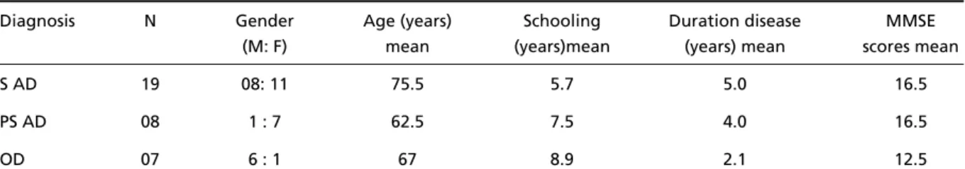
A total number of 34 individuals were included in the study. Their main demographic and clinical characte-ristics are depicted in Table 1.
The subjects evaluated were consecutively selected from the population of patients followed at the Beha-vioral and Cognitive Neurology Unit of the Hospital das Clínicas from the University of São Paulo School of Me-dicine (HCFMUSP).
All patients were submitted to a diagnostic workup investigation, including clinical history, physical and neu-rological examination, appropriate blood tests (to ex-clude other causes of dementia), CT and/or brain MRI and other complementary exams if necessary.
In every case, the clinical diagnosis was made before CSF examination. No patient was on treatment with cholinesterase inhibitors or was participating in any protocol of new drugs before the lumbar puncture.
The diagnosis of probable AD was based on the NINCDS-ADRDA criteria25. For the AD group according to DSM-III-R criteria, the severity of dementia was classi-fied as mild, moderate or severe. The diagnosis of fron-totemporal dementia (FTD) was made according to the Lund/Manchester criteria26. Vascular dementia (VD) pa-tients were selected according to probable VD criteria of NINDS-AIREN27and dementia with Lewy bodies accord-ing to McKeith et al. criteria28. Criteria used to diagno-sis of primary progressive aphasia (PPA) were those de-fined by Mesulam29.
Patients were divided in three groups: SAD (senile AD), PSAD (presenile AD) and OD (other dementias), with the latter including one patient with the diagnosis of
PPA, two cases of FTD, one with VD, one with corticobasal degeneration (CBD) and two with dementia with Lewy bodies (DLB).
CSF phosphotau analysis was performed using the kit INNOTESTTMPHOSPHO-TAU
(181P)(Innogenetics, Ghent, Belgium). The phosphotau levels from each of the patients’ groups were compared to a subset of controls (composed of 32 individuals) extracted from a previously published study.11These individuals were aged 63 ± 9 years and had no history, symptoms or signs of psychiatric or neurolog-ical disease, malignant or systemic disorders. The mean MMSE score in this group was 28.3 ± 2.7.
CSF analysis- All CSF samples were obtained by lum-bar puncture. An approximate volume of 12 ml was col-lected in polypropylene tubes and submitted to routine analysis (cytology, biochemistry and protein electrophore-sis, as well as immunology for syphilis, cysticercosis and ADA measurement), always within a six-hour interval after the collection procedure. Samples with more than 500 erythrocytes/mm3were not included. After routine analysis and centrifugation of the material at 1500 rpm for 10 minutes, the samples destined to phosphotau measurement were identified and stored at - 70oC freez-er in the CSF laboratory.
An enzymatic immunoassay in solid phase for the quantitative determination of phosphotau in human CSF standardized in the kit INNOTESTTMPHOSPHO-TAU
(181P) (Innogenetics, Ghent, Belgium) was the method used for this evaluation.
The study was approved by the Ethics Committee of the HCFMUSP and all participants signed a written infor-med consent.
Statistical analysis - The comparison between phos-photau levels in AD groups and controls from literature was made by the Student’s t test. The comparison between AD and OD groups was made by the Mann-Whitney test. In the AD group, the correlation between phosphotau levels and severity of dementia, MMSE scores and duration of the disease was made by the Spearman’s rank correlation test.
RESULTS
Due to difficulties in obtaining CSF samples from normal controls, we have been unable to
Table 1. Main demographic and clinical characteristics of the patients.
Diagnosis N Gender Age (years) Schooling Duration disease MMSE (M: F) mean (years)mean (years) mean scores mean
S AD 19 08: 11 75.5 5.7 5.0 16.5
PS AD 08 1 : 7 62.5 7.5 4.0 16.5
OD 07 6 : 1 67 8.9 2.1 12.5
establish sensitivity and specificity of the method as we could not define cut-off values for the analy-sis of the results obtained in this study. We tried to overcome this limitation by comparing our re-sults of phosphotau levels with controls described in the literature11.
The statistical analysis showed no significant dif-ference between SAD, PSAD and OD groups accord-ing to duration of the disease (p=0.644), education (p=0.830), severity of dementia (p=0.481) and MMSE scores (p=0.651). As expected, there was a significant age difference between SAD and PSAD groups (p=0.000). Significant differences were also observed between the AD and OD groups for age (p=0.013), which was lower in the latter, and gen-der (p=0.035) with women predominating in the AD group and men in OD.
Using the mean of phosphotau levels of con-trol individuals obtained from a previous study (32.8 pg/ml)11, we obtained a significant
differ-ence (p=0.013) between AD patients and controls. The mean phosphotau concentration in the AD group was 50.4 pg/ml. No differences were obser-ved between phosphotau levels of SAD and PSAD groups (p=0.549) as it is depicted in Table 2.
There was no significant correlation, in the AD group, between levels of phosphotau and severi-ty of dementia (r=-0.082), duration of disease (r=0.015) and MMSE scores (r=-0.020).
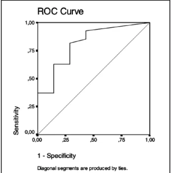
A significant difference was found between phosphotau levels of AD and OD groups (p= 0.023). Plotting the values of phosphotau levels from the AD and OD groups in a Receiving Operator Characteristics (ROC) curve we found that phospho-tau levels differentiated AD from OD patients, what was confirmed with an area under the curve (AUC) of 0.812 (Fig 1).
DISCUSSION
In the present study CSF levels of phosphotau were increased in AD patients when compared to
normal controls from the literature and to patients with OD, a finding that is similar to previous re-ports11,20,21,23,24. However, It is important to
reinfor-ce that phosphotau levels in a normal range do not exclude AD. The absence of a correlation between phosphotau levels and severity of dementia sug-gests that this elevation is an early event in AD pa-thogenesis. Indeed, some studies demonstrated the clinical value of this biomarker in the early stages of AD11,30.
Two patients with other dementias in the pres-ent study prespres-ented high levels of phosphotau (one case with FTD and one case with DLB). Although it is not possible to exclude the hypoth-esis of diagnostic errors, the long follow-up of all these patients, before and after the CSF analysis, is a feature that certainly increases the diagnostic confidence within this sample.
The utilization of additional biomarkers, such as the combination Aβ1-42protein with phospho-tau, and its correlation with the clinical presenta-tion, certainly would have given a higher diagnos-tic sensitivity and specificity. This fact was recent-ly confirmed in a meta-anarecent-lysis that included all the studies that evaluated the combination of bio-markers in an expressive number of patients and using an adequate diagnostic method of the cause of dementia14. Unfortunately, it has not been
pos-sible to verify the diagnostic values of this combina-tion in the present study.
Table 2. Mean (± SD) levels of phosphotau.
Levels of CSF phosphotau (pg/ml) Mean (± SD)
S AD 49.78 (± 36.09)
PS AD 44.06 (± 33.29)
OD 15.01 (± 27.05)
S AD, senile Alzheimer’s disease; PS AD, presenile Alzheimer’s disease; OD, other dementias.
Additional studies are necessary to establish a methodological standardization of CSF immunoas-says between the research centers and to observe if they represent a very high specificity for AD diag-nosis, mainly in relation to forms of dementia that present an important overlap with AD either on clinical and on pathological examination (such as DLB and VD). Moreover, the conflicting findings from the literature, correlating CSF biomarkers with clinical severity measures of dementia, sug-gest the need of larger samples to establish a con-fident statistical significance. The reason for this variability is probably due to the fact that a single marker is not able to reflect precisely the central pathological process of each stage of the disease.
Maybe a potential utility of such biomarkers is in the follow-up of individuals at risk of develop-ing AD in prospective studies. However, much work is still necessary to standardize assay methods be-fore the determination of the prognostic value attributable to these biomarkers. With the defini-tion of well standardized values, in large popula-tions, it is possible that gradual alterations in these levels can be interpreted as suggestive evidences of incipient AD. To test this hypothesis, longitudi-nal studies including large samples of elderly indi-viduals are necessary.
In conclusion, the present study found that CSF levels of phosphotau analysis is a good biomarker for AD and is able to help in differentiating AD from other dementias, independently of the age of onset, severity of dementia or MMSE scores.
REFERENCES
1. Herrera E Jr, Caramelli P, Silveira AS, Nitrini R. Epidemiologic survey of dementia in a community-dwelling Brazilian population. Alzheimer Dis Assoc Disord 2002;16:103-108.
2. Mirra SS, Heyman A, McKeel D et al. The Consortium to Establish a Registry for Alzheimer’s Disease (CERAD): part II. Standardization of the neuropathologic assessment of Alzheimer’s disease. Neurology 1991;41:479-486.
3. Wisniewski HM, Robe A, Zigman W, Silverman W. Neuropathological diag-nosis of Alzheimer disease. J Neuropathol Exp Neurol 1989;48:606-609. 4. Arai H, Clark CM, Ewbank DC, et al. Cerebrospinal fluid tau protein
as a potential diagnostic marker in Alzheimer’s disease. Neurobiol Aging 1998;19:125-126.
5. Blennow K, Wallin A, Agren H, Spenger C, Siegfried J, Vanmechelen E. Tau protein in cerebrospinal fluid: a biochemical marker for axonal degen-eration in Alzheimer disease? Mol Chem Neuropathol 1995;26:231-245. 6. Galasko D, Hansen L Vigo-Pelfrey C, et al. Antemortem CSF tau is
relat-ed to neuronal pathology at autopsy in Alzheimer´s disease. Soc Neurosci Abstr 1995;581.
7. Bramblett GT, Goedert M, Jakes R, Merrick SE, Trojanowski JQ, Lee VM. Abnormal tau phosphorylation at Ser396 in Alzheimer’s disease reca-pitulates development and contributes to reduced microtubule bind-ing. Neuron 1993;10:1089-1099.
8. Vigo-Pelfrey C, Seubert P, Barbour R, et al. Elevation of microtubule-associated protein tau in the cerebrospinal fluid of patients with Alzheimer’s disease. Neurology 1995;45:788-793.
9. Consensus report of the Working Group on: “Molecular and Biochemical Markers of Alzheimer’s Disease”. The Ronald and Nancy Reagan Research Institute of the Alzheimer’s Association and the National Institute on Aging Working Group. Neurobiol Aging 1998;19:109-116. 10. Andreasen N, Minthon L, Vanmechelen E, et al. Cerebrospinal fluid tau and Abeta42 as predictors of development of Alzheimer’s disease in patients with mild cognitive impairment. Neurosci Lett 1999;273:5-8. 11. Andreasen N, Vanmechelen E, Vanderstichele H, et al. Cerebrospinal fluid levels of total-tau, phospho-tau and AB42 predicts development of Alzheimer´s disease in patients with mild cognitive impairment. Acta Neurol Scand. 2003;179:(Suppl)47-51.
12. Hu YY, He SS, Wang XC, et al. Elevated levels of phosphorylated neu-rofilament proteins in cerebrospinal fluid of Alzheimer disease patients. Neurosci Lett 2002;320:156-160.
13. Shoji M, Matsubara E, Kanai M, et al. Combination assay of CSF tau, A beta 1-40 and A beta 1-42(43) as a biochemical marker of Alzheimer’s disease. J Neurol Sci 1998;158:134-140.
14. Sunderland T, Linker G, Mirza N, et al. Decreased B-amyloid and increased tau levels in cerebrospinal fluid of patients with Alzheimer’s disease. JAMA 2003;289:2094-2103.
15. Kanai M, Matsubara E, Isoe K, et al. Longitudinal study of cerebrospinal fluid levels of tau, A beta1-40, and A beta1-42(43) in Alzheimer’s dis-ease: a study in Japan. Ann Neurol 1998;44:17-26.
16. Kapaki E, Kilidireas K, Paraskevas GP, Michalopoulou M, Patsouris E. Highly increased CSF tau protein and decreased beta-amyloid (1-42) in sporadic CJD: a discrimination from Alzheimer’s disease? J Neurol Neurosurg Psychiatry 2001;71:401-403.
17. Riemenschneider M, Wagenpfeil S, Diehl J, et al. Tau and Abeta42 pro-tein in CSF of patients with frontotemporal degeneration. Neurology 2002;58:1622-1628.
18. Vandermeeren M, Mercken M, Vanmechelen E, et al. Detection of tau proteins in normal and Alzheimer’s disease cerebrospinal fluid with a sensitive sandwich enzyme-linked immunosorbent assay. J Neurochem 1993;61:1828-1834.
19. Ishiguro K, Ohno H Arai H, et al. Phosphorylated tau in human cere-brospinal fluid is a diagnostic marker for Alzheimer’s disease. Neurosci Lett 1999;270:91-94.
20. Vanmechelen E, Van Kerschaver E, Blennow K, et al. CSF-phosphotau (181P) as a promising marker for discriminating Alzheimer´s disease from dementia with Lewy bodies. In Iqbal K, Sisodia SS, Winblad B (eds). Alzheimer’s disease: advances in ethiology, pathogenesis and therapeu-tics. Chichester: John Willey Sons; 2001:285-291.
21. Parnetti L, Lanari A, Amici S, Gallai V, Vanmechelen E, Hulstaert F. CSF phosphorylated tau is a possible marker for discriminating Alzheimer’s disease from dementia with Lewy bodies. Phospho-Tau International Study Group. Neurol Sci2001;22:77-78.
22. Kopke E, Tung YC, Shaikh S, Alonso AC, Iqbal K, Grundke-Iqbal I. Microtubule-associated protein tau: abnormal phosphorylation of a non- paired helical filament pool in Alzheimer disease. J Biol Chem 1993;268:24374-24384.
23. Sjogren M, Davidsson P, Tullberg M, et al. Both total and phosphory-lated tau are increased in Alzheimer’s disease. J Neurol Neurosurg Psychiatry 2001;70:624-360.
24. Vanmechelen E, Vanderstichele H, Davidsson P, et al. Quantification of tau phosphorylated at threonine 181 in human cerebrospinal fluid: a sandwich ELISA with a synthetic phosphopeptide for standardiza-tion. Neurosci Lett 2000;285:49-52.
25. McKhann G, Drachman D, Folstein M, Katzman R, Price D, Stadlan EM. Clinical diagnosis of Alzheimer’s disease: report of the NINCDS-ADR-DA Work Group under the auspices of Department of Health and Human Services Task Force on Alzheimer’s Disease. Neurology 1984;34:939-944.
26. Clinical and neuropathological criteria for frontotemporal dementia. The Lund and Manchester Groups. J Neurol Neurosurg Psychiatry 1994;57:416-418.
27. Erkinjuntti T. Clinical criteria for vascular dementia: the NINDS-AIREN criteria. Dementia 1994;5:189-192.
28. McKeith IG, Galasko D, Kosaka K, et al. Consensus guidelines for the clinical and pathologic diagnosis of dementia with Lewy bodies (DLB): report of the consortium on DLB international workshop. Neurology 1996;47:1113-1124.
29. Mesulam M, Mesulam M. Clinical Neurology. International practice and research: unusual dementias. Guest Edition. MN Rossor, 1992. 30. Buerger K, Teipel SJ, Zinkowski R, et al. CSF tau protein