www.bjorl.org
Brazilian Journal of
OTORHINOLARYNGOLOGY
1808-8694/$ - see front matter © 2014 Associação Brasileira de Otorrinolaringologia e Cirurgia Cérvico-Facial. Published by Elsevier Editora Ltda. All rights reserved.
DOI: 10.5935/1808-8694.20140028
ORIGINAL ARTICLE
Auditory pathways’ maturation after cochlear implant via cortical
auditory evoked potentials
,
Liliane Aparecida Fagundes Silva
a, Maria Inês Vieira Couto
b, Robinson Koji Tsuji
c,
Ricardo Ferreira Bento
d, Carla Gentile Matas
e, Ana Claudia Martinho de Carvalho
e,*
a Rehabilitation Sciences, Faculdade de Medicina da Universidade de São Paulo (USP), São Paulo, SP, Brazil
b Department of Science Experimental Pathophysiology, Faculdade de Medicina da Universidade de São Paulo (USP), São Paulo, SP, Brazil c Division of Otolaryngology, Hospital das Clínicas da Faculdade de Medicina da Universidade de São Paulo (USP), São Paulo, SP, Brazil
d Departament of Ophtalmology and Otorhinolaryngology, Faculdade de Medicina da Universidade de São Paulo (USP), São Paulo, SP, Brazil
e Department of Physiotherapy, Communication Science & Disorders, Faculdade de Medicina da Universidade de São Paulo (USP),
São Paulo, SP, Brazil
Received 17 May 2013; accepted 7 November 2013
KEYWORDS
Cochlear implants; Electrophysiology; Hearing;
Neuronal plasticity; Child
Abstract
Introduction: Evaluation of cortical auditory evoked potentials in children with cochlear im-plants has been proven to be an effective method for assessing cortical maturation after elec-trical stimulation.
Objective: To analyze the changes in latency values of cortical auditory evoked potentials be-fore and three months after cochlear implant use.
Material and methods: This was a case-control study with a group of ive children using cochle -ar implant awaiting activation of the electrodes, and a control group composed of ive nor -mal-hearing children. Auditory electrophysiological assessment was performed by the testing of the cortical auditory evoked potentials at two different periods: prior to cochlear implant activation and after three months of cochlear implant use.
Results: A signiicant decrease in the latency time of the P1 component was observed after
three months of stimulation via cochlear implant, whose values were higher than those from the control group. The younger the child was at electrode activation, the greater the reduction in latency of the P1 component.
Conclusion: Changes in the characteristics of cortical auditory evoked potentials can be ob-served in children who receive cochlear implants; these changes are related to the age of in-tervention, suggesting a rapid maturation of the auditory pathways after electrical stimulation. © 2014 Associação Brasileira de Otorrinolaringologia e Cirurgia Cérvico-Facial. Published by Elsevier Editora Ltda. All rights reserved.
Please cite this article as: Silva LAF, Couto MIV, Tsuji RK, Bento RF, Matas CG, Carvalho ACM. Auditory pathways’ maturation after cochlear
implant via cortical auditory evoked potentials. Braz J Otorhinolaryngol. 2014;80:131-7.
Study conducted at work performed in the Departament of Phisotherapy, Phonoaudiology and Occupational Therapy and in Departament of Otorhinolaryngology, Faculdade de Medicina, Universidade de São Paulo (FM-USP), São Paulo, SP, Brasil.
* Corresponding author.
Introduction
Hearing loss in early life can affect the communication development and interfere in several other aspects, such as cognitive, psychosocial, and academic, among others.1,2 The consensus is that proper auditory
stimula-tion from the beginning of life is necessary for the normal development of speech.3-6
The full reception of the acoustic signal by the au-ditory cortex allows it to constantly changes due to the phenomenon of neuronal plasticity. These changes and reorganizations enable the development of the ability to discriminate sounds that reach the central auditory
ner-vous system (CANS), thus enabling the gradual learning of
oral language.7-9
Given the damage caused by bilateral severe-to-pro-found sensorineural hearing loss in children during the development of auditory skills and oral language, there is a need to provide the ability to know and recognize the sound world.
For hearing impaired children who did not experience
signiicant beneits with the use of a hearing aid (HA), the cochlear implant (CI) has been shown to be an effective cli -nical resource for intervention. This electronic device aims to partially replace the function of the ear through direct
stimulation of the auditory nerve ibers, improving the qua -lity of life of adults and children.10,11
In order to provide maximum beneit to children -
allowing for the development of auditory skills and oral language - it is important that CI stimulation is initia-ted within a sensitive period, preferably up to 3 years
of age, so that the maturation of the CANS can properly
occur.12-14 Some authors explain this phenomenon by ar
-guing that although the deeper layers of the cortex un-dergo maturation processes even in the absence of
sti-mulation, the most supericial layers of the cortex need
stimulation to properly develop.15
After this sensitive period, appreciable alterations in relation to synaptic plasticity may occur, resulting in an ab-normal connectivity among neuronal cells, functional disin-tegration and immaturity of auditory cortical areas, as well as the possibility that some auditory areas develop non-au-ditory functions, causing abnormalities of cognitive function restructuring.16
To verify the changes in the CANS throughout its deve
-lopment, objective techniques are currently capable of ac
-curately demonstrating the beneits of effective use of CI in
the process of cortical maturation. Cortical auditory evoked
potentials (CAEP) have emerged as a procedure capable of objectively measuring the degree of development and li -mits of plasticity of the central auditory pathway through the analysis of changes in morphology and latency values of
P1-N1-P2 components.13,17
The P1 wave of CAEP has been established as a bio -marker to evaluate the maturation of the central auditory system in children. Thus, these measures can assist in veri-fying the effectiveness of auditory rehabilitation in children using HAs or CIs.17
Considering that the development and organization of central auditory pathways in children is closely related to an effective and appropriate auditory experience, the
ef-fective use of CAEP as a procedure capable of relecting
mainly the activity of thalamic and cortical regions appears to be potentially valid for determining the integrity of the auditory pathway and monitoring neurophysiological
chan-PALAVRAS-CHAVE
Implante coclear;
Eletroisiologia;
Audição;
Plasticidade neuronal;
Criança
Estudo da maturação das vias auditivas pós-implante coclear por meio dos potenciais evocados auditivos de longa latência
Resumo
Introdução: A avaliação de potenciais evocados auditivos de longa latência em crianças usuárias de implante coclear tem se mostrado um método eicaz para avaliar a maturação cortical após estimulação elétrica.
Objetivo: Analisar as modiicações nos valores de latência do potencial evocado auditivo corti -cal antes e três meses após o uso do implante coclear.
Material e método: Estudo de caso-controle em um grupo de cinco crianças usuárias do implan-te coclear que aguardavam a ativação dos eletrodos, e um grupo controle constituído por cinco crianças ouvintes. A avaliação eletroisiológica da audição foi realizada por meio do registro dos potenciais evocados auditivos corticais em duas diferentes etapas: anterior à ativação do implante coclear e após três meses de adaptação.
Resultados: Os resultados demonstraram diminuição signiicativa em relação ao tempo de la -tência do componente P1 no grupo estudo, cujos valores foram maiores daqueles do grupo controle. Quanto menor a idade na ativação, maior a redução no tempo de latência do com-ponente P1.
Conclusão: Modiicações nas características dos potenciais evocados auditivos corticais podem
ser observadas em crianças que recebem o implante coclear e estas modiicações têm uma relação com a idade de intervenção, sugerindo uma rápida maturação das vias auditivas após estimulação elétrica.
ges in the population with hearing loss after the interven-tion and auditory stimulainterven-tion by CI.18-22
Many studies combined the results of CAEP electrophy -siological tests with behavioral assessments, which indicates
that the decrease in latency time of P1 is correlated with the improvement of communicative behaviors (vocalization),4
speech perception,11 and also with speech and language
skills of children.20
However, it is not clear how these changes occur in
CANS. It is uncertain whether there are changes in latency time values of the P1 component after a short period of
auditory stimulation via CI.
Objective
To assesss central auditory pathway maturation in chil-dren with hearing loss after three months of auditory stimu-lation via CI.
Methods
Study design and Ethical aspects
This investigation consisted of a case-control study with he-aring impaired children who were CI users. The study group
(SG) was composed of ive children with bilateral sensori -neural hearing loss who received surgical indication for the use of CI as an intervention in the process of auditory
ha-bilitation. The control group (CG) consisted of ive normal hearing children age-matched to the children in the SG.
The study was approved by the ethics committee of the institution under the process No. 0319/11. The procedures were performed after the parents or tutors signed the infor-med consent.
Sample characteristics
Study group
The inclusion criteria established for SG were: maximum
age of 4 years; severe/profound bilateral sensorineural
hearing loss; without beneits from HA; included in the CI
program; device properly functioning (according to
ma-pping registration); full electrode insertion; daily use of
the device for eight hours or longer; not using HA in the contralateral ear. Exclusion criteria were: children with neurological or cognitive impairment, or any other impair-ment that might compromise auditory or language deve-lopment.
The SG comprised ive children with cochlear implants (three males and two females) with a mean age at the time
of CI activation of 2 years and 3 months (minimum 9 months
and a maximum of 3 years and 6 months). The children were residents of São Paulo, Brazil, and all had bilateral severe --to-profound sensorineural hearing loss, and were awaiting activation of CI electrodes in the period between May and
September of 2012.
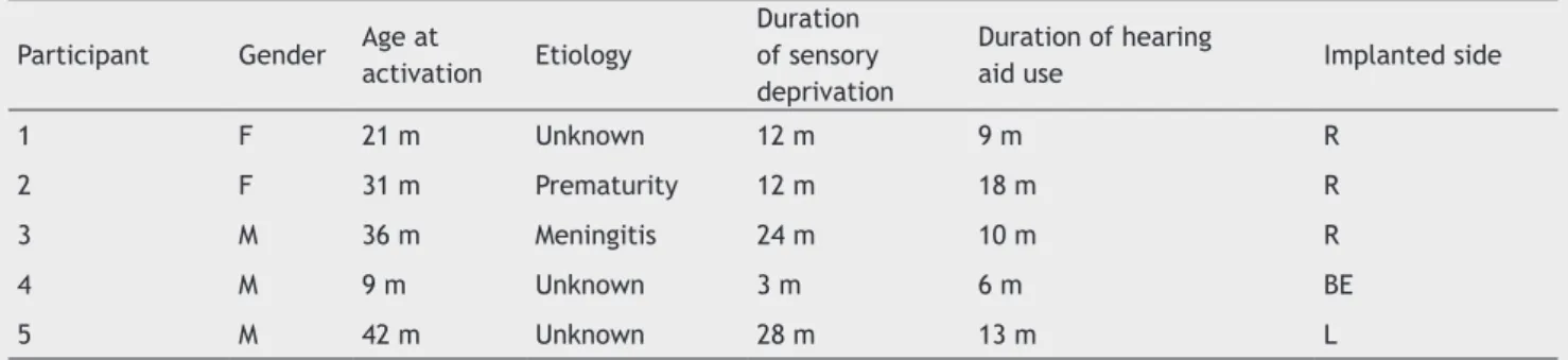
Table 1 shows the characteristics and speciications of each participant of SG.
The children from SG were age-matched to those of CG,
so that the results obtained after three months of CI use could be compared with those observed in normal hearing children at the same age range. The criteria used to pair children was a difference of 4 months of age between chil-dren of the study group and the control group.
Statistical analysis showed no difference for age and
gender. The Mann-Whitney test indicated that there was no
difference in age mean between groups (mean SG = 27.8 ± 13.03; mean CG = 27.8 ± 12.60; U = 12.0, z = -0.104, p = 0.917). Fisher’s exact test indicated no difference on gender distribution between groups (p = 0.524), although there were more males in the SG (60%) and females in the CG (80%).
Control group
Inclusion criteria established for CG were: maximum age of 4 years; without hearing loss (tympanometric curve
type A, present relexes and auditory and speech reception thresholds less than or equal to 15 dB); and without impair -ments in neurological, motor, or language development.
The CG comprised ive children (one male and four fe
-Table 1 Sample characterization regarding gender, age, etiology, degree of hearing loss, duration of sensory deprivation, duration of hearing aid use, implanted side, and cochlear implant.
Participant Gender Age at
activation Etiology
Duration of sensory deprivation
Duration of hearing
aid use Implanted side
1 F 21 m Unknown 12 m 9 m R
2 F 31 m Prematurity 12 m 18 m R
3 M 36 m Meningitis 24 m 10 m R
4 M 9 m Unknown 3 m 6 m BE
5 M 42 m Unknown 28 m 13 m L
males) with a mean age of 2 years and 3 months (minimum 11 months and maximum of 3 years and 2 months). The par -ticipants of the CG are characterized in Table 2.
Children of this group underwent hearing assessment prior to electrophysiological evaluation to discard any type of hearing impairment. This consisted of: pure tone
audiome-try, speech audiometry (speech reception threshold - SRT), as well as immittance measures with acoustic relex research.
Procedures
Children were invited to participate in the study by an in-vitation letter delivered to their parents or guardians or by telephone.
An interview was conducted with the parents or
guar-dians of the SG at the Audiology Clinic of the Department
in order to obtain information on: age, education, side of implantation, HAs, etiology of hearing loss, and results of the last audiometry performed at the institution.
The evaluation of the CAEP was performed in an acous -tically treated room with the child in an alert state, sitting comfortably in a reclining chair. They were instructed and encouraged to watch a puppet theater or movie with no sound during the procedure. Before starting the procedure,
it was veriied that the CI was functioning: battery, pro -gram, and microphone.
The Smart EP USB Jr Intelligent Hearing Systems (IHS 5020), a device that provides two channels of stimulation, was used. Channel A was intended to capture the CAEP in
the right ear, and channel B, in the left ear. In both chan-nels, the active electrode was placed at Cz connected to
the input (+) of the pre-ampliier, and the reference elec -trode was placed on the earlobe of the CI side and
connec-ted to the input (-). The ground electrode was positioned at
Fpz and connected to the ground position.
The electrodes were placed with conductive paste for
electroencephalogram (EEG) from Tem 20TM after cleaning
the skin with an EEG abrasive gel from NUPREP. The impe -dance level of electrodes that was accepted for the
proce-dure was between 1 and 3 K ohms.
The acoustic stimulation was presented by a sound ield
system with speakers positioned at an angle of 90° azimuth and 40 cm away from the implanted side of children from
the SG. The same procedure was used with children
from CG; the stimuli presentation was performed on the
side with better audiometric thresholds. For subjects with
symmetric audiometric thresholds, stimuli was presented at the right side. Two samples were collected from each
sub-ject to conirm the results.
Regarding the parameters of stimulation, the CAEP were
recorded with the speech stimulus of the syllable /ba/, pre-sented with inter-stimulus intervals of 500 ms, at an inten-sity of 70 dBNA and a presentation rate of 1.9 stimuli per second. The parameters described below were also used
during registration: bandpass ilter from 1 to 30 Hz, gain of
100,000, averaging 512 stimuli, and response analysis win-dow of 100 ms pre-stimulus and 500 ms post-stimulus.
Data analysis consisted of an assessment of the latency
times of the P1 component, represented in milliseconds, before and after three months of CI use. The indings were
compared to those obtained with children from the CG.
Results
Control group
The values of the latency time of the P1 component from CG
children ranged between 123 ms and 140 ms (children aged
35 months and 10 months, respectively) (Table 3 and Fig. 1).
Study group
After three months of CI use, a reduction in latency of the
CAEP in all participants from the SG was observed.
The irst child assessed was diagnosed at 1 year of age
and used bilateral HAs for nine months. Hearing thresholds
obtained in free ield with the device were 85 and 80 dB for frequencies between 250 and 500 Hz, respectively. Three
months after surgery, the audiometric thresholds in the
im-planted ear ranged between 55 and 70 dB at frequencies
Table 2 Characterization of the control group regarding gender and age.
Participant 1 2 3 4 5
Gender F F M F F
Age at first
assessment 19 m 35 m 37 m 10 m 38 m
F, female; M, male; m, months.
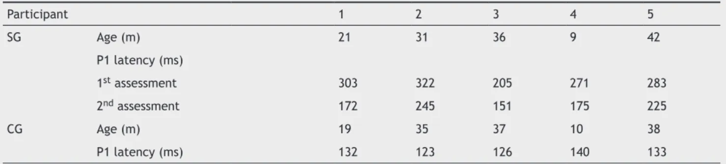
Table 3 Description of age and latency values obtained with cortical auditory evoked potential for the study group and control group.
Participant 1 2 3 4 5
SG Age (m) 21 31 36 9 42
P1 latency (ms)
1st assessment 303 322 205 271 283
2nd assessment 172 245 151 175 225
CG Age (m) 19 35 37 10 38
P1 latency (ms) 132 123 126 140 133
from 0.25 to 2 KHz. In the CAEP record prior to activation, the P1 wave latency was observed at 303 ms. After three months of CI use, a decrease in P1 component latency was
observed, which was registered at 172 ms.
The second child assessed was diagnosed at 3 months
and received bilateral HAs at 1 year of age. Pre-surgical pure tone audiometry in free ield with HAs revealed res
-ponses between 70 and 100 dB for frequencies from 0.25 to 2 KHz in the right ear. Three months after surgery, the
audiological data showed responses in the implanted ear
between 45 and 65 dB at frequencies from 0.25 to 4 kHz.
At the evaluation of long latency potentials, there was a decrease in latency, with values of 322 and 245 ms in the pre- and post-activation periods, respectively.
The third child was diagnosed at 1 year and 8 months after meningitis at 11 months, and used bilateral HAs for 10 months. The audiometric results with the HA in the left ear
showed responses of 80 and 90 dB for frequencies of 250 and
500 Hz, respectively. Three months after surgery, the pa-tient detected speech sounds at average intensity. The data
initially showed the CAEP P1 component at 205 ms. After
stimulation via CI, this component was observed at 151 ms. The fourth child was diagnosed at 1 month of age with unknown etiology. After using bilateral HAs for six months, the child underwent CI surgery at 9 months. The results of
audiometry in free ield with HAs were 80 dB for the fre
-quency of 250 Hz bilaterally. After three months of CI use, these values were between 30 and 50 dB for frequencies
from 500 to 4,000 Hz in the left ear. In the evaluation of long latency potential, there was a decrease in latency, with values of 271 and 175 ms in the pre- and post-activa-tion period, respectively.
The last child was diagnosed at 2 years of age without
deined etiology of hearing loss. This child had used HAs
bilaterally for one year and one month when he unde-rwent the surgery. The audiometric results with the HA
in free ield showed responses in the left ear from 55 to for frequencies from 0.25 to 1 KHz. Three months after
surgery, responses were observed at 50 and 60 dB for
fre-quencies 250 and 500 Hz, respectively. The CAEP records initially showed the P1 component at 283 ms. After three
months of CI use, the latency of this component was
ob-served at 225 ms.
A decrease in the latency values of the P1 component
was observed in all participants of this study after three
months of stimulation via CI. The P1 latency values were
closer to values found in normal hearing children of the same age, as shown in Table 3 and Fig. 1. Children from the CG were also reassessed after an interval of three
mon-ths from the irst evaluation; however, there were no diffe -rences in latency between the two assessments.
Discussion
Considering the maturation of the CANS in children who re -ceive CI, this study aimed to investigate the changes in
la-tency values of the P1 component observed in CAEP before
and three months after electrode activation.
The P1 component, a positive wave generated by thala -mic cortical activity upon sound stimulation, is a measure
capable of relecting changes in the CANS arising from neu -ronal plasticity, an essential phenomenon for the develop-ment of auditory skills and language.16-18
In children with CI, these changes can be observed, as the electrical stimulation provides better functionality of synaptic connections with gradual increase in the rate of synaptic neural transmission and synchronization. These cortical changes allow for a gradual decrease in the latency
of P1 component - a phenomenon observed in this study - corroborating the indings described in the literature that
evaluated children in similar conditions.5,16,19,23
There are several published studies that suggest a rapid change of this latency time, especially in children who are
early-implanted. Some authors observed that these children reached the latency values of P1 component expected for
their respective age three months after implantation;5,23
others have reported these changes between three and six months;21 other studies have shown changes after four
mon-ths of CI use;22 and still others have observed changes after
six to eight months.16,19 In the latter, the results
demons-trated that children who receive CI early exhibit a rapid
development of CANS, with changes in waveform morpho
-logy as well as in latency of P1 component: one week after
Figure 1 P1 component latency values before and after activation of cochlear implant electrodes of the study group compared to con -trol group.
350 300 250 200 150 100 50 0
P1
L
at
en
cy
(
m
s)
Before activation After activation Control group
1 2 3 4 5
implantation, the latency of P1 decreased approximately
100 ms, generating results similar to those of normal he-aring newborns, and after six to eight months of use, this value reached the normal range for children at the same age. The results also demonstrated that children who are late-implanted present abnormal waveform morphology and slow decrease in latency time.19
This reduction in latency time of the P1 component in
relation to age at activation is related to the existence of a sensitive period in which the auditory stimulation should be initiated for obtaining a higher degree of clinical effective-ness. Thus, as seen in the literature, children who receive
stimulation via CI before three years and ive months of age quickly reach the expected values of normality. Those who receive CI later present less development of the CANS than
that observed in normal hearing children.11,19,23,24
In the present study, it was not possible to follow-up
the modiications of the CANS for a period exceeding three
months of CI use in a larger group of children. However, a longitudinal assessment for a period exceeding 12 months,
with more subjects, would demonstrate how CANS struc
-tures are modiied after a longer period of CI use. It would
also be possible to verify the alignment of registry
parame-ters of the CAEP found in implanted children compared to
those observed in normal hearing children of the same age. Considering that the analysis of this component appears to be related to the results of speech perception obtained after activation of CI, the use of these electrophysiologi-cal measures, associated with behavioral assessment of au-ditory skills, can contribute to a better understanding of
intervention results. Several studies highlight the P1 com -ponent analysis as a biomarker, which can provide informa-tion about the evoluinforma-tionary process of rehabilitainforma-tion when
associated with behavioral tests, and, consequently, suggest
prognosis.5,16,20,21,22,25
In this sense, the longitudinal assessment of these
chil-dren is fundamental, so that the changes in the CANS of
children with CI can be assessed in the long term and
com-pared to the development of the CANS of normal hearing children. Studies with larger sample sizes and with longer
observation time are necessary for a better understanding
of CANS changes.
Therefore, it was veriied that CAEP may be used as a bio
-marker for the CANS, which is able to register changes caused
by electric stimulation via CI after three months of its use.
Conclusion
The results of this case-control study indicated a decrease
in the P1 latency values of CAEP in children under the age
of 4 years after three months of CI activation.
The values of the latency time of the P1 component from
children who have used a CI for more than three months are higher than those from normal hearing children.
Funding
This study was supported by Fundação de Amparo à Pesquisa do Estado de São Paulo (FAPESP).
Conlicts of interest
The authors declare no conlicts of interest.
References
1. Mendes BCA, Barzaghi L. Percepção e produção da fala e dei -ciência auditiva. In: Bevilacqua MC. [et al.]. Tratado de audio -logia. São Paulo: Santos; 2011. p.653-69.
2. Bento RF, Brito Neto R, Castilho AM, Gómez VG, Giorgi SB, Gue -des MC. Resultados auditivos com o IC multicanal em pacientes submetidos a cirurgia no Hospital das Clínicas da Faculdade de Medicina da Universidade de São Paulo. Rev Bras Otorrinolarin -gol. 2004;70:632-7.
3. Maurer J, Collet L, Pelster H, Truy E, Gallégo S. Auditory late cortical response and speech recognition in digisonic cochlear implant users. Laryngoscope. 2002;112:2220–4.
4. Boéchat EM. Plasticidade e ampliicação. In: Fernandes FDM, Mendes BCA, Nava, ALPGP, eds. Tratado de fonoaudilogia.2nd edition. São Paulo: Roca; 2010. p.160-8.
5. Sharma A, Tobey E, Dorman M, Bharadwaj S, Martin K, Gilley P, et al. Central auditory maturation and babbling development in infants with cochlear implants. Arch Otolaryngol Head Neck Surg. 2004;130:511-6.
6. Thai-Van H, Veuillet E, Norena A, Guiraud J, Collet L. Plasti -city of tonotopic maps in humans: inluence of hearing loss, hearing aids and cochlear implants. Acta Oto-Laryngologica. 2010;130:333–7.
7. Moret ALM, Bevilacqua MC, Costa AO. IC: audição e linguagem em crianças deicientes auditivas pré-linguais. Pró-Fono Rev Atual. Cient. 2007;19:295-304.
8. Bevilacqua MC, Costa AO, Carvalho ACM, Moret ALM. IC. In: Fernandes FDM, Mendes BCA, Nava ALPGP, eds. Tratado de fo -noaudilogia. 2nd edition. São Paulo: Roca; 2010. p. 220-31. 9. Clark GM. Cochlear implants: fundamentals & applications.
New York: Springer; 2003. p. 830.
10. Sharma A, Dorman M. Central auditory development in chil -dren with cochlear implants: clinical implications. Møller A, ed. Cochlear and brainstem implants. Adv Otorhinolaryngol. 2006;64:66–88.
11. Sharma A, Nash AA, Dorman M. Cortical development, plasti -city and re-organization in children with cochlear implants. J Comm Disord. 2009;42:272-9.
12. Fallon JB, Irvine DRF, Shepherd RK. Neural prostheses and brain plasticity. J Neural Eng. 2009;6:065008.
13. Eggermont JJ, Ponton CW. Auditory-evoked potential studies of cortical maturation in normal hearing and implanted chil-dren: correlations with changes in structure and speech per-ception. Acta Otolaryngol. 2003;123:249-52.
14. Kral A, Sharma A. Developmental neuroplasticity after cochle -ar implantation. Trends Neurosci. 2012;35:111-22.
15. Nash A, Sharma A, Martin K, Biever A. Clinical applications of the p1 cortical auditory evoked potential (CAEP) biomarker. In: Seewald R, Bamford J, eds.; 2007. p. 43-9.
16. Sharma A, Dorman M, Spahr J. Rapid development of cortical auditory evoked potentials after early cochlear implantation. NeuroReport. 2002;13:1365-8.
17. Jang JH, Jang HK, Kim SE, Oh SH, Chang SO, Lee JH. Analysis of P1 latency in normal hearing and profound sensorineural hearing loss. Clin Exp Otorhinolaryn. 2010;3:194-8.
19. Sharma A, Dorman M, Kral A. The inluence of a sensitive period on central auditory development in children with unilateral and bilateral cochlear implants. Hear Res. 2005a;203:134-43. 20. Sharma A, Martin K, Roland P, Bauer P, Sweeney MH, Gilley P,
et al. P1 latency as a biomarker for central auditory develop -ment in children with hearing impair-ment. J Am Acad Audiol. 2005b;16:564-73.
21. Bauer PW, Sharma A, Martin K, Dorman M. Central auditory development in children with bilateral cochlear implants. Arch Otolaryngol Head Neck Surg. 2006;132:1133-6.
22. Dorman MF, Sharma A, Gilley PM, Martin K, Roland P. Cen -tral auditory development: evidence from CAEP measure -ments in children it with cochlear implants. J Comm Disord. 2007;40:284-94.
23. Sharma A, Gilley PM, Martin K, Roland P, Bauer P, Dorman M. Simultaneous versus sequential bilateral implantation in young children: effects on central auditory system development and plasticity. Audiol Med. 2007;5:218-23.
24. Gilley PM, Sharma A, Dorman MF. Cortical reorganization in children with cochlear implants. Brain Res. 2008;1239:56–65. 25. Dinces E, Chobot-Rhodd J, Sussman E. Behavioral and electro