The aim of this study was to evaluate in vitro and in situ the effects of two bleaching
treatments on human enamel surface microhardness. Sixty enamel slabs from recently extracted thirty molars were used. The specimens were polished with sandpapers under water-cooling. The enamel samples were randomly divided in four groups, treated with 10% hydrogen peroxide (HP) or Whitening Strips (WS) containing 10% hydrogen peroxide and using two conditions: in vitro or in situ model. For in situ condition, six volunteers wore an intra-oral appliance containing enamel slabs, while for in vitro condition the
specimens were kept in deionized water after the bleaching protocols. The bleaching treatments were applied one-hour daily for 14 days. Similar amounts of bleaching agents were used in both conditions. Before and after bleaching treatments, microhardness was measured. Statistical analysis (ANOVA and Tukey test) showed that in the in situ condition
there was no statistically significant microhardness reduction in the bleached enamel (p>0.05). Significant decrease in hardness was observed for enamel slabs bleached with both treatments in the in vitro condition (p<0.05). Regarding the bleaching agents, in situ
results showed no difference between HP and WS, while in vitro WS produced the lowest
hardness value. It could be concluded that there was no deleterious effect on enamel produced by any of the bleaching protocols used in the in situ model. The reduction of
hardness was only observed in vitro.
I n S i t u
a n d
I n V i t r o
E f f e c t s o f
T w o B l e a c h i n g T r e a t m e n t s
o n H u m a n E n a m e l H a r d n e s s
Sandrina Henn-Donassollo1, Cristiane Fabris1, Morgana Gagliola1, Ícaro Kerber1, Vinícius Caetano1, Vitor Carboni1, Mabel Miluska Suca Salas2, Tiago Aurélio Donassollo1, Flávio Fernando Demarco2
1School of Dentistry, FASURGS
– Faculdade Especializada na Área de Saúde do Rio Grande do Sul, Passo Fundo, RS, Brazil 2Post-Graduate Program in Dentistry,
UFPel – Universidade Federal de Pelotas, Pelotas, RS, Brazil
Correspondence: Prof. Dr. Flávio Fernando Demarco, Rua Gonçalves Chaves, 457, 96015-568 Pelotas, RS, Brasil. Tel: +55-53-3222-6690. e-mail: [email protected]
Key Words: tooth bleaching, hardness, hydrogen peroxide,
in situ, in vitro.
Introduction
There is a high demand in dental offices nowadays for treatments that improve the aesthetic appearance. Face aesthetics harmony has been associated with perfect smiles and satisfaction with dental appearance. In this context, tooth color plays an important role in the perceptions of aesthetics and satisfaction with dental appearance (1,2). Tooth bleaching has become a popular treatment, with a large variability of commercial presentations that may be applied in the dental office, at-home under the supervision of the dentist or using over-the-counter (OTC) products without professional supervision (3). At-home bleaching treatment is the preferred treatment protocol for vital teeth according to the Brazilian dentists, using low concentration gels with mouthguards, while OTC products are used/ recommended by 5% of these dentists (4).
Vital tooth bleaching is a non-invasive treatment that can be used to improve tooth color. Depending on the used kind of product and application, the color improvement may be clinically relevant and even maintained for long periods (up to two years) (1). Usually the bleaching treatments are well accepted by patients (5) and could have a positive impact in the individuals’ oral health quality of life (2). While there is some sound scientific evidence regarding
the effectiveness from at-home and in office treatments, the over-the-counter products, despite their increased use, still present little evidence of their bleaching effect, except for hydrogen-containing bleaching strips (3,6).
One of the potential adverse effect related to vital bleaching is hypersensitivity (6), which depends upon the used bleaching concentration. Usually, sensitivity is mild to moderate but transient, and could be treated with remineralizing agents or interrupting the treatment (7). Sensitivity may have a negative impact on patients (2). The sensitivity observed during the bleaching treatment has been related to the removal of minerals, resulting in increased porosity on the enamel surface and subsurface (8). The decrease in enamel hardness has been used frequently as an indicator of the mineral loss following bleaching procedures (9). Most in vitro studies demonstrated a
significant reduction in the enamel hardness (10,11). However, few studies simulated the conditions observed in the oral cavity, as in such conditions the decrease in hardness could hardly be seen, due to the remineralizing effect of human saliva (9).
Therefore, the aim of this study was to evaluate the microhardness of human enamel using in vitro and in situ models and two different bleaching protocols (at
ISSN 0103-6440
Braz Dent J 27(1) 2016
57
Hardness of bleached enamel, in vitro and in situ results
home and OTC products). The tested hypothesis was that when bleaching protocols were performed under in situ conditions in the oral cavity, the harmful effect of mineral loss would not be observed, despite the bleaching protocol.
Material and Methods
Ethical AspectsThe Ethics Research Committee of the University of Cruz Alta, Cruz Alta, RS, Brazil approved this clinical trial (protocol number 462.123). Based on pre-established criteria, six volunteers agreed to participate in the in situ
part and signed an informed consent form. Human teeth were obtained from the tooth bank and consent terms for donations were also obtained.
Preparation of Enamel Specimens
Thirty third molars freshly extracted for orthodontic reasons were used in this study. . All teeth were examined under magnification (40×) to detect defects on the surface. The crowns of the selected tooth were cut at the CE-junction and the pulp was removed. The crowns were sectioned longitudinally from the middle third of buccal and lingual surfaces (5 mm x 5 mm x 2 mm) (9).
Sixty enamel slabs were obtained. The samples were embedded in 1% chloramine and stored at 5 °C until their use. Before the hardness measures, all specimens’ surfaces were prepared. The specimens were included in a metallic matrix and the enamel surface polished for 40 s under water-cooling with sandpapers of decreasing grits (400, 600 and 1200) to obtain flat standardized enamel surfaces.
Initial Measurements
Knoop microhardness test was used. Before bleaching treatments, the hardness of the samples was obtained using a micro hardness-testing machine (Buehler, Model 1600, Lake Buff, IL, USA). Three indentations on each specimen were made using a 50 g load for 10 s. The indentations were performed at a 100 µm distance between them to avoid any interference between indentations.
The enamel slabs were then randomly assigned to four different groups (n=15) considering the type of condition (in situ or in vitro condition) and the used bleaching agent
(10% hydrogen peroxide gel – HP, or 10% hydrogen peroxide
in strips – WS). Composition and details of the bleaching agents used in the study are in Table 1.
In Situ Conditions
Six undergraduate dental students volunteered for the
in situ experiment. First, full-arch maxillary impressions and
stone cast models based in the impressions were obtained. Six intra-oral acrylic appliances were prepared and thirty enamel slabs were randomly allocated and included in these appliances as follows: three appliances with 4 specimens and three appliances with 6 specimens each.
The volunteers were instructed to remove the appliances from the oral cavity once a day to perform the bleaching. The enamel slabs were covered with 0.05 mL of 10% hydrogen peroxide (HP) and with a piece of 5 mm x 5 mm of whitening strips (WS) for one hour. After this period, the bleaching gel or the strip was removed from enamel surface and the volunteers placed the appliances again in their oral cavities, for more 23 h to simulate clinical conditions. The bleaching protocols were conducted for 14 days.
In Vitro Conditions
The enamel slabs received the application of 0.05 mL from either 10% HP gel or WS, which remained in place for one hour. After removal of the bleaching agent, the enamel slabs were placed individually in containers with deionized water for the next 23 h. The bleaching protocols were performed for 14 days.
Final Measurements
Twenty-four hours after bleaching conclusion in the tested conditions, the enamel slabs were removed from the appliances or the containers, included and fixed in previously manufactured acrylic resin matrices. The bleached enamel surfaces were submitted to Knoop microhardness test using the same protocol as for the initial measurements.
Statistical Analysis
Statistical analysis was performed using the Sigma Stat 3.5 statistical software package (Informer Technologies, Inc., San Jose, CA, USA). Equality of variance (p>0.05) was first performed to see if the values showed a normal
distribution. Mean values were obtained for each specimen considering all the three measurements. Three factors were under investigation: study method (in vitro and in situ), time (before and
after bleaching) and type of bleaching product (10% HP or WS). Analysis of variance (ANOVA) for repeated measures was used. An additional Tukey’s post-hoc test was carried out to identify differences among the groups. All analyses were
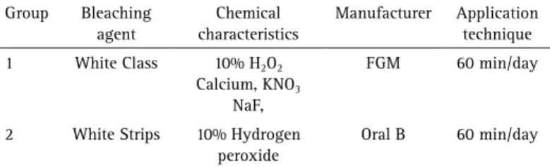
Table 1. Characteristics of the tested bleaching agents
Group Bleaching agent
Chemical characteristics
Manufacturer Application technique
1 White Class 10% H2O2
Calcium, KNO3 NaF,
FGM 60 min/day
2 White Strips 10% Hydrogen
peroxide
Braz Dent J 27(1) 2016
58
S. Henn-Donassollo et al.
carried out with a confidence level of 5% (p<0.05).
Results
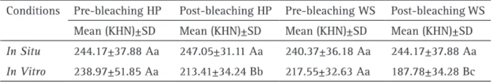
Knoop hardness values exhibited a normal distribution in the study. Table 2 shows the results from hardness measurements considering the different tested conditions. In the in situ condition, no statistically significant difference
in enamel hardness could be observed for both bleaching protocols. Significant decrease in enamel hardness was only observed for the specimens treated with HP or WS in the
in vitro condition (p<0.05). Comparing the two bleaching
protocols (HP or WS), only in the in vitro condition, a lower
harness was observed for WS compared with the enamel treated with HP. The results from the in situ model showed
no statistical difference between the bleaching protocols.
Discussion
The hypothesis tested in the present study was accepted. Despite the bleaching protocol (10% HP gel or 10% HP whitening strips), no significant difference in the enamel hardness was observed, different from the in vitro condition,
which showed a significant decrease of hardness for both bleaching agents.
Previous reports have demonstrated that the use of hydrogen peroxide or carbamide peroxide over enamel surface produced a decrease in hardness when applied
in vitro (12,13). In this in vitro condition, bleaching
agents may significantly alter the enamel surface and the composition could also be modified, due to the removal of some components such as calcium and phosphate, and in consequence, the hardness could be reduced (13,14). On the other hand, when peroxide agents are applied in conditions resembling the oral cavity like in the in situ methods, usually the decrease in hardness is reduced or avoided due to the human saliva action, and even with the mineral loss occurred the remineralizing effect of saliva (9,15). A recent study (16)showed microhardness decreasing immediately after bleaching; however, after 7 days post- bleaching, microhardness values were the same regardless the type of treatments. These findings reinforce the importance of the remineralizing potential of saliva (16). Saliva protects from demineralization by the presence
of the salivary pellicle, providing calcium and phosphorous, its buffer capacity able to neutralize the environment and due to its clearance potential (17). In situ studies showed that the salivary pellicle formed over enamel could protect the enamel surface within a period of 3 min from citric acid and reduce de effects of erosive acids for 2 h (18,19). Thus, it was speculated that the presence of the salivary pellicle could be responsible for the lack of changes in enamel microhardness(15), demonstrating that the loss of mineral tissue observed in vitro following bleaching treatments is not prone to happen in clinical situations. Other in situ
studies corroborate these findings, as enamel hardness was not affected by acidic challenge in the presence of saliva (15,20).
The type of bleaching agent and the concentration of these agents may also affect the bleaching efficacy and the potential adverse effect (2). Hydrogen peroxide is a stronger bleaching agent than carbamide peroxide. In the present study, the same concentration of hydrogen peroxide was used, but in different application methods: HP gel and whitening strips. Whitening strips are an OTC method easy to use and less expensive than at-home or in-office bleaching methods, presenting some efficacy in tooth color recovery (21,22) and these are some of the reasons for their growing popularity (4). Few studies have investigated enamel microhardness after the use of hydrogen peroxide strips and the results demonstrated no deleterious effect on the enamel microhardness (23,24). In the present study, when applied in situ, there was no
difference in enamel hardness between the enamel slabs bleached with 10% HP gel or 10% HP WS. In the in vitro
condition, a higher decrease in hardness was observed for WS group compared to HP gel group. A previous study has shown that enamel microhardness after the application of 10% carbamide peroxide gel and 6.5% hydrogen peroxide strips was significantly reduced in vitro (24).
These results may be explained by calcium in the 10% HP gel (White Class). The manufacturer suggests that calcium is intended to minimize the enamel demineralization process. Studies have reported that adding calcium into the bleaching agents prevents changes in enamel hardness and morphology without reducing bleaching efficacy, in
vitro (25).
There are some limitations to be considered. The difference between products is one of the points to be highlighted. The 10% HP (White Class-FGM), besides hydrogen peroxide, has also potassium nitrate, sodium fluoride and calcium in its composition. The white strips do
Table 2. Means (SD) of micro hardness (KHN) pre and post bleaching for different conditions (in situ and in vitro) and different products (hydrogen peroxide - HP and Whitening Strips - WS)
Conditions Pre-bleaching HP Post-bleaching HP Pre-bleaching WS Post-bleaching WS Mean (KHN)±SD Mean (KHN)±SD Mean (KHN)±SD Mean (KHN)±SD
In Situ 244.17±37.88 Aa 247.05±31.11 Aa 240.37±36.18 Aa 244.17±37.88 Aa
Braz Dent J 27(1) 2016
59
Hardness of bleached enamel, in vitro and in situ results
not present calcium and this could influence the results. Another fact is that samples were stored in deionised water in the in vitro essay. However, the study of Parreira et al,
found no different microhardness results using saliva or distilled water in vitro (12).
This study highlights the importance of study design in the evaluation of adverse effects following bleaching treatments. It seems that the results pointing for adverse effects on enamel surface after bleaching observed by in vitro studies should be considered with caution, since the in situ models have demonstrated that they could not
occur in the clinical condition.
Within the limitations of this study, it could be concluded that there is no deleterious effect of bleaching protocols using 10% HP or WS when using a model that simulates the oral cavity and the reduction of hardness could only be observed in vitro.
Resumo
O objetivo deste estudo foi avaliar in vitro e in situ os efeitos de dois tratamentos clareadores sobre a microdureza do esmalte dental humano. Sessenta blocos de esmalte obtidos de trinta molares recentemente extraídos foram utilizados. Os espécimes foram polidos com lixas sob refrigeração com água. As amostras de esmalte foram divididas aleatoriamente em quatro grupos e tratadas com 10% de peróxido de hidrogênio (HP) ou fitas de clareamento (WS) contendo 10% de peróxido de hidrogênio, testadas em duas condições experimentais: in vitro ou in situ. Para o ensaio in situ, seis voluntários foram selecionados e usaram um aparelho intra-oral contendo blocos de esmalte, enquanto que para a condição in vitro, os espécimes foram mantidos em água deionizada após os protocolos de clareamento. Os tratamentos clareadores foram aplicadas durante uma hora/dia durante 14 dias. Quantidades semelhantes de agentes de clareamento foram usadas nas duas condições. Antes e após os tratamentos de clareamento, a análise da microdureza foi realizada. A análise estatística (ANOVA e teste de Tukey) mostrou que, na condição in situ não houve redução estatisticamente significante na microdureza do esmalte clareado (p>0,05). Diminuição significativa na dureza foi observada nos blocos de esmalte clareados, em ambos os tratamentos, na condição in vitro (p <0,05). Em relação aos agentes de clareamento, os resultados in situ não mostraram nenhuma diferença entre a HP e WS, enquanto que in vitro, WS produziu o menor valor de dureza. Conclui-se que não houve nenhum efeito deletério na dureza do esmalte, em nenhum dos protocolos de clareamento ao usar um modelo in situ. A diminuição da dureza foi observada somente na condição in vitro.
Acknowledgements
The authors are grateful to the TWAS/CNPQ (process 83903402087/ 190268/2010-7) for the full-time Postgraduate Fellowship provided to one of the co-authors.
References
1. Meireles SS, Santos IS, Bona AD, Demarco FF. A double-blind randomized clinical trial of two carbamide peroxide tooth bleaching agents: 2-year follow-up. J Dent 2010;38:956-963.
2. Meireles SS, Goettems ML, Dantas RV, Bona ÁD, Santos IS, Demarco FF. Changes in oral health related quality of life after dental bleaching in a double-blind randomized clinical trial. J Dent 2014;42:114-121. 3. Demarco FF, Meireles SS, Masotti AS. Over-the-counter whitening
agents: a concise review. Braz Oral Res 2009;23 Suppl 1:64-70. 4. Demarco FF, Conde MC, Ely C, Torre EN, Costa JR, Fernández MR, et
al.. Preferences on vital and nonvital tooth bleaching: a survey among dentists from a city of Southern Brazil. Braz Dent J 2013; 24:527-531. 5. Meireles SS, Heckmann SS, Leida FL, Santos Ida S, Della Bona A, Demarco
FF. Efficacy and safety of 10% and 16% carbamide peroxide tooth-whitening gels: a randomized clinical trial. Oper Dent 2008;33:606-612. 6. Swift EJ, Jr., Heymann HO, Wilder AD, Jr., Barker ML, Gerlach RW. Effects
of duration of whitening strip treatment on tooth color: a randomized, placebo-controlled clinical trial. J Dent 2009;37 Suppl 1:e51-e56. 7. Armenio RV, Fitarelli F, Armenio MF, Demarco FF, Reis A, Loguercio AD.
The effect of fluoride gel use on bleaching sensitivity: a double-blind randomized controlled clinical trial. J Am Dent Assoc 2008;139:592-597. 8. Berger SB, Pavan S, Dos Santos PH, Giannini M, Bedran-Russo AK. Effect of bleaching on sound enamel and with early artificial caries lesions using confocal laser microscopy. Braz Dent J 2012;23:110-115. 9. Justino LM, Tames DR, Demarco FF. In situ and in vitro effects of
bleaching with carbamide peroxide on human enamel. Oper Dent 2004;29:219-225.
10. Soares DG, Ribeiro AP, Sacono NT, Loguércio AD, Hebling J, Costa CA. Mineral loss and morphological changes in dental namel induced by a 16% carbamide peroxide bleaching gel. Braz Dent J 2013;24:517-521. 11. Silva AF, Demarco FF, Meereis CT, Cenci MS, Piva E. Light-activated
bleaching: effects on surface mineral change on enamel. J Contemp Dent Pract 2014;15:567-572.
12. Parreiras SO, Vianna P, Kossatz S, Loguercio AD, Reis A. Effects of light activated in-office bleaching on permeability, microhardness, and mineral content of enamel. Oper Dent 2014;39:E225-E230.
13. Salomao D, Santos D, Nogueira R, Palma-Dibb R, Geraldo-Martins V. Acid demineralization susceptibility of dental enamel submitted to different bleaching techniques and fluoridation regimens. Oper Dent 2014;39:E178-E185.
14. Ferreira SS, Araujo JL, Morhy ON, Tapety CM, Youssef MN, Sobral MA. The effect of fluoride therapies on the morphology of bleached human dental enamel. Microsc Res Tech 2011;74:512-516.
15. Sa Y, Chen D, Liu Y, Wen W, Xu M, Jiang T, et al.. Effects of two in-office bleaching agents with different pH values on enamel surface structure and color: an in situ vs. in vitro study. J Dent 2012;40Suppl 1:e26-e34. 16. Borges AB, Guimaraes CA, Bresciani E, Ramos CJ, Borges AL, Rocha Gomes
Torres C. Effect of incorporation of remineralizing agents into bleaching gels on the microhardness of bovine enamel in situ. J Contemp Dent Pract 2014;15:195-201.
17. Hara AT, Lussi A, Zero DT. Biological factors. Monogr Oral Sci 2006;20:88-99.
18. Hannig M, Fiebiger M, Güntzer M, Döbert A, Zimehl R, Nekrashevych Y. Protective effect of the in situ formed short-term salivary pellicle. Arch. Oral Biol. 2004;49:903-910.
19. Hannig M, Hess NJ, Hoth-Hannig W, De Vrese M. Influence of salivary pellicle formation time on enamel demineralization--an in situ pilot study. Clin Oral Investig 2003;7:158-161.
20. Sa Y, Wang Z, Ma X, Lei C, Liang S, Sun L, et al.. Investigation of three home-applied bleaching agents on enamel structure and mechanical properties: an in situ study. J Biomed Opt 2012;17:035002.
21. Hasson H, Ismail AI, Neiva G. Home-based chemically-induced whitening of teeth in adults. Cochrane Database Syst Rev 2006:Cd006202. 22. Karadas M, Duymus ZY. In vitro evaluation of the efficacy of different
over-the-counter products on tooth whitening. Braz Dent J 2015;26:373-377. 23. Duschner H, Gotz H, White DJ, Kozak KM, Zoladz JR. Effects of
hydrogen peroxide bleaching strip gels on dental restorative materials in vitro: surface microhardness and surface morphology. J Clin Dent 2004;15:105-111.
24. Soldani P, Amaral CM, Rodrigues JA. Microhardness evaluation of in situ vital bleaching and thickening agents on human dental enamel. Int J Periodontics Restorative Dent 2010;30:203-211.
25. Alexandrino L, Gomes Y, Alves E, Costi H, Rogez H, Silva C. Effects of a bleaching agent with calcium on bovine enamel. Eur J Dent 2014;8:320-325.