PONTIFÍCIA UNIVERSIDADE CATÓLICA DO RIO GRANDE DO SUL FACULDADE DE ODONTOLOGIA
PROGRAMA DE PÓS-GRADUAÇÃO EM ODONTOLOGIA DOUTORADO EM DENTÍSTICA
CAROLINA FRANÇA DE MEDEIROS MELO
RUGOSIDADE E MICRODUREZA DO ESMALTE CLAREADO COM PERÓXIDO DE CARBAMIDA A 10% E ESCOVADO COM DIFERENTES
DENTIFRÍCIOS: ESTUDO IN VITRO E IN SITU
Profa. Dra. Ana Maria Spohr Orientadora
CAROLINA FRANÇA DE MEDEIROS MELO
RUGOSIDADE E MICRODUREZA DO ESMALTE CLAREADO COM PERÓXIDO DE CARBAMIDA A 10% E ESCOVADO COM DIFERENTES
DENTIFRÍCIOS: ESTUDO IN VITRO E IN SITU
Tese apresentada como parte dos requisitos obrigatórios para a obtenção do grau de Doutor em Odontologia, curso de Pós- graduação em Odontologia, área de Concentração em Dentística, pela Faculdade de Odontologia da Pontifícia Universidade Católica do Rio Grande do Sul. Orientadora: Profa. Dra. Ana Maria Spohr
CAROLINA FRANÇA DE MEDEIROS MELO
RUGOSIDADE E MICRODUREZA DO ESMALTE CLAREADO COM PERÓXIDO DE CARBAMIDA A 10% E ESCOVADO COM DIFERENTES
DENTIFRÍCIOS: ESTUDO IN VITRO E IN SITU
Tese apresentada como parte dos requisitos obrigatórios para a obtenção do grau de Doutor em Odontologia, curso de Pós- graduação em Odontologia, área de Concentração em Dentística, pela Faculdade de Odontologia da Pontifícia Universidade Católica do Rio Grande do Sul.
Aprovada em 05 de março de 2010.
BANCA EXAMINADORA
Orientadora: Profa. Dra. Ana Maria Spohr (PUCRS)
Prof. Dr. Alex José Souza dos Santos (UFRN)
Prof. Dr. Marcos Antonio Japiassú Resende Montes (UPE)
Profa. Dra. Julieta Gomes Tavares (PUCRS)
Dedico este trabalho...
Ao meu marido, Márcio, meu incentivador! Por estar sempre ao meu lado, tão
dedicado, me ajudando, apoiando, orientando. Pelo entusiasmo com que participa da minha vida! Por me encher de amor, carinhos, atenção e mimos. Por acreditar em mim! Por realizar nosso sonho... Vítor, nosso elo mais forte, que nascerá em breve e me presenteará com um mais novo título, o de mãe.
Aos meus pais, Jovany e Gracinha, por tudo que sou. Por não medirem
esforços para me ajudar a crescer, tanto pessoalmente como profissionalmente. Pela dedicação e amor infinito. Obrigada!
Especialmente, agradeço...
À professora Dra. Ana Maria Spohr, pela oportunidade que me deu de ser sua
AGRADECIMENTOS
À Pontifícia Universidade Católica do Rio Grande do Sul, na pessoa do diretor
da Faculdade de Odontologia, Prof. Marcos Túlio Mazzini Carvalho e
vice-diretora Profa. Angélica Maria Genehr Fritscher.
Ao Prof. José Antônio Poli de Figueiredo coordenador do Programa de
Pós-Graduação em Odontologia.
À Profa. Nilza Pereira da Costa pelo seu exemplo de força e dedicação.
À Maria Paula Paranhos, amiga admirável, responsável, sincera, de
personalidade forte e singular. Você é tão especial que mesmo longe consegue me ajudar como se estivesse perto. Obrigada por todos os momentos que passamos juntas, pelas dicas de como se preparar para o frio de Porto Alegre, pela companhia maravilhosa e especialmente pela sua amizade.
À Perpétua Freitas pela disponibilidade para me ajudar a fazer os grampinhos
da pesquisa, pelos materiais emprestados e pelas boas conversas que tivemos no laboratório.
À Helena Carracho por todo apoio e disponibilidade prestada na pesquisa.
Às colegas da graduação, Débora, Natália, Nathalie, Tassiane e Valesca,
sem a ajuda de vocês não teria conseguido, obrigada de verdade!
Aos meus colegas de turma Alexandre Heredia, Gustavo, Regênio,
Maurício, Rogério, Grazi, Micéli, Sávio, Tatiana, Carlos Alexandre, Luiz Felipe e Luis André e Marcel. Especialmente Gustavo Frainer, pela sua
amizade, atenção e confiança.
Aos colegas da turma do Mestrado em Dentística, Paula, Rosana, Patrícia, Dani, Guilherme, Ruy e Marcelo. Especialmente à Paula e à Rosana pelo
auxílio prestado para a conclusão deste trabalho, pelo carinho e amizade e
também à Patrícia pelas longas conversas e por ter me ajudado em todos os
momentos de que precisei.
Ao Prof. Luiz Henrique pela participação na banca de qualificação e
sugestões dadas para melhorar a qualidade do meu trabalho e pela orientação durante o estágio docente na clínica da Dentística.
Ao Prof. Hugo Oshima pela participação na banca da qualificação e suas
sugestões que só enriqueceram a qualidade do trabalho.
Ao Prof. Ari Tietzmann pelos momentos mais que agradáveis recheados de
Aos professores Eduardo Mota, Rosemary Shinkai, Antônio Oliveira e Luciana Hirakata que de alguma maneira contribuíram para a realização
desse trabalho.
Ao professor Hélio Bittencourt pela estatística do trabalho.
Aos meus padrinhos e sogros, Fátima e Aluísio, pela atenção, apoio, incentivo
e carinho.
Ao meu irmão, Emanoel, meu consultor e “resolvedor” de problemas
tecnológicos, sou dependente de você.
Ao professor Alex Santos, meu primeiro orientador e sempre incentivador. Que
estando sempre presente, acreditou em mim e me ajudou a chegar até aqui. Obrigada pela sua amizade!
Aos funcionários da Secretaria de Pós-Graduação: Ana, Carlos, Davenir e
Marcos.
Às funcionárias da limpeza Angelita, Regina e Flávia por estarem sempre a
disposição.
Aos amigos, Klerysson, Germana, Camila, Bruna que apesar de distantes me
ajudaram bastante!
À CAPES pela bolsa de estudos concedida.
A todos que direta ou indiretamente contribuíram para a realização deste trabalho.
RESUMO
Foram realizados dois estudos, in vitro e in situ para avaliar a rugosidade e microdureza do esmalte clareado com peróxido de carbamida a 10% (PC10) e escovado com dentifrícios. Fragmentos de esmalte foram obtidos a partir de dentes humanos. A rugosidade e microdureza iniciais foram medidas. Para o estudo in vitro, estes fragmentos foram divididos em seis grupos (n=12): G1 –
PC10 e escovado com um dentifrício regular (R); G2 – PC10 escovado com dentifrício clareador (W); G3 – PC10 escovado com dentifrício clareador com bicarbonato de sódio e peróxido de cálcio (BS); G4 – Placebo (PLA) + R; G5 –
De acordo com a ANOVA GLM, apenas os dentifrícios foram significantes (p=0,037) para a rugosidade superficial. No estudo in vitro, o agente PLA apresentou um aumento significativamente maior que o PC10 na microdureza do esmalte, enquanto que para o teste de Bonferroni, o dentifrício BS apresentou uma rugosidade significativamente maior que o W (p<0,05). Em relação a microdureza, com o PC10, os dentifrícios não diferiram entre si. Pode-se concluir, para o estudo in vitro, que a associação do PC10 com dentifrícios R, W e BS por 21 dias não alterou significativamente a rugosidade, mas diminuiu significativamente a microdureza do esmalte. Para o estudo in situ, a associação do peróxido de carbamida a 10% com os dentifrícios BS, W e R causou um aumento significativo na rugosidade e uma diminuição significativa na microdureza superficial do esmalte. O dentifrício BS apresentou a maior rugosidade, seguido por W e R.
LISTA DE FIGURAS, TABELAS E GRÁFICOS
Capítulo 1 – ROUGHNESS AND MICROHARDNESS OF ENAMEL BLEACHED WITH 10% CARBAMIDE PEROXIDE AND BRUSHED WITH DIFFERENT TOOTHPASTES: IN VITRO STUDY.
TABLE 1: COMPOSITION AND MANUFACTURER OF EACH TREATMENT AGENT
AND TOOTHPASTES. ... 39
FIGURE 1: SCHEMATIC ILLUSTRATION OF THE EXPERIMENTAL SEQUENCE. .. 40
FIGURE 2: MEAN VALUES OF ENAMEL SURFACE ROUGHNESS (µM) ... 41
FIGURE 3: MEAN VALUES OF ENAMEL VICKERS MICROHARDNESS (VHN). ... 42
TABLE 2: FINAL SURFACE ROUGHNESS MEAN VALUES. ... 43
TABLE 3: FINAL VICKERS MICROHARDNESS MEAN VALUES. ... 44
Capítulo 2 – ROUGHNESS AND MICROHARDNESS OF ENAMEL BLEACHED WITH 10% CARBAMIDE PEROXIDE AND BRUSHED WITH DIFFERENT TOOTHPASTES: IN SITU STUDY. FIGURE 1: SCHEMATIC DRAWING OF THE EXPERIMENTAL DESIGN ... 67
TABLE 1: COMPOSITION AND MANUFACTURER OF EACH TREATMENT AGENT AND TOOTHPASTES. ... 68
FIGURE 2 – MEAN VALUES OF ENAMEL SURFACE ROUGHNESS (µM). ... 69
LISTA DE ABREVIAÇÕES, SIGLAS E SIGNIFICADOS
PUCRS Pontifícia Universidade Católica do Rio Grande do Sul
Toothpaste R Colgate máxima proteção anticáries
Toothpaste W Colgate Total 12 Whiteness gel
Toothpaste BS Colgate Whitening Oxygen Bubbles Fluoride Toothpaste
PC10 Peróxido de carbamida a 10%
PLA Placebo
VHN Vickers Hardness Number
µm Micrometro
µg Micrograma
mm Milímetro
mm2 Milímetro quadrado
cm Centímetros
ANOVA GLM Análise de variância: Modelo linear Geral
α
Nível de significânciaP Valor de probabilidade
et al. Abreviação de et alii (e outros)
SUMÁRIO
INTRODUÇÃO GERAL ... 13
CAPÍTULO 1 ... 16
ROUGHNESS AND MICROHARDNESS OF ENAMEL BLEACHED WITH 10% CARBAMIDE PEROXIDE AND BRUSHED WITH DIFFERENT TOOTHPASTES: IN VITRO STUDY... 17
SUMMARY ... 18
INTRODUCTION ... 19
METHODS AND MATERIALS... 21
Experimental design... 21
Preparation of the enamel fragments ... 21
Experimental phase – In vitro conditions ... 22
Microhardness test ... 24
Surface roughness test ... 24
Statistical Analysis ... 24
RESULTS ... 25
DISCUSSION ... 26
CONCLUSIONS ... 32
REFERENCES ... 33
CAPÍTULO 2 ... 45
ROUGHNESS AND MICROHARDNESS OF ENAMEL BLEACHED WITH 10% CARBAMIDE PEROXIDE AND BRUSHED WITH DIFFERENT TOOTHPASTES: IN SITU STUDY ... 46
ABSTRACT ... 47
1. INTRODUCTION ... 48
2. MATERIALS AND METHODS ... 50
2.1. Selection of volunteers ... 50
2.2. Experimental design... 50
2.3. Preparation of the enamel fragments ... 51
2.4. Intraoral appliance preparation and mounting of the fragments ... 52
2.5. Experimental phase - In situ conditions ... 52
2.6. Microhardness test ... 53
2.7. Surface roughness test ... 54
2.8. Statistical analysis ... 54
3. RESULTS ... 54
4. DISCUSSION ... 55
5. CONCLUSIONS ... 61
REFERENCES ... 62
DISCUSSÃO GERAL ... 71
CONCLUSÕES ... 81
REFERÊNCIAS ... 83
INTRODUÇÃO GERAL
A introdução do peróxido de carbamida como agente clareador caseiro (HAYWOOD; HEYMANN, 1989) causou um grande impacto na Odontologia estética, uma vez que esse tratamento oferece um caminho atrativo para embelezar os dentes de forma simplificada, com economia de tecido dental sadio e, muitas vezes, sem a necessidade de tratamento restaurador adicional (GOLDSTEIN, 1997). Por estas características, a técnica de clareamento caseiro ficou popular entre pacientes e dentistas. A técnica original envolve a aplicação do agente clareador em uma moldeira de acetato por 6 a 8 horas, à noite, durante 2 a 6 semanas (HAYWOOD; HEYMANN, 1989).
Por se tornar uma técnica segura e efetiva (HAYWOOD, 2003), fabricantes vem colocando no mercado uma grande variedade de novos produtos, desde géis clareadores a dentifrícios que prometem deixar os dentes mais claros.
O hábito de escovar os dentes é a prática de higiene bucal mais comum para evitar o aparecimento de lesões de cáries. A escovação normal com o uso de dentifrícios produz mínimo desgaste na superfície do esmalte que não vai causar danos significantes durante a vida (ADDY; HUNTER, 2003). A adição de agentes abrasivos aos dentifrícios é importante para facilitar a remoção de depósitos encontrados na superfície dos dentes (DAWSON et al., 1998). Os dentifrícios clareadores encontrados no mercado possuem partículas abrasivas em sua composição, como carbonato de cálcio, sílica hidratada, bicarbonato de sódio e óxido de alumínio (MEYERS et al., 2000). A adição de substâncias clareadoras, como os peróxidos, proporciona um efeito clareador moderado devido à liberação de oxigênio (RITTER, 2002).
Assim, dentifrícios que prometem ação clareadora estão sendo usados como agentes potencializadores do tratamento clareador. Diante da grande variedade de dentifrícios ao alcance dos pacientes, é importante saber se o tipo de dentifrício causará algum prejuízo ao esmalte dental durante o tratamento clareador caseiro.
morfológicas severas na superfície do esmalte como aumento da rugosidade dessa estrutura (PIMENTA et al., 2003), as quais podem ser responsáveis pela diminuição da microdureza do esmalte (RODRIGUES et al., 2001).
Estudos in vitro têm relatado que o clareamento com peróxido de carbamida a 10% não alterou a microdureza (MCCRACKEN; HAYWOOD, 1995), nem a rugosidade superficial do esmalte humano (ÇOBANKARA et al., 2004). No entanto, quando esse agente clareador está associado a dentifrícios abrasivos através de escovações simuladas, houve um aumento significativo da rugosidade superficial (WORSCHECH, 2006).
A interação in vivo entre saliva e esmalte é um fator que, geralmente, não é incorporado em pesquisas in vitro (CIMILLI; PAMEIJER, 2001). A saliva tem o poder de limpeza, capacidade tampão e habilidade de remineralização (THYLSTRUP; FEJERSKOV, 1998) que pode prevenir os efeitos de desmineralização dos agentes clareadores (JUSTINO; TAMES; DEMARCO, 2004). Trabalhos in situ têm sido feitos para simular condições de efeito real dos tratamentos clareadores no meio bucal, avaliando microdureza (ARAÚJO et al., 2003; MULLER ARCARI et al., 2005), rugosidade (BASTING et al., 2007) e morfologia superficial (JUSTINO; TAMES; DEMARCO, 2004), mas ainda não foi avaliado o desgaste produzido por diferentes dentifrícios durante o tratamento clareador caseiro.
ROUGHNESS AND MICROHARDNESS OF ENAMEL BLEACHED WITH 10%
CARBAMIDE PEROXIDE AND BRUSHED WITH DIFFERENT
TOOTHPASTES: IN VITRO STUDY
“ALTERATIONS IN BLEACHED ENAMEL AFTER BRUSHING WITH THREE DIFFERENT TOOTHPASTES ” (running title)
Carolina França de Medeiros Melo, DDS, DMD, PhD Student, Department of Restorative Dentistry, School of Dentistry, PUCRS.
Address: Rua Luzitana, 441/601, Higienópolis, Porto Alegre, RS, Brazil, ZIP 90.520-080, e-mail: carolfmedeiros@gmail.com
Ana Maria Spohr*, DDS, DMD, PhD, Associate Professor, Department of Prosthodontics, School of Dentistry, PUCRS.
*Correspondence should be addressed to: Dra. Ana Maria Spohr
PUCRS - Av. Ipiranga 6681, prédio 6 – Faculdade de Odontologia Porto Alegre, RS, Brazil Zip 90619900
Fax: +55 51 3320-3626
ROUGHNESS AND MICROHARDNESS OF ENAMEL BLEACHED WITH 10%
CARBAMIDE PEROXIDE AND BRUSHED WITH DIFFERENT
TOOTHPASTES: IN VITRO STUDY
Clinical Relevance
Because of the large variety of whitening toothpastes available and knowing that the patient may use these products to potentiate the whitening treatment, dentists should be aware whether the toothpaste would be harmful to
the patient’s enamel during home bleaching treatment.
SUMMARY
measurements of each fragment were obtained. According to paired Student’s
t-test, there was a significantly decrease in microhardness for all groups (p<0.05). The final roughness was statistically higher than the initial roughness only in the group treated with G6 (p=0.001). According to ANOVA GLM and Bonferroni tests, PLA presented a significantly higher roughness than PC10, and toothpaste BS presented a statistically higher roughness (p<0.05) than W. Toothpaste R did not differ statistically from W and BS (p>0.05). For PC10, the toothpastes did not differ among them in relation to microhardness (p>0.05). According to paired Student’s t-test, toothpastes R and W were statistically different for both treatments. The association of PC10 with toothpastes R, W and BS for 21 days did not significantly alter the enamel surface roughness, but there was a significant decrease in the enamel microhardness.
Keywords: Dental bleaching, toothpastes, 10% carbamide peroxide, artificial saliva, in vitro study, enamel, microhardness, surface roughness.
INTRODUCTION
Some in vitro research pointed out no significant alterations in the enamel microhardness,2-3 or enamel roughness4. However, when 10% carbamide peroxide was associated with abrasive toothpastes by simulated tooth brushing, there was a significant increase in the enamel surface roughness. 5
The primordial role of toothpastes is to clean the largest number of tooth surfaces. This cleaning involves the elimination of plaque and food residues.6 Regular brushing with toothpastes is safe and does not harmfully affect enamel.7 Over the last few years, manufacturers have developed new products in order to meet the expectations of patients and consumers. Therefore, abrasive components such as calcium carbonate, sodium bicarbonate, peroxides, hydrated silica, and aluminum oxide have been added to the toothpastes to increase their cleaning power and to whiten the teeth.8 Whitening toothpastes that contain small quantities of abrasive particles or peroxides may abrade the enamel surface if excessively used.9
Thus, toothpastes that promise whitening action have been used as agents to potentiate bleaching treatment. In view of the large variety of whitening toothpastes available to the patient and consumers, it is important to
know whether the toothpaste would cause any harm to the patient’s enamel
when combined to nightguard vital bleaching treatment.
METHODS AND MATERIALS
Experimental design
The factors under evaluation were treatment agents at two levels (experimental and control) and fluoride toothpastes at three levels (Colgate Máxima Proteção Anticáries, Colgate Total 12 Whiteness Gel and Colgate Whitening Oxygen Bubbles Fluoride). The specifications of the materials are listed in Table 1.
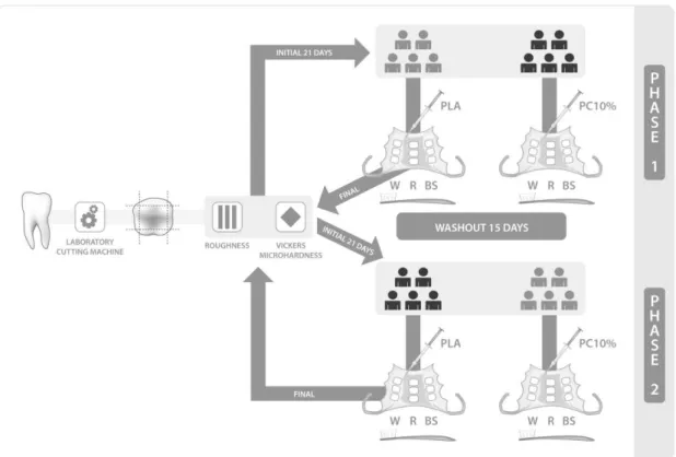
The experimental units consisted of 72 fragments of sound human enamel, randomly assigned to six different groups (n=12). The variables were surface roughness and microhardness. The schematic illustration of the experimental sequence is shown in Figure 1.
Preparation of the enamel fragments
Thirty-six freshly extracted human third molars were scraped to remove any remaining soft tissues, polished with pumice slurry and kept in 0.5% chloramine for 24 hours. After this period, the teeth were stored in deionized water at room temperature until required for use. The use of extracted human teeth followed a protocol that was reviewed and approved by the Ethics Committee of the Pontifical Catholic University of Rio Grande do Sul – PUCRS.
The root of each tooth was mounted in self-cured acrylic resin (Clássico
– Dental Products, São Paulo, SP, Brazil). Each tooth was cut along the long
diamond disc (Labcut 1010 low-speed diamond saw, Extec Corp, Enfield, CT, USA) to obtain fragments of enamel measuring approximately 16mm2 (4mm X 4mm X 2mm), without any stains or cracks. The fragments were steam sterilized for 20 minutes at 121o C.
The sectioned fragments of enamel were fixed in acrylic resin and the upper surface of the samples was then manually ground and polished with 1500-, 2000- and 2500-grit carbide abrasive papers (Carborundum, 3M do Brazil Ltda., Sumaré, SP, Brazil) to create a flat surface. They were immersed in containers with deionized water. Before the experimental phase, the initial measurements of surface roughness and microhardness of each fragment were obtained.
Experimental phase – In vitro conditions
The specimens were randomly divided into six groups (n=12): G1 – 10% carbamide peroxide (PC10) and brushed with a regular toothpaste (R); G2 –
PC10 and brushed with a whitening toothpaste (W); G3 – PC10 and brushed with a whitening toothpaste with baking soda and peroxide (BS); G4 – Placebo (PLA) and brushed with R; G5 – PLA and brushed with W; G6 – PLA and brushed with BS.
Utah, USA) was used in the first three groups and a placebo agent (Carbopol gel, Pharmacus, Porto Alegre, RS, Brazil) was used in the remaining groups. After daily treatment agent application, the specimens were rinsed under running deionized water for five seconds.
All samples from each group were individually bonded (Superbonder Loctite, Henkel Ltda., São Paulo, SP, Brazil) to the center of the acrylic plate (5.3cm X 2.5cm X 0.5cm). This set was put into an acrylic reservoir to start brushing procedures. A homogeneous solution of each toothpaste (slurry) was freshly prepared every day with a mixture containing toothpaste that was weighed on an analytical balance (AG 204, Mettler/Toledo, São Paulo, SP, Brazil), and deionized water in a ratio of 1:2.6 Sufficient slurry (20ml) was placed in the acrylic reservoir to completely cover the set of specimens.
During the remaining time, the groups were kept in containers with an artificial saliva at 37ºC until the next cycle of treatment agent and brushing procedures. The artificial saliva (Salivan®, APSEN Farmacêutica S/A, São Paulo, SP, Brazil) used was changed weekly. During the weekends these groups were kept in containers with artificial saliva at 37 ºC.
Microhardness test
The Vickers microhardness number (VHN) was measured with a microhardness tester (HMV-2, Shimadzu, Tokyo, Japan). Three indentations were made in the central region of each fragment under a 100g load for five seconds in each experimental period: initial (before the bleaching treatment) and final (after the bleaching treatment). The final VHN value of each fragment was the arithmetic mean of the three measurements.
Surface roughness test
The mean enamel roughness (Ra, μm) of each tooth was measured with a roughness tester (SL-201, Mitutoyo Surftest Analyzer, Tokyo, Japan) before and after each treatment period. In the center of the fragment surface, three different traces were recorded for each fragment with a 0.25 cut-off. The Ra value of each specimen was the arithmetic mean of the three measurements.
Statistical Analysis
The surface microhardness and roughness results were submitted to the Kolmogorov-Smirnov test to verify the normal distribution of the variables. The
roughness and microhardness values. This was followed by the Analysis of Variance (ANOVA GLM) in a 2 X 3 factorial scheme (treatment agents X toothpastes) and Bonferroni test. The initial roughness and microhardness readouts were used as covariables to verify the interference of these in the final readouts. For all tests, groups were considered statistically different at α = 0.05.
All statistical analyses were performed using SPSS version 9.0 (SPSS Inc., Chicago, IL, USA).
RESULTS
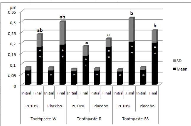
According to the Student’s t-test, the final roughness was statistically
higher than the initial roughness only in the group treated with placebo and BS toothpaste (p=0.001) (Figure 2). The final microhardness was statistically lower than the initial microhardness for all groups (p<0.05) (Figure 3).
According to ANOVA GLM, treatment factor (p=0.00) and toothpaste factor (p=0.890) were significant for surface roughness. The interaction between the factors (p=0.06) and the initial roughness (p=0.890) were not significant. According to Bonferroni, the placebo agent (0.16 μm) presented a
significantly higher roughness value than the bleaching agent (0.12 µm) (Table 2). The toothpaste BS (0.16 μm) presented a statistically higher final roughness (p<0.05) than toothpaste W (0.13 μm). Toothpaste R (0.14 μm) did not differ
statistically from toothpastes W and BS (p>0.05) (Table 2).
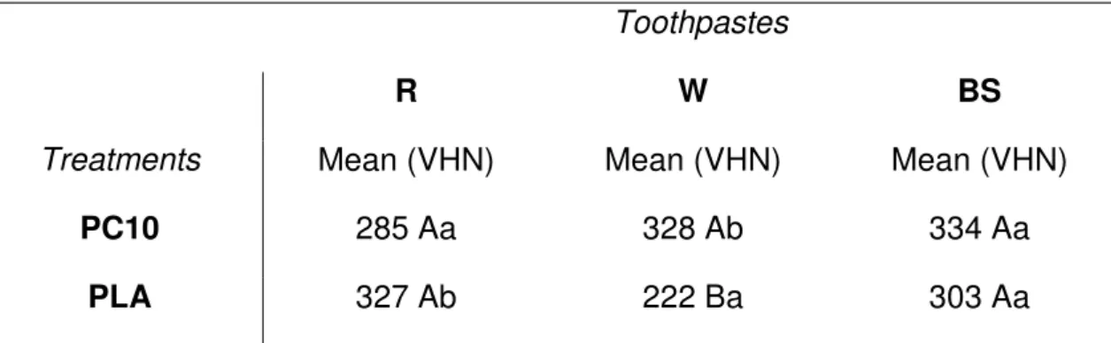
(Table 3), for the placebo treatment, the toothpastes R (327 VHN) and BS (303 VHN) presented statistically higher final microhardness (p<0.05) when compared with toothpaste W (222 VHN). For bleaching treatment, the toothpastes did not differ among them (p>0.05). As SPSS does not make multiple comparisons for the effect of the interaction, Student’s t-tests (Table 3) were performed within each toothpaste for the treatments. Toothpastes R and W were statistically different for the bleaching and placebo treatments. Toothpaste BS did not differ statistically for the treatments (p>0.05).
DISCUSSION
Whitening toothpastes are recommended for maintaining the color of the teeth after bleaching treatment because they are developed for removing stains from tooth surfaces. For this purpose, they have small quantities of peroxides or abrasive particles in their composition.9 The abrasive agents found in toothpastes are calcium carbonate, hydrated silica, sodium bicarbonate, sodium tripolyphosphate, aluminum oxide8, and a mixture of these components. The addition of whitening substances such as peroxides provides a moderate whitening effect due to the oxygen release.9 The presence of these agents in toothpastes is important to facilitate the removal of residues found on tooth surfaces.10 Nevertheless, the abrasiveness of toothpastes must be moderate to avoid the excessive wear of the enamel and dentin surfaces.11
A 50 μm deep demineralization was observed below the enamel surface
enamel roughness, but this alteration was not significant.4,13-14 On the other hand, the interaction between bleaching agents and toothpastes may exert a negative influence on the human enamel surface.
Most of the studies evaluate the separate effect of 10% carbamide peroxide2,4,14-19 and toothpastes6,8,20-22 on dental enamel. Few studies evaluate the combined effect of those two treatments to simulate realistic conditions.7,21 Whether a patient is under bleaching treatment or not, toothbrushing is considered a daily oral hygiene practice. Therefore, the aim of the present study was to focus on the combination effect of the bleaching agent with different toothpastes. Given the methodology used, it was not possible to determine, however, which factor mostly affected the surface roughness and microhardness of enamel. The goal of the present study was the combined effect of both treatments.
There were no statistical differences among the groups in the initial roughness and microhardness analyses. This result was satisfactory, because it showed homogeneity at the beginning of the experiment, as no treatment had yet been performed on the enamel surfaces.
without fluoride. Quantitative differences between the studies in regards to the enamel roughness could be due to a series of variables, such as the brushing time used in the studies. Worschech and others5 used brushing for 180 seconds daily, at a speed of 250 cycles/minute for 56 days. According to Sexton & Philips,25 for each brushing session in a certain area, the individual performs 15 brushing cycles. Thus, the present study reproduced the number of cycles daily performed by an adult. For this purpose, a simulated brushing machine was used, with a speed of 250 cycles/minute. In this sense, two daily brushing procedures resulted in 30 cycles with a total duration of 7 seconds, for 21 days.
Irrespective of the toothpaste used, the placebo agent caused a statistically higher surface roughness (0.16 µm) than carbamide peroxide (0.12 µm). In addition to the effect of the toothpastes, this increased roughness could be justified by the fact that the placebo gel was composed of carboxypolymethylene polymer (carbopol). In an in situ study by Rodrigues and others19 a significant decrease in enamel microhardness was verified when the carbopol-based placebo was applied. The authors suggested a destructive effect of carbopol on enamel surface. Although studies have correlated carbopol to enamel microhardness, it is believed that it could also influence the surface roughness of this substrate. Nevertheless, the difference of only 0.04 µm in the mean surface roughness of enamel between placebo and 10% carbamide peroxide treatments might not be clinically relevant.
contained in the placebo, and by the composition of the BS toothpaste. This toothpaste has a combination of abrasive agents (hydrated silica and sodium bicarbonate) and calcium peroxide as bleaching agent. This increased surface roughness might have influenced the statistical analyses leading to a statistical significance for the toothpaste factor.
Increased and significant enamel surface roughness was found for the toothpaste BS (0.16 µm) compared to toothpaste W (0.13 µm). Nevertheless, it is believed that a 0.03 µm difference would not be clinically relevant. Toothpaste R (0.14 µm) did not differ from the toothpastes BS and W. Toothpaste W contains hydrated silica which is an intermediate abrasive agent6 and toothpaste R has calcium carbonate which is a polishing agent with a lower abrasive power.26 The combination of abrasive agents contained in the toothpaste BS probably caused the greatest surface roughness. Moreover, no differences in surface roughness were observed among the groups treated with 10% carbamide peroxide associated with toothpastes (Figure 2). This finding highlights the similar behavior found for the toothpastes when associated with the bleaching gel.
According to Bollen, Lambrechts & Quirynen,27 surface roughness above 0.20 µm leads to dental plaque accumulation. None of the toothpastes studied presented roughness above this value.
peroxide alone (not associated with brushing procedures) did not cause changes in the enamel microhardness.14,15,28 However, there is no consense in the scientific literature regarding the harmful or beneficial effects of bleaching agents on the enamel microhardness. Attin and others29 evaluated 50 studies (with a total of 166 enamel microhardness measurements) and reported a decrease in enamel microhardness after bleaching in 51% of the measurements. No decrease in microhardness was found in 49% of the measurements.
In the 10% carbamide peroxide groups, the combination with toothpaste R yelded the lowest enamel microhardness (285 VHN) followed by the toothpaste W (328 VHN), and toothpaste BS (334 VHN). No statistical significance was found among those three groups. Within the toothpastes used in this study, only toothpaste BS contains a bleaching agent (calcium peroxide). Since bleaching agents may cause mineral loss in the enamel surface,30 it was expected that this toothpaste together with the bleaching gel would lead to a decrease in enamel microhardness. However, the toothpaste BS associated with the bleaching gel presented the highest enamel microhardness, suggesting that the bleaching agent contained in the toothpaste did not significantly affect enamel microhardness. Therefore, the different compositions of the toothpastes were not a significant factor in the enamel microhardness when associated with 10% carbamide peroxide.
polymer may cause enamel demineralization31. The abrasive agents contained in the toothpastes may also be responsible for the decrease in enamel microhardness in all groups (bleaching and placebo). The abrasive agents remove part of the mineral on the enamel surface.32,33 This mineral loss wears out the enamel leading to a proximity to dentin and, consequently, decrease in microhardness. For the placebo treatment, the toothpastes R (327 VHN) and BS (303 VHN) presented statistically higher final microhardness than the toothpaste W (222 VHN). According to the composition of toothpaste BS, which contains two abrasive agents and a bleaching agent, the lowest surface microhardness value was already expected.
treatment with 10% carbamide peroxide in comparison with placebo. It is difficult to find a plausible explanation for this result, as well as for the fact that toothpaste BS did not have a lower microhardness. Although care was taken to control the variables of simulated brushing, one cannot discard the possibility of the influence of some confounding variables. The action of the brushes, the force and speed of brushing, and the energy required to maintain the abrasive particles of the slurry in suspension are important variables that may affect the determination of dental tissue loss. In relation to these variables, the homogeneity of the slurry is considered one of the most difficult variables to control.33
Further research should focus on the association of other bleaching agents with different toothpastes since the material composition may lead to distinct results.
CONCLUSIONS
References
1. Haywood VB & Heymann HO (1989) Nightguard vital bleaching
Quintessence International20(3) 173-176.
2. McCracken MS & Haywood VB (1995) Effects of 10% carbamide peroxide on the subsurface hardness of enamel Quintessence International26(1) 21-24.
3. Lopes GC, Bonissoni L, Baratieri LN, Vieira LCC & Monteiro Jr S (2002) Effect of bleaching agents on the hardness and morphology of enamel
Journal of Esthetic and Restorative Dentistry14(1) 24-30.
4. Çobankara FK, Ünlü N, Altinöz HC & Özer F (2004) Effect of home bleaching agents on the roughness and surface morphology of human enamel and dentin International Dental Journal54(4) 211-218.
5. Worschech CC, Rodrigues JA, Martins LRM & Ambrosano GMB (2006) Brushing effect of abrasive dentifrices during at-home bleaching with 10% carbamide peroxide on enamel surface roughness The Journal of
Contemporary Dental Practice7(1) 1-9.
6. Wülknitz P (1997) Cleaning Power and Abrasivity of European Toothpastes Advances in Dental Research11(4) 576-579.
8. Meyers JA, McQueen MJ, Harbrow D & Seymour GJ (2000) The surface effect of dentifrices Australian Dental Journal45(2) 118-124.
9. Ritter AV (2002) Tooth-Whitening toothpastes Journal of Esthetic and
Restorative Dentistry14(4) 256.
10. Dawson PL, Walsh JE, Morrison T & Grigor J (1998) Dental prevention by abrasive toothpastes: A new in vitro test and its correlation with clinical observations Journal of Cosmetic Science49(4) 275-283.
11. Joiner A, Philpotts CJ, Ashcroft AT, Laucello M & Salvaderi A (2008) In vitro cleaning, abrasion and fluoride efficacy of a new silica based whitening toothpaste containing blue covarine Journal of Dentistry, 36(Supplement 1) s32-s37.
12. Efeoglu N, Wood D & Efeoglu C (2005) Microcomputerised tomography evaluation of 10% carbamide peroxide applied to enamel Journal of
Dentistry33(7) 561-567.
13. Pinto CF, Oliveira R, Cavali V & Giannini M (2004) Peroxide bleaching agent effects on enamel surface microhardness, roughness and morphology Brazilian Oral Research18(4) 306-311.
15. Lopes GC, Bonissoni L, Baratieri LN, Vieira LCC & Monteiro Jr S (2002) Effect of bleaching agents on the hardness and morphology of enamel
Journal of Esthetic and Restorative Dentistry14(1) 24-30.
16. Shannon H, Spencer P, Gross K & Tira D (2003) Characterization of enamel exposed to 10% carbamide peroxide bleaching agents
Quintessence International24(1) 39-44.
17. Justino LM, Tames DR & Demarco FF (2004) In situ and in vitro effects of bleaching with carbamide peroxide on human enamel Operative Dentistry 29(2) 219-225.
18. Araújo Jr EM, Baratieri LN, Vieira LCC & Ritter AV (2003) In Situ Effect of 10% Carbamide Peroxide on Microhardness of Human Enamel: Function of time Journal of Esthetic and Restorative Dentistry15(3) 166-174.
19. Rodrigues VER, Marchi GM, Ambrosano GMB, Heymann HO & Pimenta LA (2005) Microhardness evaluation of in situ vital bleaching on human dental enamel using a novel study design Dental Materials 21(11) 1059-1067.
20. Joiner A, Collins LZ, Cox TF, Pickles MJ, Weader E, Liscombe C & Holt JS (2005) The measurement of enamel and dentine abrasion by tooth whitening products using an in situ model International Dental Journal (Supplement 1) 194-196.
roughness of enamel and dentin Revista de Odontologia da UNESP 36(2) 121-126.
22. Joiner A, Pickles MJ, Lynch S & Cox TF (2008) The measurement of enamel wear by four toothpastes International Dental Journal58(1) 23-28.
23. Wiegand A, Otto YA & Attin T (2004) In vitro evaluation of toothbrushing abrasion of differently bleached bovine enamel American Journal of
Dentistry17(6) 412-416.
24. Faraoni-Romano JJ, Turssi CP & Serra MC (2009) Effect of a 10% carbamide peroxide on wear resistance of enamel and dentine: In situ study Journal of Dentistry 37(4) 273-278.
25. Sexson JS & Phillips RW (1951) Studies on the effects of abrasives on acrylic resins The Journal of Prosthetic Dentistry1(4) 454-471.
26. Andrade Júnior ACC, Andrade MRTC, Machado WAS & Fischer RG (1998) Estudo in vitro da abrasividade de dentifrícios Revista de
Odontologia da Universidade de São Paulo12(3) 231-236.
27. Bollen CML, Lambrechts P & Quirynen M (1997) Comparasion of surface roughness of oral hard materials to the threshold surface roughness for bacterial plaque retention: A review of the literature Dental Materials13(4) 258-269.
28. Murchison DF, Charlton DG & Moore BK (1992) Carbamide peroxide bleaching effects on enamel surface hardness and bonding Operative
29. Attin T, Schmidlin PR, Wegehaupt F & Wiegand A (2009) Influence of study design on the impact of bleaching agents on dental enamel microhardness: A review Dental Materials25(2) 143-157.
30. McCracken MS & Haywood VB (1996) Demineralization effects of 10 percent carbamide peroxide Journal of Dentistry24(6) 395-398.
31. Van der Reijden WA, Buijs MJ, Damen JJ, Veerman EC, ten Cate JM & Nieuw Amerongen AV (1997) Influence of polymers for use in saliva substitutes on de- and remineralization of enamel in vitro Caries
Research31(3) 216-223.
32. Pickles MJ, Joiner A, Weader E, Cooper YL & Cox TF (2005) Abrasion of human enamel and dentine caused by toothpastes of differing abrasivity determined using an in situ wear model International Dental Journal
(Supplement 1) 188-193.
33. Parry J, Harrington E, Rees GD, McNab R & Smith AJ (2008) Control of brushing variables for the in vitro assessment of toothpaste abrasivity using a novel laboratory model Journal of Dentistry36(2) 117-124.
34. Basting RT, Rodrigues AL Jr & Serra MC (2003) The effects of 10% carbamide peroxide bleaching agents on enamel microhardness over time Journal of the American Dental Association134(10) 1335-1342.
36. Attin T, Albrecht K, Becker K, Hannig C & Wiegand A (2006) Influence of carbamide peroxide on remineralization of bleached enamel Journal of
Table 1: Composition and manufacturer of each treatment agent and toothpastes.
Treatment Agents/Toothpaste Composition Manufacturer
Opalescence PF 10% carbamide peroxide, 0,5%
potassium nitrate, 0,11% w/w fluoride, water (pH~6.5)
Ultradent Products, Inc – Utah, USA
Carbopol Gel Carbopol Pharmacus, Porto Alegre,
RS, Brazil Regular toothpaste:
Colgate Máxima Proteção
Anticáries, identified by “R”.
Calcium Carbonate, water,
Sorbitol, Sodium Lauryl Sulfate,
Sodium Monofluorophosphate
(1450 ppm fluorine), flavor,
Cellulose gum, Tetrasodium
Pyrophosphate, Sodium Silicate,
Sodium Saccharin,
Methylparaben, Propylparaben*.
Colgate-Palmolive Indústria e Comércio Ltda., São Bernardo do Campo – SP, Brasil
Whitening fluoride toothpaste: Colgate Total 12 Whiteness Gel identified by “W”.
Water, Sorbitol, Hydrated Silica, Sodium Lauryl Sulfate, PVM/MA Copolymer, flavor, Carrageenan,
Sodium Hydroxide, Sodium
Fluoride (1450 ppm fluorine), Triclosan, Sodium Saccharin, CI 77891, CI 77019, CI 42090*.
Colgate-Palmolive Indústria e Comércio Ltda., São Bernardo do Campo – SP, Brasil
Whitening Fluoride Toothpaste with Baking Soda and Peroxide:
Colgate Whitening Oxygen Bubbles Fluoride Toothpaste identified by “BS”.
Glycerin, Hydrated Silica,
Propylene glycol, water, sodium
bicarbonate, Sodium
Monofluorophosphate (0.14% w/v
fluoride ion), Pentasodium
Triphosphate, Tetrasodium
Pyrophosphate, Sodium Lauryl Sulfate, flavor, Sodium Hydroxide, Sodium Saccharin, Carrageenan, Cellulose gum, calcium peroxide, titanium dioxide*.
Colgate/Palmolive
Company New York, NY, USA
Artificial Saliva Carmellose Sodium (10ml),
Vehicle (1ml): Sorbitol, sodium chloride, potassium chloride, calcium chloride dihydrate, magnesium chloride hexahydrate,
potassium acid fosfate,
methylparaben, water.
Salivan®, APSEN
Farmacêutica S/A, São Paulo, SP, Brazil.
Means followed by the same amount of signal (*) did not differ statistically
according to paired Student’s t-test (α = 0.05).
Means followed by the same amount of signal (*) did not differ statistically according to paired Student’s t-test (α = 0.05).
Table 2: Final surface roughness mean values.
Treatments Mean (µm) SD Toothpastes Mean (µm) SD PC10 0.12a 0.058 R 0.14ab 0.071 PLA 0.16b 0.070 W 0.13a 0.044
BS 0.16b 0.081
Table 3: Final vickers microhardness mean values.
Toothpastes
R W BS
Treatments Mean (VHN) Mean (VHN) Mean (VHN)
PC10 285 Aa 328 Ab 334 Aa
PLA 327 Ab 222 Ba 303 Aa
Different capital letters in the same LINE indicate statistically significant difference for the Bonferroni test (p <0.05). Different lowercase in the same COLUMN indicate statistically significant difference for the paired Student’s t-test (p<0.05).
ROUGHNESS AND MICROHARDNESS OF ENAMEL BLEACHED WITH 10%
CARBAMIDE PEROXIDE AND BRUSHED WITH DIFFERENT
TOOTHPASTES: IN SITU STUDY
Carolina França de Medeiros Meloa, Ana Maria Spohr b,*
a DDS, DMD, PhD Student, Department of Restorative Dentistry, School of
Dentistry, PUCRS.
b DDS, DMD, PhD , Associate Professor, Department of Restorative Dentistry,
School of Dentistry, PUCRS.
*
Correspondence should be addressed to:Dra. Ana Maria Spohr PUCRS
Av. Ipiranga 6681, prédio 6 – Faculdade de Odontologia Porto Alegre, RS, Brazil
Zip 90619900
Fax: +55 51 3320-3626
ROUGHNESS AND MICROHARDNESS OF ENAMEL BLEACHED WITH 10%
CARBAMIDE PEROXIDE AND BRUSHED WITH DIFFERENT
TOOTHPASTES: IN SITU STUDY
ABSTRACT
Objectives: This in situ study evaluated the association effect of 10% carbamide peroxide bleaching and three toothpastes used during bleaching treatment on the microhardness and surface roughness of enamel.
Results: According to the paired Student’s t-test, the final roughness was
statistically higher than the initial roughness, and final microhardness was statistically lower than the initial microhardness (p<0.05). According to ANOVA GLM, only the toothpaste factor was significant (p=0.037) for roughness. Factors and interactions were not significant for microhardness.
Conclusion: The association of PC10 with the toothpastes BS, W, and R caused a significant increase in enamel roughness and a significant decrease in enamel microhardness. Toothpaste BS presented the highest roughness, followed by W and R. PC10 associated with R, W or BS toothpastes caused equivalent decrease in the enamel microhardness.
Keywords: dental bleaching, toothpastes, 10% carbamide peroxide, in situ study, enamel, microhardness, surface roughness
1. Introduction
The introduction of 10% carbamide peroxide used in the home bleaching technique1 caused great impact on esthetic Dentistry because it was considered a safe, effective2, and economical technique. This treatment could help the patients whiten their teeth without the need for additional restorative treatment.3
by simulated tooth brushing, a significant increase in the surface roughness of enamel can be found.7
In vitro studies do not generally simulate the in vivo interaction between saliva and enamel.8 This is an important factor to be considered due to the remineralizing power of saliva that may prevent the demineralizing effect of bleaching agents.9 At present, in situ studies have been simulated the real effect of bleaching treatments on the oral environment, where the microhardness,10,11 surface roughness12 and surface morphology8,9 can be evaluated. However, the wear caused by different types of toothpastes during bleaching treatment has not yet been evaluated.
The toothbrushing routine is the most common oral hygiene practice to prevent the manifestation of caries lesions. The whitening toothpastes found on the market contain abrasive particles in their composition, which remove stains from the enamel surface. The whitening toothpastes with peroxide, however, contain a low concentration of substances capable of releasing oxygen to produce a small teeth whitening effect.13 Therefore, in view of the large variety of whitening toothpastes over the counter and having in mind that patient may use these products to potentiate the whitening treatment, it seems important to identify which type of toothpaste may be used during bleaching.
2. Materials and Methods
2.1. Selection of volunteers
Ten volunteers, females between the ages of 22 to 42 were selected from the Dental School of PUCRS, Porto Alegre, Brazil. The experimental procedures were performed with the informed consent of the subjects, after approval of the research protocol by the Research Ethics Committee of the Pontifical Catholic University of Rio Grande do Sul – PUCRS. Exclusion criteria for participating in this study were: use of any type of medication likely to interfere with salivary secretion, use of fixed or removable partial dentures or orthodontic appliances, pregnancy or breastfeeding, smokers or subjects with dentin sensitivity, bulimia and esophageal reflux.
2.2. Experimental design
This double-blind experiment was performed in two periods with a two-week (15 days) washout period. Ten volunteers were randomly divided into two groups (n=5). Each group received either the bleaching agent (10% carbamide peroxide) or the placebo agent for 21 days in different sequences and in two distinct periods in a crossover 2x3 study. The factors under evaluation were: a) treatment agents at two levels: experimental and control; b) fluoride toothpastes at three levels: Colgate Máxima Proteção Anticáries (R), Colgate Total 12 Whiteness Gel (W) and Colgate Whitening Oxygen Bubbles Fluoride (BS). The specifications of the materials are listed in Table 1.
acrylic appliances for each phase. All volunteers underwent treatment with the bleaching agent for 21 days, then for another 21 days with the placebo agent. The quantitative response variables were surface roughness and microhardness. The schematic drawing of this study is shown in Figure 1.
2.3. Preparation of the enamel fragments
Forty-five freshly extracted human third molars were scraped of any remaining soft tissues, polished with pumice slurry and kept in 0.5% chloramine for 24 hours. After this period, the teeth were stored in deionized water.
The root of each tooth was mounted in self-cured acrylic resin (Clássico
– Dental Products, São Paulo, SP, Brazil). Each tooth was cut along the long axis on the buccal, lingual, mesial and distal surfaces using a laboratory cutting machine Labcut 1010 (Extec, London, England) with a low-speed water-cooled diamond saw (Extec, London, England) to obtain enamel fragments measuring approximately 16mm2 (4mm X 4mm X 2mm), with no stains or cracks.
2.4. Intraoral appliance preparation and mounting of the fragments
Intraoral removable acrylic appliances were made using the models (Plaster type III, Rutenium, Rio de Janeiro, RJ, Brazil) obtained from alginate impressions (Jeltrate II, Dentsply Indústria e Comércio Ltda., Petrópolis, RJ, Brasil) taken from the volunteers. Each appliance received nine enamel fragments, divided into sets of three. They were fixed side by side, in the center of palate, right and left premolar areas on the lingual side of each appliance.
2.5. Experimental phase - In situ conditions
The volunteers received a “research kit” with toothbrush (REACH®
Essencial Junior, Johnson&Johnson, São José dos Campos, SP, Brazil), three
different toothpastes kept in syringes identified by letters (“R”, “W” and “BS”),
treatment agent and operating instructions. A fluoride-free toothpaste, PHILLIPS (GlaxoSmithKline do Brasil Ltda., Rio de Janeiro, RJ, Brazil), was used by the volunteers for daily oral hygiene during the experimental phase.
In the first phase (21 days), five volunteers applied the bleaching agent whereas the other five volunteers applied the placebo agent on the appliance out of the mouth for about eight hours during the night. The volunteers were blind as regards which agent they were using. In the morning and at night, they were instructed to clean the intraoral appliance under running water, followed by brushing the fragments: Triplet “R” (in the center of palate area) with toothpaste
“R”; Triplet “W” (right premolar area) with toothpaste “W” and Triplet “BS” (left
toothbrush and rubbed in back and forthmovement cycles for 30 seconds, twice a day.
After brushing, the volunteer inserted the intraoral appliance in the mouth for about 16 hours to simulate clinical conditions and the effects of saliva on bleached enamel. During this period, the appliances were allowed to be removed only during meals and for oral hygiene purposes.
After 21 days of the first experimental phase, the fragments were removed from the acrylic resin with tungsten carbide instruments (KOMET do Brazil Ltda., São Paulo, SP, Brazil). Volunteers were then submitted to a washout period of 15 days to eliminate the residual effects of the previously applied treatment. Subsequently, they received the second intraoral appliance
and a new “research kit” for experimental phase 2. This time the volunteers
used the treatment agent they had not received in experimental phase 1 for another 21 days. In the end of this last phase, the fragments were removed with tungsten carbide instruments.
2.6. Microhardness test
2.7. Surface roughness test
The mean enamel roughness (Ra, µm) of each tooth was measured with a roughness tester SL-201 (Mitutoyo Surftest Analyzer, Tokyo, Japan) before and after each treatment period. In the center of the fragment surface, three different traces were recorded for each specimen with a 0.25 cut-off. The Ra value of each specimen was the arithmetic mean of the three measurements.
2.8. Statistical analysis
The microhardness and surface roughness results were submitted to the Kolmogorov Smirnov test to verify the normal distribution of the variables. The paired Student’s t-test was performed for comparison between the initial and final roughness and microhardness values. This was followed by the Analysis of Variance (ANOVA GLM) and Bonferroni test. Each patient was considered a block, in a factorial scheme 2 X 3 (treatment agents X toothpastes). The initial roughness and microhardness readouts were used as co variables to verify the interference of these in the final readouts. For all tests, groups were considered
statistically different at α = 0.05. All statistical analyses were performed using
SPSS version 9.0 (SPSS Inc., Chicago, IL, USA).
3. Results
According to ANOVA GLM, the treatment factor (p=0.438), the interaction between the treatment and toothpaste factors (p=0.369), as well as the initial roughness (p=0.138) were not significant. The toothpaste factor (p=0.037) and the effect of patients (block) (p=0.003) were significant. According to the Bonferroni test, the toothpaste BS (0.20μm) presented statistically higher surface roughness (p<0.05) than toothpaste R (0.15 μm). Toothpaste W (0.18
μm) did not differ statistically (p>0.05) from toothpastes BS and R.
Regarding Vickers microhardness, the treatment factor (p=0.076), the toothpaste factor (p=0.070), the interaction between treatment and toothpaste factors (p=0.410), as well as the initial microhardness (p=0.06) were not significant. Only the effect of patients was significant (p=0.001).
4. Discussion
The addition of abrasive agents to toothpastes is important to facilitate the removal of debris from tooth surfaces.15 Regular brushing with the use of toothpaste is a safe oral hygiene method and does not affect human enamel.16 However, patients who are submitted to bleaching treatment may use whitening and abrasive toothpastes to increase the treatment efficacy, and this association could be harmful to dental enamel.
peroxide was chosen because it is the bleaching agent most frequently found on the market17 and has been present in the majority of publications on home bleaching over the last 20 years.18
In relation to different methodologies, in situ models have advantages in comparison with in vitro models, because the presence of human saliva and acquired pellicle provide remineralization and protection to the enamel surface.9,19 Two different methodologies using the in situ model to simulate realistic clinical conditions for nightguard vital bleaching treatments were found in the literature. The first methodology used fragments of enamel and dentin fixed on the buccal surface of sound molars and/or premolars with the aid of phosphoric acid, adhesive and composite resin, and a custom tray for bleaching agent application. Although this technique is frequently used,12,20,21 it presents some disadvantages for the volunteer, such as the use of the adhesive system and resin composite on sound enamel, and the need for polishing the enamel at the end of the experiment. Moreover, the area that receives the fragment bonding is not bleached, and it could generate a stain on the buccal surface of the tooth. In the present study, the bleaching treatment and brushing were performed on human enamel fragments fixed onto removable palatine acrylic appliances, outside the oral medium, thus being a less invasive method. These removable plates have also been used by Justino et al.9 and Araújo Jr et al.10 Furthermore, the crossover model was used20,21 because it is less expensive and statistically more powerful.22
biologic factors: salivation levels, buffer capacity and saliva composition, in addition to diet and force during brushing.21 Nevertheless, in the final roughness and microhardness analyses, the effect of patients (block) was significant. It means there was natural variability among the volunteers, even using exclusion criteria. Thus, as individuals are naturally different, it is expected that this effect would be significant. On the other hand, there were no statistical differences in both surface roughness and microhardness in the initial analyses. This result was satisfactory because it showed homogeneity in the beginning of the experiment.
The great majority of the published studies evaluate the separate effect of 10% carbamide peroxide4,5,6,8,9,10,11,23 and toothpastes24-28 on dental enamel. Few studies are realistic enough to evaluate the combined effect of those two treatments.7,21 Considering that tooth brushing is a daily oral hygiene practice, whether patient is under bleaching treatment or not, the aim of the present study was to focus only on the combination effect of the bleaching agent with different toothpastes. Given the methodology used, it was not possible to determine, therefore, which factor mostly affected the surface roughness and microhardness of enamel. The goal of the present study was the combined effect of both treatments only.
associated with toothpastes, this bleaching agent significantly increased the enamel surface roughness in an in vitro study.7
The enamel roughness was also increased for the placebo associated with toothpastes groups. The placebo gel consisted of a carboxypolymethylene polymer (carbopol). This placebo was similar to bleaching agent in appearance and consistency and could not be easily identified by the volunteers.11 The same placebo gel was used in other studies.11,12,20,21 However, it has been suggested that the carbopol contained in the placebo gel may also harmfully affect the dental enamel.11 Therefore, it can be assumed that the increased enamel surface roughness found in the placebo groups may be due to the effect of carbopol and abrasive agents contained in the toothpastes.
Toothpaste W, with hydrated silica, presented no statistical difference from toothpastes BS and R. Wülknitz24 evaluated the abrasiveness and cleaning capacity of different toothpastes. The author considered the hydrated silica an intermediate abrasiveness agent, and it was more efficient in removing stains from enamel and dentin when compared with other abrasives. The result obtained for toothpaste C may be due to the moderate abrasiveness of hydrated silica, placing this abrasive agent in an intermediate position.
In accordance with Bollen, Lambrechts e Quirynen,32 surface roughness above 0.20 µm lead to dental plaque accumulation. None of the toothpastes studied presented roughness above this value. Despite the statistical difference between the roughness created by the toothpaste BS (0.20 µm) and the toothpaste R (0.15 µm), it can be assumed that a difference of 0.05 µm would not be a clinical issue.
Several authors have demonstrated that the microhardness of enamel does not change after exposure to 10% carbamide peroxide under a variety of in vitro conditions.5,33,34 McCracken e Haywood35 reported that 10% carbamide peroxide caused a 1.06 µg/mm2 calcium loss on enamel without clinical significance. In the present study, however, the association of 10% carbamide peroxide with toothpastes caused a significant decrease in microhardness. The same decrease in microhardness could be observed in the placebo groups.
present study contained fluoride. Watanabe et al.36 evaluated whitening toothpastes with and without fluoride, and non-fluoride toothpastes exhibited a great mineral loss, while the fluoride toothpastes presented the cariostatic effect expected due to the remineralization process. Furthermore, brushing with fluoride toothpaste helps to prevent the decrease in surface microhardness during bleaching treatment.37 It could be assumed that the absence of fluoride in toothpastes could result in lower surface microhardness as well as higher roughness values of enamel. Besides that, the presence of fluoride in the 10% carbamide peroxide gel could have had a positive influence on the result of this research. Attin et al.38 revealed that the contact of fluoride with the tooth surface was important for the enamel remineralization process and the increase in surface microhardness.
In this in situ study, the presence of saliva may have played an important role on the results. The combination of an in situ and in vitro study to evaluate the effect of three 10% carbamide peroxide agents on the microhardness of enamel indicated that the effect of bleaching gel may be modified by the action of human saliva on enamel remineralization.8 Some studies suggested that saliva could revert some mineral loss caused by bleaching treatment.5,8,11
Within the clinical significance of the present study is the evidence that enamel microhardness and surface roughness are altered when 10% carbamide peroxide bleaching is associated with toothbrushing using the toothpastes BS, R, and W. Moreover, it can be assumed that an individual could use the toothpastes BS, R, and W while bleaching his teeth with 10% carbamide peroxide with no clinical significance on enamel microhardness and surface roughness related to the choice of the toothpaste. It means that one could use a whitening toothpaste (BS and W) instead of a regular toothpaste (R) in a regular basis.
5. Conclusions
10% carbamide peroxide associated with toothpastes BS, W and R caused a significant increase in enamel surface roughness and a significant decrease in enamel microhardness;
Toothpaste BS presented the highest surface roughness, followed by W and
R;
10% carbamide peroxide associated with R, W or BS toothpastes caused
Acknowledgements
The authors would like to thank all the volunteers that took part of this study and Professor Hélio Bittencourt for his help with statistical analyses. This research was supported by CAPES (Coordenação de Aperfeiçoamento de Pessoal de Nível Superior).
REFERENCES
1. Haywood VB; Heymann HO. Nightguard vital bleaching. Quintessence International 1989; 20:173-176.
2. Haywood VB. New bleaching considerations compared with at-home bleaching.
Journal of Esthetic and Restorative Dentistry 2003; 15:184-87.
3. Goldstein RE. Bleaching of vital teeth. Quintessence International 1997; 28 :422-24.
4. McCracken MS, Haywood VB. Effects of 10% carbamide peroxide on the subsurface hardness of enamel. Quintessence International 1995; 26:21-24. 5. Lopes GC, Bonissoni L, Baratieri LN, Vieira LCC, Monteiro Jr S. Effect of
bleaching agents on the hardness and morphology of enamel. Journal of Esthetic and Restorative Dentistry 2002; 14:24-30.
6. Çobankara FK, Ünlü N, Altinöz HC, Özer F. Effect of home bleaching agents on the roughness and surface morphology of human enamel and dentin.
7. Worschech CC, Rodrigues JA, Martins LRM, Ambrosano GMB. Brushing effect of abrasive dentifrices during at-home bleaching with 10% carbamide peroxide on enamel surface roughness. The Journal of Contemporary Dental Practice
2006; 7:1-9.
8. Shannon H, Spencer P, Gross K, Tira D. Characterization of enamel exposed to 10% carbamide peroxide bleaching agents. Quintessence International 1993; 24:39-44.
9. Justino LM, Tames DR, Demarco FF. In situ and in vitro effects of bleaching with carbamide peroxide on human enamel. Operative Dentistry 2004; 29: 219-225.
10. Araújo Jr EM, Baratieri LN, Vieira LCC, Ritter AV. In Situ Effect of 10% Carbamide Peroxide on Microhardness of Human Enamel: Function of time.
Journal of Esthetic and Restorative Dentistry 2003; 15:166-174.
11. Rodrigues VER, Marchi GM, Ambrosano GMB, Heymann HO, Pimenta LA. Microhardness evaluation of in situ vital bleaching on human dental enamel using a novel study design. Dental Materials 2005; 21:1059-67.
12. Basting RT, Rodrigues AL, Serra MC. Micromorphology and surface roughness of sound and demineralized enamel and dentin bleached with a 10% carbamide peroxide bleaching agent. American Journal of Dentistry 2007; 20: 97-102. 13. Ritter AV. Tooth-Whitening toothpastes. Journal of Esthetic and Restorative
Dentistry 2002; 14:256.
15. Dawson PL, Walsh JE, Morrison T, Grigor J. Dental prevention by abrasive toothpastes: A new in vitro test and its correlation with clinical observations.
Journal of Cosmetic Science 1998; 49:275-283.
16. Addy M, Hunter ML. Can tooth brushing damage your health? Effects on oral and dental tissues. International Dental Journal 2003; 53:177-186.
17. Haywood VB. History, safety, and effectiveness of current bleaching techniques and applications of the nightguard vital bleaching technique. Quintessence International 1992; 23:471-489.
18. Kihn PW. Vital tooth withening. The Dental Clinics of North America 2007; 51:319-331.
19. Hannig M. The protective nature of the salivary pellicle. International Dental Journal 2002; 52:417-423.
20. Basting RT, Rodrigues AL, Serra MC. The effect of 10% carbamide peroxide bleaching material on microhardness of sound and demineralized enamel and dentin in situ. Operative Dentistry 2001; 26:531-539.
21. Faraoni-Romano JJ, Turssi CP, Serra MC. Effect of 10% carbamide peroxide on wear resistance of enamel and dentine: In situ study. Journal of Dentistry
2009; 37:273-278.
22. Cleophas TJ, Vogel EM. Crossover studies are better format for comparing equivalent treatments than parallel-group studies. Pharmacy World & Science
1998; 20:113-117.