AMYLOIDOTIC MUSCLE PSEUDOHYPERTROPHY
Case report
Rosana Herminia Scola
3, Lineu Cesar Werneck
1, Cássio Slompo Ramos
4,
Ricardo Pasquini
1, Hans Graf
3, Walter Olesko Arruda
2ABSTRACT The authors report one case of amyloidosis associated with muscular pseudohypertrophy in a 46-year-old woman, who developed weakness, macroglossia and muscle hypertrophy associated with primary systemic amyloidosis. Electromyography showed a myopathic pattern and bilateral carpal tunnel syndrome. The muscle biopsy presented with a type I and II fiber hypertrophy and infiltration of amyloid material in the interstitious space and artery walls. She underwent bone marrow transplantation with stabilization and subjective improvement of the clinical picture.
KEY WORDS: muscular hypertrophy, muscle biopsy, amyloidosis.
Pseudo-hipertrofia muscular associada com amiloidose: relato de caso
RESUMO - Descreve-se um caso de pseudo-hipertrofia muscular associada a amiloidose primária em uma paciente do sexo feminino, com 46 anos, que apresentava astenia e macroglossia. O estudo eletromiográfico mostrou padrão miopático e síndrome do túnel do carpo, bilateral. A biópsia muscular revelou hipertrofia de fibras tipo I e II, com infiltração de material amilóide no interstício e parede dos vasos, principalmente arteriais. A paciente foi submetida a transplante autólogo de medula óssea, evoluindo com estabilização do quadro e um sentimento subjetivo de melhora.
PALAVRAS-CHAVE: hipertrofia muscular, biópsia muscular, amiloidose.
Serviço de Doenças Neuromusculares da Disciplina de Neurologia e de Propedêutica Médica do Departamento de Clínica Médica do Hospital de Clínicas da Universidade Federal do Paraná (UFPR) Curitiba PR, Brasil: 1Professor Titular; 2Professor Assistente; 3Professor Adjunto; 4Médico Residente em Clínica Médica.
Received 27 October 2000, received in final form 7 March 2001. Accepted 13 March 2001.
Dra. Rosana Herminia Scola Serviço de Doenças Neuromusculares, Hospital de Clínicas da UFPR - Rua General Carneiro 181 / 3° andar - 80060-900 Curitiba PR - Brasil. FAX: 041- 264 3606 E-mail: scola@hc.ufpr.br
Amyloidosis is a rare and fatal disease caused by deposition of sulfated mucopolissacarides in many organs and tissues and can be classified as primary, which includes the familial and atypical forms as well as the form associated with multiple myeloma, and secondary, which is associated with chronic diseases1-3. Neuromuscular involvement in amyloidosis can be primary or secondary, and usually takes the form of carpal tunnel syndrome4,5. It is very rare for amyloi-dosis to be characterized by muscle pseudohypertro-phy and/or generalized weakness of skeletal muscle4. We hereby report a case of muscle pseudohyper-trophy associated with primary amyloidosis in which the diagnostic methods and treatment are empha-sized.
CASE
A 46 years of age, woman, reported swelling of lower limbs for the past 2 years ago, that worsened at the end
of the day. At that time, she was diagnosed as having iron deficiency anemia and hypothyroidism, and treated with L-thyroxine 150 mg/day and iron reposition. Approximately 1 year later, the patient showed a progressive muscle en-largement associated with weakness, bradikynesia and muscle pain. The patient presented dysarthria, difficulty in swallowing, paresthesia from the second to the fourth fingers, and numbness in her arms at night. The muscle hyperthrophy gave her an athletic appearance, like that of a person who was attending muscle building sessions. In fact, the patient was asked many times about the place and the program she was having her workouts.
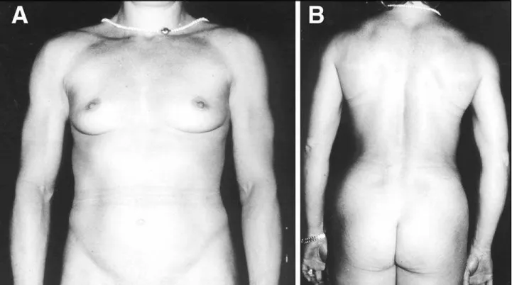
abdo-men was flat, flaccid and no pain occurred on superficial and deep palpation, and there was no visceromegally. There was muscle hypertrophy. (Fig 1 A and B) Upon neu-rological examination, the cranial nerves were normal. Proximal muscle strength in the lower and upper limbs was grade IV, and distal muscles were grade V (MCRM). Tonus, gait, coordination, superficial and deep sensitivity were normal. There was absence of deep tendon reflexes.
Hemogram, ESR, sodium, potassium, calcium, mag-nesium, phosphorus, blood sugar, creatinine, serum aldo-lase, lactic dehydrogenase, oxalacetic glutamic transami-nase, total cholesterol, HDL cholesterol, anti-nuclear fac-tor, triglycerides, C reactive protein, prolactin, estradiol, insulin, pro-thrombin time, thrombin time, X factor activ-ity, somatomedin, T3, anti-thyroglobulin antibody and antimicrosomal antibody were normal. T4 3.8 (Normal (N): 4,5-12,5), TSH 0.04 (N: 0,43-3,8), serum creatine kinase 201 IU (N:up to 70 UI), serum immunoglobulins IgA 664 mg/dl (N: 60-400), IgM 18,8 mg/dl (N: 60-300), IgG 322 mg/dl (N: 700-1500), total proteins 6,21 g/dl (N: 6,3-7,9) [albumin 51,04%, alpha-1 globulin 5,15%, alpha-2 globu-lin 14,65%, beta globuglobu-lin 24,95%, gamma globuglobu-lin 4,18%], urinary protein/osmolarity 0,27 (N: < 0,12), and total protein in 24 hour urine 101 mg/24 hs (N: 27-93). The thyroid scintillography showed a two hour test below normal. Thorax X-ray, abdominal echography and sella tursica magnetic resonance imaging were normal. The echocardiogram showed an ejection fraction of 68% and normal diastolic function . Serum immunofixation showed a monoclonal protein IgA kappa. Urine immunoelectro-phoresis and immunofixation revealed a monoclonal
pro-tein IgA kappa. Bone marrow biopsy and aspiration showed 35% of plasmatic cells. Stain to amyloid (red Congo) in the bone marrow was negative, but positive in the adipose tissue.
The electromyography showed a myopathic pattern in the left biceps, in left and right anterior tibial, and border-line myopathy in the right biceps and the left deltoid. The nervous conduction study showed elevated distal motor and sensory latencies in the median nerve, without decrement, compatible with bilateral carpal tunnel syndrome. The po-tential amplitude was reduced in the lower limbs (edema).
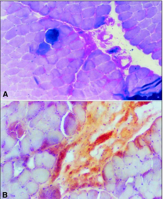
The muscle biopsy revealed a deposit of homogeneous, hyaline material in the interstitious, which stained pink in Haematoxylin-Eosin and slightly green in Gomori, with-out the typical aspect of fibrosis. There was no fatty infil-tration. There was presence of dense wall vessels only in the capillaries, which suggested a deposit of hyaline ma-terial. Hypertrophy of type 1 and 2 fiber was found. Cresil violet and crystal violet demonstrated a metachromatic material, red, with an amyloid aspect, which also involved the vessel walls (Fig 2A). Red Congo showed stained de-posits in conjunctive tissue, related to the perimysium and endomysium of some vessels and, also, stained deposits around the muscle fiber (Fig 2B).
The electronic microscopy revealed interstitious and muscle fiber with sarcoplasmatic accumulation of a fila-mentarous material with amyloid characteristics (Fig 3).
The patient underwent autologous bone marrow trans-plantation without progression of the disease in the fol-low-up.
DISCUSSION
Amyloidosis occurs in 6 to 15 percent of patients with myeloma, mainly in the form of light chain myeloma2,6. The amyloid tissue, which infiltrates and weakens the muscular tissue is rare7. Lubarsh made the first description of the presence of macroglossia and firm muscles with a wooden consistency in 1929.
Macroglossia, difficulty in swallowing, difficulty in speech, pseudohypertrophy of skeletal muscles, hy-pertrophy of small joints and gastrointestinal symp-toms are common findings in most patients with
pseudohypertrophy associated with amyloidosis5.
The etiology of primary amyloidosis is unknown. The protein deposits are fibrils, which are similar to
the immunoglobulins variable portion, which repre-sent an excessive deposit or a deficient removal5,8. The extracelular fibril deposits are insoluble, gener-ally resistant to proteolyses, and cause a disorgani-zation of the tissular architecture. This material is recognized immunohistochemically by green bire-fringence when stained with red Congo and exam-ined under polarized light9,10.
The pathogenesis of myopathy is also unknown. The muscular contraction could be mechanically impaired by the inefficient muscular elasticity caused by an amyloid deposit. Perimisial infiltration could cause muscular atrophy. The perivascular and peri-misial connective tissue with amyloid deposits could interfere with normal muscular nutrition as well as with the exchange of waste products, and the perivascular infiltration most likely causes muscular ischemia7,8.The amyloid infiltration can also inter-fere in the propagation of action potentials along
the sarcolemmal membrane8. Although accurate
me-chanisms of the motor deficit are not known; the accumulation of amyloid tissue in the muscle is the
most probable cause of muscular weakness4,6.
Despite the fact of its being a very rare disease,
with only 10 related cases (Lubarsh 1929; Osserman et al 1964; Martin et al 1970; Ikeda et al 1973; Miya-zaki et al 1973, Whitaker et al 1977; Terashima et al 1978; Miyasaki et al 1979; Komiyama et al 1996), the clinical features are uniform, with macroglossia, difficulty in swallowing and in speech, weakness, pseudohypertrophy of skeletal muscles with an ath-letic appearance. Other disorders are also common such as the carpal tunnel syndrome,
gastrointesti-nal symptoms and cardiomyopathy9. The reported
case showed the clinical features commonly obser-ved, the exception being cardiomyopathy. One clini-cal finding was absence of tendon reflexes, which is not common in such cases.
The electrophysiological studies usually reveal a myopathic pattern with normal nerve conduction. In this patient, the motor and sensory distal laten-cies in the median nerve were elevated and the nerve conduction velocity was decreased in the left mo-tor; there was no conduction in the right sensory compatible with bilateral carpal tunnel syndrome. The association between muscular pseudohypertro-phy with carpal tunnel syndrome is frequent11. The finding of a myopathic pattern is not specific, becau-se it can be found in becau-several types of myopathies.
The muscular biopsies described are usually a vari-able degree of muscular atrophy, mainly type II fi-bers, and amyloid deposits in the muscle are prima-rily in the vessels8. In our case, we found amyloid deposits in the vessel walls, mainly in the arteries, although there was hypertrophy of type I and II fi-bers, which was possibly associated with muscle overwork.
Currently, the treatment of primary amyloidosis with high doses of bussulphan and melphalan, or only melphalan associated with autologous marrow infusion and peripheric blood stem cell has been of paramount importance. Treatment with dimetilsul-foxide (DMSO), which, in vitro, promotes the solubi-lization of amyloid tissue, did not show effective re-sults in primary amyloidosis. However, when DMSO is associated with plasmapheresis, there is improve-ment in motor, respiratory and renal functions, yet the results are moderate and of limited duration4. Therapies which involved prednisone, melphalan and colchicine did not show any significant inprovement, however the association of these drugs is better than the use of colchicine alone12,13 . The impossibility of quantifying the total amount of amyloid tissue in the organism makes the therapy results difficult to evaluate14,15.
Since the majority of amyloid associated pseudo-hypertrophies were diagnosed by autopsy, there are no current data in an ideal treatment for this type of amyloidosis. Our patient was the first case of amy-loid associated pseudohypertrophy treated with melphalan associated with autologous bone mar-row transplantation. After the procedure, the pa-tient reported a subjective sense of well being, but a longer follow up period is needed to verify the rate of success in this type of treatment.
The case described shows that, despite its being a rare disease, the clinical features are characteris-tic, nevertheless there must be confirmation by mus-cle biopsy. Current treatments are ineffective as far as cure is concerned, but they can contribute to a higher rate of survival of these patients, whose prog-nostic is limited. These reasons explain the relevance of an early diagnosis, as the damage caused by the illness is still reversible in its early stages16,17.
REFERENCES
1. Azevedo EM, Scaff M, Canelas HM, Spina-França. Type I primary neu-ropathic amyloidosis. Arq Neuropsiquiatr 1975;33:105-118. 2. Elomaa I, Ekblom P, Somer T. Amyloid-associated muscle
pseudohy-pertrophy and multiple myeloma in a man with hypernephroma. Acta Med Scand 1983;214:87-91.
3. Pruzanski W, Katz A. Clinical and laboratory findings in primary gen-eralized and multiple-myeloma-related amyloidosis. CMA 1976:14; 906-909.
4. Komiyama A, Kijima M, Takahashi M, Ishida S. Amyloid associated muscle pseudohypertrophy: amelioration of motor dysfunction with plasmapheresis and dimethylsulphoxide. J Neurol Neurosurg Psychiatr 1996;60:591-592.
5. Whitaker JN, Hashimoto K, Quinone M. Skeletal muscle pseudohy-pertrophy in primary amyloidosis. Neurology 1977;27:47-54. 6. Jennekens FGI, Wooke JHJ. Proximal weakness of the extremities as a
main feature of amyloid myopathy. J Neurol Neurosurg Psychiatry 1987;50:1353-1358.
7. Roke ME, Brow WFE, Boughner D. Myopathy in primary systemic amy-loidosis. Can J Neurol Sci 1988;15:314-316.
8. Li K, Hizawa K, Numomura S, Morizumi H. Systemic amyloid my-opathy: light- microscopic and fine structural study of the skeletal muscles with histochemical and immunohistochemical study of amy-loid. Acta Neuropathol (Berl) 1984;64:114-121.
9. Kyle RA, Gertz MA. Primary sistemic amyloidosis: clinical and labora-tory features in 474 cases. Semin Hematol 1995;32:45-59.
10. Santiago RM, Scharnhorst D, Ratkin G, Crouch E. Respiratory muscle weakness and ventilatory failure in AL amyloidosis with muscular pseudohypertrophy. Am J Med 1987;83:175-178.
11. Raymond A, Fraschini G, Smith L. Amyloidosis in multiple myeloma or without apparent cause. Arch Intern Med 1984; 144:2158-2160. 12. Friman C, Pettersson T. Amyloidosis. Curr Opin Rheumatol
1996;8:62-71.
13. Kyle RA, Greipp PR, Garton JP, Gertz MA. Primary systemic amyloi-dosis: comparison of melphalan/prednisone versus colchicine. Am J Med 1985;79:708-716.
14. Kyle R, Gertz MA, Greipp PR. A trial of three regimens for primary amyloidosis: colchicine alone, melphalan and prednisone, and melphalan, prednisone, and colchicine. N Engl J Med 1997;336:1202-1207.
15. Comenzo RL, Vosburgh E, Simms RW. Dose-intensive melphalan with blood stem cell support for the treatment of AL amyloidosis: one-year follow-up in five patients. Blood 1996;88:2801-2806.
16. Majolino I, Marcenò R, Pecoraro G. High-dose therapy and autologous transplantation in amyloidosis-AL. Haematologica 1993;78:68-71. 17. Merlini G. Treatment of primary amyloidosis. Semin Hematol .