267
Radiol Bras. 2017 Jul/Ago;50(4):266–276Letters to the Editor
http://dx.doi.org/10.1590/0100-3984.2015.0122 bladder and another at the left ureterovesical junction,
contrib-uting to the selection of appropriate practice and the success of the surgical procedure. In our review of the literature, we found no other reports of a ureteral calculus in a patient with inguino-scrotal hernia of the bladder.
REFERENCES
1. Levine B. Scrotal cystocele. J Am Med Assoc. 1951;147:1439–41. 2. Fisher PC, Hollenbeck BK, Montgomery JS, et al. Inguinal bladder hernia
masking bowel ischemia. Urology. 2004;63:175–6.
3. Huerta S, Fairbanks T, Cinat M. Incarcerated vesicoinguinal hernia pre-senting with gross hematuria. J Am Coll Surg. 2005;201:992–3. 4. Kraft KH, Sweeney S, Fink AS, et al. Inguinoscrotal bladder hernias:
report of a series and review of the literature. Can Urol Assoc J. 2008;2: 619–23.
5. Ng AC, Leung AK, Robson WL. Urinary bladder calculi in a sliding
ves-Jose Domingos Contrera1, Francisco Teixeira Cardoso Sobrinho2
1. IDI – Instituto de Diagnóstico por Imagem, Ribeirão Preto, SP, Brazil. 2. Centro de Diagnóstico por Imagem, Parintins, AM, Brazil. Mailing address: Dr. Jose Do-mingos Contrera. Rua Pau Brasil, 432, Jardim Recreio. Ribeirão Preto, SP, Brazil, 14040-220. E-mail: [email protected].
ical-inguinal-scrotal hernia diagnosed preoperatively by plain abdominal radiography. Adv Ther. 2011;24:1016–9.
6. Bacigalupo LE, Bertolotto M, Barbiera F, et al. Imaging of urinary bladder hernias. AJR Am J Roentgenol. 2005;184:546–51.
7. Burkhardt JH, Arshanskiy Y, Munson JL, et al. Diagnosis of inguinal re-gion hernias with axial CT: the lateral crescent sign and other key ind-ings. Radiographics. 2011;31: E1–12.
8. Resende DAQP, Souza LRMF, Monteiro IO, et al. Scrotal collections: pictorial essay correlating sonographic with magnetic resonance imaging indings. Radiol Bras. 2014;47:43–8.
Primary tracheobronchial amyloidosis
Dear Editor,
A 58-year-old male sought treatment complaining of dyspnea on exertion, together with cough and occasional mucus secre-tion. He reported having been treated for asthma 14 years prior, as well as having used bronchodilators and inhaled corticoste-roids, although he stated that he had experienced no asthma symptoms in childhood.
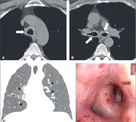
Computed tomography (CT) was performed (Figures 1A, 1B and 1C), after which the patient was submitted to bronchoscopy (Figure 1D) with biopsy. The CT showed concentric thickening
of the walls of the trachea, as well as of those of the main, lobar, segmental, and subsegmental bronchi, with small calciications. The bronchoscopy showed diffuse, concentric iniltration of the mucosa, the iniltrate having a grayish-yellow appearance. The histopathological study showed deposition of amorphous material, whose characteristics were compatible with amyloid deposits.
Amyloidosis encompasses a set of diseases characterized by deposition and abnormal accumulation of protein material in organs and tissues(1). Depending on the anatomical distribution, amyloidosis can be classiied as systemic (involving multiple or-gans) or localized (involving a single organ). In the biochemical
Figure 1.A,B: Axial CT scan of the chest (mediastinal window), without contrast administration, at the level of the proximal segment of the tra-chea (A) and below the carina (B),
showing signiicant concentric thick
-ening of the wall and small calciica -tions in the trachea and bronchi (ar-rows). C: Coronal CT scan of the chest (lung window) showing homogeneous
wall thickening affecting the segmen -tal and subsegmen-tal bronchi (ar-rowheads). Signs of volumetric loss
in the middle lobe (asterisk) due to
narrowing of the lumen of respective lobar bronchus (not shown). D: Bron-choscopic image showing narrowing of the bronchial lumen by concentric,
diffuse iniltration by grayish-yellow
mucous, resulting in enlargement of the secondary carina. LULB, left up-per lobe bronchus; LLLB, left lower lobe bronchus.
A
B
C
D
LLLB
268
Radiol Bras. 2017 Jul/Ago;50(4):266–276 Letters to the Editorclassiication, based on the type of ibrillar component in amy-loid deposits, there are innumerable subtypes. In the vast major-ity of cases, light-chain amyloid ibrils and serum amyloid A are identiied(1).
In the thoracic compartment, amyloidosis typically affects the heart but can also involve the pulmonary parenchyma, pleura, lymph node chains, tracheobronchial tree, and other sites(1,2). Pulmonary involvement is rare, reported as tracheobronchial, diffuse/alveolar-septal, or nodular manifestations, the irst being the most common(2–4).
The tracheobronchial manifestation of amyloidosis is charac-terized by the deposition of amyloid material in the trachea and main bronchi, resulting in thickening of the walls, narrowing of the lumina, and consequent airway obstruction, as well as consolidations, atelectasis, pulmonary hyperinlation, and bron-chiectasis(3).
Clinically, amyloidosis-related tracheobronchial impairment can be asymptomatic or can manifest as dyspnea, wheezing, hemoptysis, cough, or recurrent pneumonia(4,5). The symptoms can be similar to those of bronchial diseases that are more com-mon, including bronchial asthma(5).
Chest CT has been shown to be the imaging exam of choice for the evaluation of thoracic diseases(6–9), as well as for that of diseases of the tracheobronchial tree(10–12). In individuals with amyloidosis, a CT scan can reveal smooth or irregular/ nodular thickening of the tracheal wall and bronchi, which can be accompanied by calciied nodules in the submucosa(4). The differential diagnoses of diffuse tracheobronchial diseases in-clude vasculitis (Wegener’s granulomatosis), tracheobronchial papillomatosis, infectious involvement (rhinoscleroma, caused by infection with Klebsiella rhinoscleromatis), tracheopathia osteochondroplastica, and relapsing polychondritis(13). Unlike tracheal involvement in tracheopathia osteochondroplastica or relapsing polychondritis, tracheobronchial amyloidosis involves the posterior membranous wall of the trachea(4,13).
In individuals with amyloidosis, bronchoscopy usually shows thickening of the walls of the trachea and bronchi, with lat, multifocal, grayish-yellow plaques in the trachea and bronchi. In rare cases, amyloid pseudotumors can be seen(5,13). Histopatho-logical indings of the disease include amyloid thickening of the submucosa, in nodular masses or laminae, showing apple-green birefringence after staining with Congo red(14). There is also a re-duction in the number of submucosal glands, together with cal-ciications and foci of bone metaplasia in the upper airways(14).
In patients suspected of having bronchial asthma who present with atypical symptoms and respond poorly to clinical treatment, various differential diagnoses should be considered(15). The pa-tient in question was initially diagnosed with asthma but did not
respond to treatment, and the deinitive diagnosis of primary tra-cheobronchial amyloidosis was made after a directed follow-up assessment. We can conclude that, albeit rare, tracheobronchial amyloidosis should be considered in such patients.
REFERENCES
1. Czeyda-Pommersheim F, Hwang M, Chen SS, et al. Amyloidosis: mod-ern cross-sectional imaging. Radiographics. 2015;35:1381–92. 2. Marchiori E, Souza Jr AS, Ferreira A, et al. Amiloidose pulmonar:
aspec-tos na tomograia computadorizada. Radiol Bras. 2003;36:89–94. 3. Lee AY, Godwin JD, Pipavath SN. Case 182: pulmonary amyloidosis.
Radiology. 2012;263:929–32.
4. Ngo AV, Walker CM, Chung JH, et al. Tumors and tumorlike conditions of the large airways. AJR Am J Roentgenol. 2013;201:301–13.
5. Serraj M, Kamaoui I, Znati K, et al. Pseudotumoral tracheobronchial am-yloidosis mimicking asthma: a case report. J Med Case Rep. 2012;6:40. 6. Francisco FAF, Rodrigues RS, Barreto MM, et al. Can chest high-resolu-tion computed tomography indings diagnose pulmonary alveolar micro-lithiasis? Radiol Bras. 2015;48:205–10.
7. Batista MN, Barreto MM, Cavaguti RF, et al. Pulmonary artery sarcoma mimicking chronic pulmonary thromboembolism. Radiol Bras. 2015; 48:333–4.
8. Torres PPTS, Moreira MAR, Silva DGST, et al. High-resolution com-puted tomography and histopathological indings in hypersensitivity pneumonitis: a pictorial essay. Radiol Bras. 2016;49:112–6.
9. Mogami R, Goldenberg T, Marca PGC, et al. Pulmonary infection caused by Mycobacterium kansasii: indings on computed tomography of the chest. Radiol Bras. 2016;49:209–13.
10. Ribeiro GMR, Natal MRC, Silva EF, et al. Tracheobronchopathia osteo-chondroplastica: computed tomography, bronchoscopy and histopatho-logical indings. Radiol Bras. 2016;49:56–7.
11. Barbosa BC, Amorim VB, Ribeiro LFM, et al. Tuberculosis: tracheal in-volvement. Radiol Bras. 2016;49:410–1.
12. Barbosa AGJ, Penha D, Zanetti G, et al. Foreign body in the bronchus of a child: the importance of making the correct diagnosis. Radiol Bras. 2016;49:340–2.
13. Prince JS, Duhamel DR, Levin DL, et al. Nonneoplastic lesions of the tracheobronchial wall: radiologic indings with bronchoscopic correla-tion. Radiographics. 2002;22 Spec No:S215–30.
14. Kurtz KA, Kirby PA. Pathologic quiz case: a 49-year-old man with chron-ic cough and a left lung hilar mass. Tracheobronchial amyloidosis. Arch Pathol Lab Med. 2003;127:e420–2.
15. Tilles SA. Differential diagnosis of adult asthma. Med Clin North Am. 2006;90:61–76.
Pedro Paulo Teixeira e Silva Torres1, Matheus Rabahi2, Sebastião Alves Pinto3, Karla Cristina de Morais Arantes Curado4, Marcelo Fouad Rabahi3
1. Multimagem Diagnósticos, Goiânia, GO, Brazil. 2. Pontifícia Universidade Católica de Goiás (PUC Goiás), Goiânia, GO, Brazil. 3. Universidade Federal de Goiás (UFG), Goiânia, GO, Brazil. 4. Hospital e Maternidade Jardim América, Goiânia, GO, Brazil. Mailing address: Dr. Pedro Paulo Teixeira e Silva Torres. Rua 9, nº 326, Residencial Amaury Menezes, ap. 1502, Setor Oeste. Goiânia, GO, Brazil, 74110-100. E-mail: [email protected].
http://dx.doi.org/10.1590/0100-3984.2015.0177
Dear Editor,
A seven-year-old female patient with acute appendicitis underwent an emergency appendectomy. During the procedure, as incidental indings, a bulky bladder and a probable collec-tion adhered to the wall were observed. Cystoscopy revealed an enlarged bladder with diffuse thickening of its walls. Subse-quently, computed tomography of the abdomen showed a well-deined, hypointense collection, with cystic attenuation, with regular contours, showing no enhancement and in contact with the right lateral wall of the bladder (Figure 1). An investigation
of pathological antecedents revealed that the patient had expe-rienced recurrent episodes of cystitis in the last year. The deci-sion was made to perform laparoscopic surgery, during which a small communicating oriice was identiied (between the lesion and the interior of the bladder) and partial cystectomy was per-formed. Histopathological analysis demonstrated ibroadipose tissue exhibiting a xanthogranulomatous reaction (characterized by the presence of xanthomatous macrophages), together with a giant-cell reaction, cholesterol crystals, and mild chronic inlam-matory iniltrate. A similar macrophage reaction was observed in the lymph node (Figure 2). In view of those indings, the main diagnostic hypothesis was xanthogranulomatous cystitis.