effusion, nephrotic syndrome, Hodgkin’s lymphoma, follicular dendritic cell sarcoma, POEMS syndrome, temporal arteritis, myasthenia gravis (MG), etc., might be associated with Castleman’s disease.(3)
Despite the well-known association between Castleman’s disease and follicular dendritic cell sarcoma, there are few reported cases of sarcoma arising as a complication of preexisting
Introduction
Castleman’s disease is an atypical lymphoproliferative disorder and usually has a benign clinical course.(1) Although its etiology remains unclear, factors such as overproduction of IL-6, infection with human herpes virus 8, and autoimmunity have been implicated in the genesis of lymphoid proliferation.(2) Various other clinical conditions, such as paraneoplastic pemphigus, Kaposi’s sarcoma, recurrent pleural
Castleman’s disease associated with follicular
dendritic cell sarcoma and myasthenia gravis*
Doença de Castleman associada a sarcoma de células dendríticas foliculares e miastenia gravis
Fernando Luiz Westphal, Luís Carlos de Lima, Luiz Carlos Lopes Santana, José Corrêa Lima Netto, Vanise Campos Gomes Amaral, Márcia dos Santos da Silva
Abstract
Castleman’s disease is an atypical lymphoproliferative disorder of unknown etiology, which might be associated with various clinical conditions, including autoimmune diseases and malignant neoplasms. We report the case of a 72-year-old female patient who was referred to the thoracic surgery department of Getúlio Vargas University Hospital, in the city of Manaus, Brazil, for the resection of a posterior mediastinal tumor. Three months prior, the patient had been admitted to the ICU with signs of severe dyspnea, at which time she was diagnosed with myasthenia gravis. After the resection of the mediastinal tumor, the histopathological examination revealed hyaline vascular-type Castleman’s disease, complicated by follicular dendritic cell sarcoma. At this writing, the patient was being treated with an anticholinesterase agent and corticosteroids for the control of myasthenia gravis.
Keywords: Giant lymph node hyperplasia; Dendritic cell sarcoma, follicular; Myasthenia gravis;
Mediastinal diseases.
Resumo
A doença de Castleman é um distúrbio linfoproliferativo atípico, de etiologia desconhecida, que pode estar associada a uma série de condições clínicas, inclusive doenças de caráter autoimune e neoplasias malignas. No presente relato, uma paciente de 72 anos foi encaminhada ao serviço de cirurgia torácica do Hospital Universitário Getúlio Vargas, localizado na cidade de Manaus (AM) para a ressecção de um tumor de mediastino posterior. Três meses antes, havia sido internada em UTI com um quadro de dispneia intensa, ocasião na qual foi diagnosticada miastenia gravis. Após a ressecção da massa mediastinal, a análise histopatológica revelou doença de Castleman hialino-vascular complicada por sarcoma de células dendríticas foliculares. Até o momento da redação deste estudo, a paciente utilizava um anticolinesterásico e corticoides para o controle da miastenia gravis.
Descritores: Hiperplasia do linfonodo gigante; Sarcoma de células dendríticas foliculares; Miastenia gravis;
Doenças do mediastino.
* Study carried out at the Getúlio Vargas University Hospital, Federal University of Amazonas, Manaus, Brazil.
Correspondence to: Fernando Luiz Westphal. Hospital Universitário Getúlio Vargas, Coordenação de Ensino e Pesquisa, Avenida Aripuanã, 4, Praça 14 de Janeiro, CEP 69020-170, Manaus, AM, Brasil.
Tel 55 92 234-6334. E-mail: [email protected] Financial support: None.
receptor antibody levels, which were found to be 10.63 nmol/L (reference, 0-0.2 nmol/L). These findings, together with the clinical profile, corroborated the diagnosis of MG.
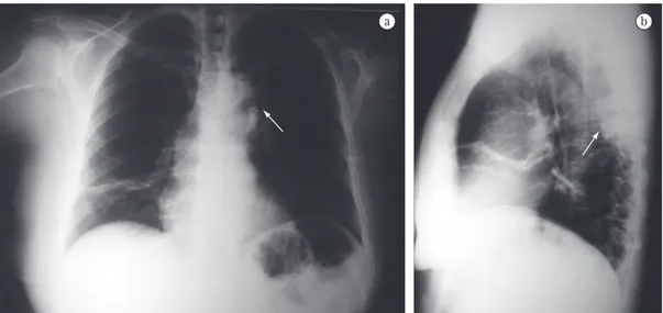
A chest X-ray revealed a mass in the region of the posterior mediastinum, which was confirmed by a preoperative chest CT scan, which identified a heterogeneous mass with dense calcification in the region of the left costovertebral gutter (Figures 1 and 2). The mass in the posterior mediastinum was resected together with the adjacent lung parenchyma. The histopathological examination revealed a lymph node whose architecture was partially distorted by spindle cell proliferation in close association with an inflammatory infiltrate, composed predominantly by lymphocytes. We identified characteristics that were definitely consistent with hyaline vascular-type Castleman’s disease: follicles showing slight germinal center regression; a concentric arrangement of small lymphocytes; and collagenous stroma. The proliferating spindle cells formed spiral fascicles and matrices, with cells with ill-defined borders, vesicular nuclei, and distinct nucleoli, with little variation in nucleus size, as well as varying degrees of atypia, morphologically consistent with follicular dendritic cell sarcoma. Immunohistochemical analysis was negative for the following markers: 40-, 48-, 50-, and 50.6-kDa cytokeratins; CD30; CD246; CD21; CD23; CD4RB; CD68; smooth muscle actin; and Castleman’s disease.(4) Cases of Castleman’s
disease associated with MG are equally rare. (5) The objective of this study was to report a
case of MG associated with Castleman’s disease, complicated by follicular dendritic cell sarcoma.
Case report
A 72-year-old female patient was referred to the Department of Thoracic Surgery of the Portuguese Beneficent Society Getúlio Vargas University Hospital for the resection of a posterior mediastinal tumor.
The patient had a seven-month history of fluctuating muscle weakness associated with eyelid ptosis and diplopia. In addition, three months prior, she had been admitted to the ICU with signs of respiratory failure, requiring mechanical ventilation. During the ICU stay, the patient underwent pharmacologic testing with edrophonium, and an immediate response was obtained, with an improvement in muscle strength and a reduction in eyelid ptosis. Subsequently, the patient underwent five sessions of plasmapheresis, showing a significant improvement of the signs and symptoms and being weaned from mechanical ventilation at the third session. After hospital discharge, the patient underwent electroneuromyography with repetitive nerve stimulation test, which revealed a 12% reduction in the action potentials, and underwent determination of anti-acetylcholine
Figure 1 - Chest X-rays: anteroposterior (in a) and lateral (in b), revealing a mass projecting into the left hilar
changes. When these changes are found concomitantly with plasma cell proliferation, Castleman’s disease is classified as mixed or intermediate.(8)
The clinical manifestations of Castleman’s disease range from asymptomatic forms or forms with mild localized lymphadenopathy to recurrent forms of generalized lymphadenopathy with severe systemic symptoms.(9) In the localized form of the disease, there is lymphadenopathy, usually located in the mediastinum or abdomen, although the disease can also manifest as a pulmonary nodule or a nodule in the pelvis, axilla, or neck.(10,11) Symptoms can result from compression by the lesion, or Castleman’s disease can be an incidental finding in imaging studies in asymptomatic patients.(7,9) In this patient, Castleman’s disease manifested as a single mass in the posterior mediastinum, without systemic symptoms.
The systemic form manifests as generalized lymphadenopathy associated with nonspecific systemic symptoms, such as fever, night sweats, weight loss, weakness, nausea, and anorexia. The plasmacytic variant, even in the localized form of the disease, includes elevated erythrocyte sedimentation rate, anemia, neurological disorders, hypergammaglobulinemia, and hepatosplenomegaly.(7,9,10)
Other immune system disorders might be associated with Castleman’s disease, especially in its systemic form. In the present case report, the patient, in addition to presenting with MG, also developed follicular dendritic cell sarcoma as a complication of Castleman’s disease.
Follicular dendritic cell sarcoma is a rare neoplasm that affects the lymph nodes, although it can occur in extranodal sites such as the liver, spleen, and breast, with low to intermediate risk of metastasis or recurrence.(12) In 10-20% of cases, it is associated with Castleman’s disease, usually hyaline vascular-type Castleman’s disease, which can be concurrent with or precede the sarcoma.(13) It has been proposed that Castleman’s disease-related hyperplasia acts on the expression of the p53 gene, leading to follicular dendritic cell dysplasia in the germinal center of the lymphoid follicle affected. Over time, the dysplasia extends into the interfollicular area until it affects the entire lymph node and causes sarcoma. Therefore, Castleman’s disease might S-100 protein. The final diagnosis was hyaline
vascular-type Castleman’s disease, complicated by follicular dendritic cell sarcoma.
There was no recurrence of the resected mass, and a chest CT scan performed eight months after the surgical procedure revealed no abnormalities.
At this writing, the patient was being treated with an anticholinesterase agent and a low-dose corticosteroid, which resulted in good clinical control of MG.
Discussion
Initially, Castleman’s disease was described as benign mediastinal lymph node hyperplasia, histologically similar to a thymoma.(6) Currently, Castleman’s disease has three histological variants and two clinical presentations. From a histological standpoint, Castleman’s disease is classified as hyaline vascular, plasmacytic, or mixed. Clinically, the disease can be localized or systemic.(7)
In the localized form of the disease, approximately 90% of cases are of the hyaline vascular type, which is characterized by lymphoid follicles with prominent germinal centers containing vascular proliferation and hyalinized vessels, as observed in this patient. When Castleman’s disease presents in its systemic form, histological analysis typically reveals the plasmacytic variant, which is characterized by diffuse proliferation of plasma cells in the interfollicular tissues, without hyaline vascular
in this case (myofibroblastic tumor, Langerhans cell sarcoma, and reticular cell sarcoma), because the morphological pattern found was hardly suggestive of these neoplasms. Regarding MG, the complementary test results and the lack of improvement after resection of the tumor mass ruled out the hypothesis of paraneoplastic syndrome.
In conclusion, we emphasize the importance of including Castleman’s disease in the differential diagnosis of mediastinal masses, because, despite being rare, Castleman’s disease can present very similarly to diseases considered more common, especially neoplasms.
References
1. Chagas JF, Bezerra TP, Bombini JR, Del’Arco AL, Silenci RM, Santos RC, et al. Doença de Castleman: relato de caso e revisão de literatura. Arq Inter Otorrinolaringol [serial on the Internet]. 2004 Oct/Dec [cited 2009 Dec 17]; 8(4):[about 9 p.] Available from: http://www. arquivosdeorl.org.br/conteudo/acervo_port.asp?id=296 2. Martino G, Cariati S, Tintisona O, Veneroso S, De Villa F,
Vergine M, et al. Atypical lymphoproliferative disorders: Castleman’s disease. Case report and review of the literature. Tumori. 2004;90(3):352-5.
3. Olak J. Benign lymphnode disease involving the mediastinum. In: Shields TW, Lo Cicero J, Ponn PB, editors. General Thoracic Surgery. Philadelphia: Lippincott Williams & Wilkins; 2000. p. 2251-5. 4. Chan AC, Chan KW, Chan JK, Au WY, Ho WK,
Ng WM. Development of follicular dendritic cell sarcoma in hyaline-vascular Castleman’s disease of the nasopharynx: tracing its evolution by sequential biopsies. Histopathology. 2001;38(6):510-8.
5. Day JR, Bew D, Ali M, Dina R, Smith PL. Castleman’s disease associated with myasthenia gravis. Ann Thorac Surg. 2003;75(5):1648-50.
6. Castleman B, Iverson L, Menendez VP. Localized mediastinal lymphnode hyperplasia resembling thymoma. Cancer. 1956;9(4):822-30.
7. Mendonça C, Rios E, Reis C, Santos A, Silva PS. Doença de Castleman – a propósito de um caso clínico. Rev Soc Port Med Interna. 2008;15(4):249-53.
8. Yamashita T, Mattos AC, Ferreira MC, Alvarenga M. Doença de Castleman: hiperplasia com aspectos de neoplasia. Rev Cienc Med. 2006;15(2):173-7.
9. Pinheiro VG, Fernandes GH, Cezar LC, Alves Nde A, de Menezes DB. Castleman’s disease accompanied by pleural effusion. J Bras Pneumol. 2008;34(8):626-30. 10. Yeh CM, Chou CM, Wong LC. Castleman’s disease
mimicking intrapulmonary malignancy. Ann Thorac Surg. 2007;84(2):e6-7.
11. Krawczun GA, Garcia Cde M, Ito K, Ferreira Filho OF, Thomson JC. Castleman’s disease or angiofollicular hyperplasia as a solitary pulmonary nodule: case report. J Bras Pneumol. 2007;33(2):226-8.
12. Puerto IM, Martinez IB, Ochoa MC. Follicular dendritic cell sarcoma arising from nodal hyalinevascular Castleman’s disease: a case report. Haema. 2003;6(1):77-80. act as a precursor lesion to follicular dendritic
cell sarcoma.(4,14)
The combination of Castleman’s disease and MG is extremely rare and particularly interesting, because, in addition to the fact that 65% of patients with MG have follicular hyperplasia in the thymus, it is important to remember that Castleman’s disease was initially described as a lesion very similar to a thymoma. Therefore, it is possible that the concomitance of Castleman’s disease and MG is not just a coincidence and that some other mechanism is involved.(5,15)
In cases of localized disease, surgical excision is the procedure of choice, with a low recurrence rate. However, due to the risk of malignant neoplasms, long-term follow-up is required.(16) When surgery in contraindicated, radiotherapy is an option.(17,18)
Although the treatment of the systemic form remains controversial, it is mainly based on chemotherapy, albeit with a worse prognosis due to the higher rate of association with malignancy.(19) Treatments with corticosteroids, immunomodulators, and monoclonal antibodies have been used, mainly for the control of any accompanying immunological disorders, because surgery does not affect the prognosis.(19,20)
17. Muhsein KA, Liew NC, Shaker AR, Shahrin IA. Localized Castleman’s disease presenting as a vascular right iliac fossa mass. Asian J Surg. 2004;27(1):54-7.
18. Oliveira CV, Gonçalves CE, Almeida VF, Oliveira AM, Pimenta FC. Doença de Castleman localizada abdominal. Rev Bras Hematol Hemoter. 2005;27(2):133-7. 19. Bowne WB, Lewis JJ, Filippa DA, Niesvizky R, Brooks
AD, Burt ME, et al. The management of unicentric and multicentric Castleman’s disease: a report of 16 cases and a review of the literature. Cancer. 1999;85(3):706-17. 20. Ocio EM, Sanchez-Guijo FM, Diez-Campelo M, Castilla
C, Blanco OJ, Caballero D, et al. Efficacy of rituximab in an aggressive form of multicentric Castleman disease associated with immune phenomena. Am J Hematol. 2005;78(4):302-5.
13. Swerdlow SH, International Agency for Research on Cancer; World Health Organization. WHO classification of tumours of haematopoietic and lymphoid tissues. Lyon: International Agency for Research on Cancer, 2001.
14. Rodríguez Silva H, Buchaca Faxas E, Machado Puerto I, Pérez Román G, Pérez Caballero D. Castleman’s disease. Review of five cases [Article in Spanish]. An Med Interna. 2005;22(1):24-7.
15. Emson HE. Extrathoracic angiofollicular lymphoid hyperplasia with coincidental myasthenia gravis. Cancer. 1973;31(1):241-5.
16. Vasef M, Katzin WE, Mendelsohn G, Reydman M. Report of a case of localized Castleman’s disease with progression to malignant lymphoma. Am J Clin Pathol. 1992;98(6):633-6.
About the authors
Fernando Luiz Westphal
Adjunct Professor. Department of Thoracic Surgery, Federal University of Amazonas, Manaus, Brazil.
Luís Carlos de Lima
Physician-in-Chief. Department of Thoracic Surgery, Getúlio Vargas University Hospital, Federal University of Amazonas, Manaus, Brazil.
Luiz Carlos Lopes Santana
Attending Physician. Department of Clinical Medicine, Getúlio Vargas University Hospital, Federal University of Amazonas, Manaus, Brazil.
José Corrêa Lima Netto
Attending Physician. Department of Thoracic Surgery, Getúlio Vargas University Hospital, Federal University of Amazonas, Manaus, Brazil.
Vanise Campos Gomes Amaral
Attending Physician. Department of Neurology, Getúlio Vargas University Hospital, Federal University of Amazonas, Manaus, Brazil.
Márcia dos Santos da Silva