Braz Dent J 17(3) 2006
GAPO Syndrome 259
Braz Dent J (2006) 17(3): 259-262
Dental Findings in GAPO Syndrome: Case Report
Heloísa Emília Dias da SILVEIRA¹ Onofre Francisco QUADROS² Reni Raymundo DALLA-BONA¹ Heraldo Luis Dias da SILVEIRA1
Guilherme Genehr FRITSCHER4
¹Department of Surgery and Orthopedics, School of Dentistry, Federal University of Rio Grande do Sul, Porto Alegre, RS, Brazil ²Department of Otorhinolaryngology and Division of Stomatology, Clinics Hospital of Porto Alegre; Department of Conservative Dentistry, School of Dentistry, Federal University of Rio Grande do Sul, Porto Alegre, RS, Brazil
4 Department ofSurgery,School of Dentistry, Pontifical Catholic University, Porto Alegre, RS, Brazil
This article reports the case of a young female adult with GAPO syndrome who presented as a peculiar dental finding unerupted primary and permanent dentitions, which resembled total anodontia on clinical examination. A cephalometric analysis was performed to investigate the alterations in facial bone development. This is the 9th GAPO syndrome case reported in a Brazilian patient.
Key Words: GAPO syndrome, pseudoanodontia, alopecia, growth retardation.
Correspondence: Dra. Heloísa Emília Dias da Silveira, Faculdade de Odontologia, Univesidade Federal do Rio Grande do Sul, Rua Ramiro Barcelos, 2492, 90035-003 Porto Alegre, RS, Brasil. Tel: +55-51-3316-5199. Fax: +55-51-3316-5032. e-mail [email protected] ISSN 0103-6440
INTRODUCTION
GAPO syndrome is a very rare genetic disorder with approximately 30 cases reported worldwide. GAPO is the acronymic designation for a complex of growth retardation, alopecia, pseudoanodontia (failure of tooth eruption) and progressive optic atrophy. It seems to have an autosomal recessive pattern of inheritance but its etiology is not yet well understood (1). This condition was first described by Andersen and Pindborg (2) in 1947, but it was named GAPO syndrome only in 1984 by Tripton and Gorlin (3).
Patients who suffer from GAPO syndrome may have several abnormalities including wide anterior fon-tanel, bossed forehead, prominent scalp veins, increased intracranial pressure, micrognathia, protruding lips and auricles, depressed nasal bridge, altered ability to sweat, skin redundancy, hyperextensible joints, hyperconvex nails, umbilical hernia, delayed bone maturation, hepatomegaly, hypoplasia of the genitalia and mammary glands, and irregular gonadal function.
Eight cases have been described in Brazil. This paper reports the dental findings in a Brazilian young adult diagnosed with GAPO syndrome.
CASE REPORT
Braz Dent J 17(3) 2006
260 H.E.D. Silveira et al.
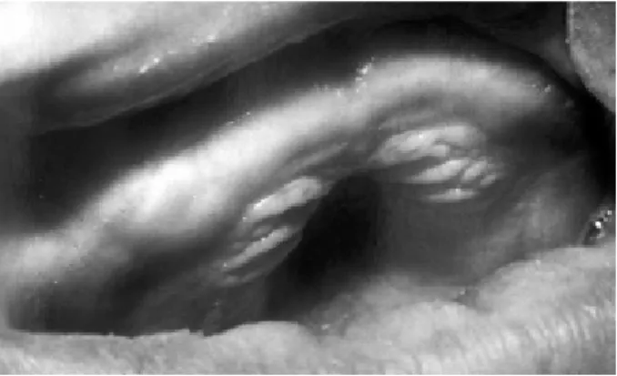
Figure 2. Thick upper alveolar ridge without any erupted tooth.
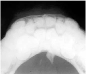
Figure 4. Panoramic radiograph revealing the presence of several impacted primary and permanent teeth.
Figure 3. Intraoral view showing ower alveolar ridge without any erupted tooth present.
Ricketts and McNamara cephalometric com-puted analyses (Fig. 5; Tables 1 and 2) were performed to investigate the relation of the facial bones with the anterior skull base (ASB) and evaluate the syndrome-related alterations. Some measurements could not be made because some of the cephalometric points could not be marked. The maxilla was prominent compared to the ASB and the patient presented a reduced maxillary height. Both the ASB and the mandible were shorter than normal in length. The mandible seemed to have a normal relation with the ASB and the lower segment of the face was higher than normal. The upper lip was larger than normal, and the lower lip was prominent.
The need for a multidisciplinary approach
involv-ing Surgery, Prosthodontics and Oral Rehabilitation was discussed with the patient and the placement of a complete denture was presented as the best treatment option for her case. However, the patient refused to undergo the surgical intervention required to allow denture installation and gave up the treatment.
She returned only 5 years later complaining of increased volume in the sublingual region and pain every time she ate. The occlusal radiographic examination revealed the presence of a small salivary calculus (Fig. 6) close to the exit of Wharton’s duct orifice, which could be easily removed due to its location. It was also observed the root of a tooth projected out of the cortical bone in the lingual surface of the mandible. However, the patient denied any painful symptomatology. After removal of the salivary calculus, the patient had total resolution of the symptoms.
Braz Dent J 17(3) 2006
GAPO Syndrome 261
The patient has been followed up by physicians from different medical specialties for management of the complications arising from GAPO syndrome.
DISCUSSION
In 1977, Epps et al. (4) described the case of two siblings of consanguineous parents who were mistak-enly diagnosed as having Rothmund-Thompson syn-drome. Five years later, the same case was reported by Wajntal et al. (5) now as a new syndrome because some of the typical findings of Rothmund-Thompson syn-drome had not been found in those patients. In 1990, Wajntal et al. (6) finally reported this case as being GAPO syndrome, based on the short stature, alopecia and pseudoanodontia present in both patients. In 1984, Gagliardi et al. (7) documented the case of 3 Brazilian
siblings that had also consanguineous parents. Silva (8) presented a female patient whose parents were unaf-fected and apparently nonconsanguineous. These Bra-zilian cases seemed to belong to four distinct families. In 2005, Rim and Marques-de-Faria (9) described the ophtalmic aspects of two sisters with GAPO syndrome from consaguineous parents.
The patient described in this article had the typical signs and abnormalities of GAPO syndrome. Alopecia was observed in her eyelids, eyebrows and head (Fig. 1).
The dental findings observed in this patient were quite interesting. The pseudoanodontia that resulted from the unerupted primary and permanent teeth caused an increase in bone ridge volume in a buccolingual direction, as shown in the occlusal radiograph. This condition hindered the installation of dentures. Surgical removal of the teeth, even if partial, was deemed too complicated because the teeth were ankylosed (10). In
Figure 6. Occlusal radiograph showing a salivary calculus. Figure 5. Lateral cephalometric radiograph.
Table 1. Ricketts landmarks used for cephalometric analysis.
Lower lip position 7.87 mm
Upper lip length 45.60 mm
Maxillary depth 98.56°
Maxillary height 44.17°
Palatal Plan 5.93°
Mandibular body length 58.96 mm
Anterior cranial length 49.56 mm
Table 2. McNamara landmarks used for cephalometric analysis.
A-N Pep 5.41 mm
Prn.(Sn-Ls) 123.87°
Co-A 65.72 mm
ENA-Me 72.48 mm
Braz Dent J 17(3) 2006
262 H.E.D. Silveira et al.
addition, in spite of all efforts, the patient strongly refused to have her case studied by a multidisciplinary team, which could have improved her esthetic and functional condition. The major complain was the volume increase and pain in the sublingual region caused by a salivary calculus, which was resolved.
A cephalometric analysis was performed be-cause none of the previous reports have done so, probably because there has not been too much interest in investigating the orodental manifestations of this syndrome. Cephalometric analysis should be conducted in further GAPO syndrome cases in order to address new characteristic of this disorder.
RESUMO
Este artigo relata o caso de um jovem paciente, gênero feminino, portadora da síndrome de GAPO, apresentando impacções dos dentes decíduos e permanentes, sugerindo anodontia total no exame clínico. Foi realizada uma análise cefalométrica para investigar as alterações no desenvolvimento ósseo facial. Este é o nono caso descrito no Brasil.
REFERENCES
1 . Sandgren G. GAPO syndrome, a new case. Am J Med Genet 1995;58:87-90.
2 . Andersen TH, Pindborg JJ. El tifaelde at total “pseudoanodonti” inforbindelse med kranie deformitet, dvaergvaekst og ektodermal displasi. Odontol Tilster 1947;55:484-493.
3 . Tripton RE, Gorlin RJ. Growth retardation, alopecia, pseudoanodontia, and optic atrophy - The GAPO syndrome. Am J Med Genet 1984;19:209-216.
4 . Epps DR, Mendonça BB, Olazabal LC, Billerbeck AEC, Wajntal A. Familial congenital poiquilodermia. (Rpthmund-Thompson Syndrome). Cienc Cult 1977;29(Suppl):740. 5 . Wajntal A, Epps DR, Mendonca BB, Billerbeck AEC. New
Sydrome of ectodermal dysplasia: dwarfism, alopecia, an-odontia and cutis laxa. Cienc Cult 1982;3(Suppl):705. 6 . Wajntal A, Koiffmann CP, Mendonca BB, Epps-Quaglia D,
Sotto MN, Rati PB, Opitz JM. GAPO syndrome (McKusick 23074), a connective tissue disorder: Report on two affected sibs and on the pathologic findings in the older. Am J Med Genet 1990;37:213-223.
7 . Gagliardi ART, Gonzalez CH, Pratesi R. GAPO syndrome: Report of three affected brothers. Am J Med Genet 1984;19:217-223.
8 . Silva EO. Dwarfism, alopecia, pseudoanodontia and other anomalies. Report of a case. Rev Bras Genet 1984;7:743-747. 9 . Rim PH, Marques-de-Faria AP. Ophtalmic aspects of GAPO syndrome: case report and review. Ophtalmic Genet 2005;26:143-147.
10. Bacon W, Hall RK, Roset JP, Boukari A, Tenenbaum H, Walter B. GAPO syndrome: a new case of this rare syndrome and a review of the relative importance of different pheno-typic features in diagnosis. J Cranifac Genet Dev Biol 1999;19:189-200.