Assessing the growth and recovery of
Salmonella
Enteritidis SE86 after sodium
dichloroisocyanurate exposure
Fernanda Stoduto Ferreira, Mariana Bandeira Horvath, Eduardo Cesar Tondo
Laboratório de Microbiologia e Controle de Alimentos, Instituto de Ciência e Tecnologia de Alimentos, Universidade Federal do Rio Grande do Sul, Porto Alegre, RS, Brazil.
Submitted: May 2, 2012; Approved: November 13, 2012.
Abstract
The objective of the present study was to assess the growth and the recovery of Salmonella(S.) Enteritidis SE86 in different diluents, culture media and using different plating methods after the ex-posure to 200 mg/kg sodium dichloroisocyanurate (NaDCC). Before and after NaDCC exex-posure, SE86 was cultured at 30 °C and 7 °C in the following diluents: Peptone water (P), Saline solution (SaS), Peptone water+Saline solution (P+SaS), Peptone water+Tween 80+Lecithin+Sodium thio-sulfate (P+N) and Saline solution+Tween 80+Lecithin+Sodium thiothio-sulfate (SaS+N). The SaS dilu-ent was chosen because it was able to maintain cells viable without growth and was further used for plating SE86 on non selective medium (Tryptic Soy Agar-TSA) and on selective media (Mannitol Lysine Crystal Violet Brilliant Green Agar-MLCB; Brilliant Green Agar-BGA;Salmonella Shigella Agar-SS and Xylose Lysine Dextrose–XLD). The Thin Agar Layer method (TAL)i.e., selective me-dia overlayed with non selective TSA was also evaluated. Results indicated that SE86 not exposed to NaDCC was able to grow in P, P+N, SaS+N and P+SaS, but not in SaS, that was able to maintain cells viable. SE86 exposed to NaDCC demonstrated similar counts after dilution in SaS and the plating on non selective TSA, selective media MLCB, BGA, SS and XLD and on TAL media. SE86, S. Typhimurium andS. Bredeney, exposed or not exposed to NaDCC, showed no significant differ-ences in counts on TSA, XLD and XLD overlayed with TSA, suggesting that all those media may be used to quantify NaDCC-exposedSalmonellaby plating method.
Key words:SalmonellaEnteritidis SE86, diluents, plating methods, stressed cells, sodium dichloroi-socyanurate.
Introduction
Salmonellosis is one of the most important public health problems worldwide (Greig and Ravelet al., 2009; Tondo and Ritter, 2012). In Brazil,Salmonella spp. was identified as the main etiological agent causing foodborne diseases in the period of 1999 to 2011, being responsible for 1660 foodborne outbreaks investigated by the Brazilian In-spection Services (Brasil, 2011). Using pheno and genotyp-ing methods (Oliveriaet al., 2012; Geimbaet al., 2004), a specific strain ofS.Enteritidis (named SE86) was identified in more than 95% of the salmonellosis notified in State of Rio Grande do Sul (RS), southernmost State of Brazil, in the last years (Oliveiraet al., 2012), arising an expressive
interest in the investigation of this pathogen. Further stud-ies demonstrated that SE86 presents high capability for thermal and acid adaptation after its exposure to sub-lethal pH and this capability was not verified in other Sal-monellaserovars (Malheiroset al., 2009). SE86 was also more resistant than otherSalmonellaserovars to 200, 400 and 800 mg/kg sodium hypochlorite and was able of form-ing biofilms on stainless steel and polyethylene (Tondoet al., 2010).
In the State of RS, between 2000 and 2002, insuffi-cient cleanliness of utensils and equipments and cross con-tamination, together, were responsible for 14.15% of the foodborne outbreaks occurred in food services, indicating a
Send correspondence to E.C. Tondo. Laboratório de Microbiologia e Controle de Alimentos, Instituto de Ciência e Tecnologia de Alimentos -ICTA/UFRGS, Universidade Federal do Rio Grande do Sul. Av. Bento Gonçalves 9500, Prédio 43212, Campus do Vale, Agronomia, Caixa Postal 15090, 91501-970 Porto Alegre, RS, Brasil. E-mail: tondo@ufrgs.br.
possible deficiency in the hygiene of surfaces that come into contact with foods (Cunhaet al., 2008). Microbiologi-cal analysis of utensil and equipment surfaces is an impor-tant tool for verifying the effectiveness of cleaning and disinfection procedures (Moore and Griffith, 2007), and the swab technique is the most suitable method for that (APHA, 2001). After disinfection of surfaces, many micro-organisms are inactivated, but a part of the bacterial popula-tion may survive and remain in the VNC (Viable but Not Cultivable) condition. These bacteria may have been af-fected by the action of disinfectants, losing the ability to form colonies on selective media, however, they still be able to reproduce in non-selective media, being called “stressed” or “injured” (Wu and Fung, 2006; Wu, 2008). Stressed bacterial cells have great relevance to the food in-dustry and food services, since they can not be detected in the microbiological analysis, but may be able to cause out-breaks or food quality problems, after their recovery. In this sense, the methods for analysis should allow recovery of bacterial cells, in order to obtain the number of microorgan-isms that are actually present in sample or on a surface (Jassonet al., 2007).
In food services and food industries, the chlorine compounds are among the most commonly disinfectants used for disinfection of surfaces (Penget al., 2002; Rossoni and Gaylarde, 2000). In Brazil, probably the most used chlorine compounds are 200 mg/kg sodium hypochlorite or sodium dichloroisocyanurate, which can injure microor-ganisms present on food contact surfaces, avoiding their detection. Therefore, evaluating the effect of these sani-tizers on important food pathogens such asS. Enteritidis SE86 is of great importance.
The aim of this study was to assess the growth and the recovery ofS.Enteritidis SE86 in different diluents, media and by plating methods, after sodium dichloroisocyanurate exposure, aiming to identify adequate procedures to be used in swab sampling methods.
Materials and Methods
Bacterial strains
Strains of Salmonella enterica of three different serovars were used in this study.S. Enteritidis SE86 was isolated from a cabbage involved in a salmonellosis out-break occurred in the State of RS, in 1999. This microor-ganism presents the same genotypic pattern ofS.Enteritidis involved in more than 95% of the salmonellosis outbreaks occurred in the State of RS during the period of 1999 to 2006 (Oliveira et al., 2012). S. Typhimurium and S. Bredeney strains were isolated from pig fecal samples also in the State of RS. These strains were provided by Prof. Dr. Marisa Ribeiro de Itapema Cardoso of the Preventive Vet-erinary Department of Universidade Federal do Rio Grande do Sul (UFRGS) and there is no reports indicating the in-volvement of them with foodborne diseases in the State of
RS. Before experiments, the strains were stored at -18 °C in 30% (v/v) glycerol, and working cultures were kept at 4 °C on Brain Heart Infusion Agar (BHI; Oxoid). Before use in the experiments, strains were activated in BHI broth at 37 °C for 18-24h, reaching a concentration of approxi-mately 108cfu.mL-1. The initial concentration of cells was measure by plating the inoculum on BHI agar.
Evaluation of growth of S. Enteritidis SE86 in different diluents
Cultures ofS.Enteritidis SE86 were activated in 9 mL of BHI at 37 °C for 18-24 h, reaching a concentration of ap-proximately 109 cfu.mL-1. Two experiments were con-ducted in parallel: one withS.Enteritidis SE86 not exposed to the disinfectant (Control) and other withS. Enteritidis SE86 exposed to 200 mg/kg NaDCC (Trade mark Kaly-clean S313, Kalykim, Brazil). The exposure to NaDCC was performed by inoculating 1 mL of bacterial culture in 9 mL of 200 mg kg-1NaDCC, for five minutes. This exposure time was used because it was able to reduce but not inacti-vate all the inoculatedSalmonella. The exposure time and bacterial concentrations were assessed previously to the ex-periments of multiplication. The exposed and not exposed bacterial cultures were diluted until a concentration of 108cfu mL-1in five different diluent solutions: 0.1% Pepto-ne water (P); 0.85% SaliPepto-ne solution (SaS); 0.85% SaliPepto-ne so-lution + 0.1% Peptone water (P + SaS); 0.1% Peptone water + 0.5% Tween 80 + 0.07% Lecithin + 1% Sodium Tios-sulfate (P + N) and 0.85% Saline Solution + 0.5% Tween 80 + 0.07% Lecithin + 1% Sodium Tiosulfate (SaS + N). The diluent solutions containing exposed and not exposedS. Enteritidis SE86 were incubated at 30 °C and 7 °C for 1, 2, 3, 4, 5 and 6 hours. These incubation times were chosen in order to evaluate regular transport times that generelly are lesser than 6 hours. These temperatures were chosen as-suming that they represent temperatures easily performed under controlled (7 °C) and uncontrolled (30 °C) condi-tions, during the transport of swab samples. The number of colony forming units (cfu) of exposedS.Enteritidis SE86 was quantified by the drop technique (Silvaet al., 1997). To quantify not exposedSalmonella, 0.1 mL of each dilution was cultured on the surface of BHI plates. Before incuba-tion for 18-24 hours at 37 °C, the plates were left to dry at room temperature, inside a laminar flow hood (LabConco, Kansas city, Missouri), previously sterilized by 15 min of UV exposure. The plates were quantified and the counts were expressed in UFC.mL-1. Each measurement was per-formed in duplicate and each experiment was repeated at least twice.
Evaluation of culture media and plating methods
Crystal Violet Brilliant Green Agar (MLCB; Oxoid); Bril-liant Green Agar (BGA; Oxoid),Salmonella ShigellaAgar (SS; Oxoid) and Xylose Lysine Dextrose (XLD; Oxoid) and on selective media (MLCB, BGA, SS and XLD) over-layed with TSA (TAL method). The cell concentration was determinated by a growth curve. The TAL plates were pre-pared overlaying 14 mL of nonselective medium (TSA) onto 25 mL of each selective medium. The plates were in-cubated at 37 °C for 18-24 hours and bacterial colonies were quantified and expressed in cfu mL-1.
Evaluation of growth and recovery ofS.Enteritidis SE86,S.Typhimurium andS.Bredeney on XLD by different plating methods and culture media
After the choose of the best diluent and the best plat-ing method for the growth of exposed and not exposedS. Enteritidis SE86, other two serovars of Salmonella (S. Typhimurium and S. Bredeney) were tested by the same techniques, aiming to compare their growth. All the bacte-ria were individually exposed to NaDCC and diluted as de-scribed previously. After, 0.1 mL of the dilutions was plated in duplicate on the surface of TSA, XLD and XLD overlayed with TSA (TAL). The plates were incubated for 24 hours at 37 °C. After incubation, the UFC.mL-1were quantified and the results of each serovar in each medium was evaluated.
Statistical analysis
All experiments performed to evaluate the growth of microorganisms were carried out at least twice, and all re-sults from counts were made in duplicates. The mean val-ues were calculated and the analysis of variance (ANOVA) and a Tukey Test were carried out to compare the differ-ences between the mean values. The differdiffer-ences were con-sidered significant with p values were less than 0.05.
Results
Multiplication and recovery ofS.Enteritidis SE86 in different diluents
The differences between counts of not exposed and exposed S. Enteritidis SE86 were approximately 2 log cfu.mL-1, demonstrating the effect of NaDCC on the bacte-rial populations.
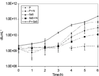
S.Enteritidis SE86 not exposed to NaDCC (Control) showed no significant multiplication (p < 0.05) in any of the diluents tested in the first three hours of experiment. However, the diluents P + SaS, P, SaS + N and P + N sus-tained significant multiplications (more than 1.5 log) after four, five, five and six hours at 30 °C, respectively. In oppo-site,S.Enteritidis SE86 did not multiply in the diluent SaS, at 30 °C, during the six hours of incubation, but the cells re-mained viable. P + SaS was the diluent that sustained higher counts of S. Enteritidis SE86, increasing 2.0 log UFC.mL-1after six hours of incubation at 30 °C (Figure 1).
S.Enteritidis SE86 exposed to NaDCC showed to be stressed because no significant multiplication (p < 0.05) in any of the diluents were observed during the six hours at 30 °C, but cells still viable.S.Enteritidis SE86 exposed and not exposed to NaDCC showed no significant growth in the diluents (p < 0.05), during the six hours of incubation at 7 °C, however cells remained viable.
Evaluation of culture media and plating methods
S. Enteritidis SE86 not exposed and exposed to NaDCC showed no significant differences (p < 0.05) among counts on non selective media (TSA), on selective media (BGA, SS XLD and MLCB) and on selective media XLD, SS, BGA overlayed with TSA (TAL media). TSA counts were higher than counts observed on selective me-dia and TAL meme-dia, but this difference was not significant. MLCB overlayed with TSA demonstrated lower counts when compared with other TAL media.
Recovery ofS.Enteritidis SE86,S.Typhimurium andS.Bredeney grown in XLD by different plating methods
The three serovars S. Enteritidis SE86, S. Typhimurium andS.Bredeney, not exposed (Figure 2) and exposed (Figure 3) to NaDCC showed no significant differ-ence in counts on non selective TSA, on selective medium XLD and XLD overlayed with TSA (TAL).
Discussion
In the present study,S.Enteritidis SE86 was able to grow at 30 °C in all diluents tested, except SaS. In order to quantify microorganisms present in a diluted sample, an ap-propriate diluent should not promote bacterial multiplica-tion or death, should be ease to prepare, should present low Figure 1- Multiplication ofSalmonellaEnteritidis SE86 not exposed to NaDCC at 30 °C in Peptone water (¨), Saline solution (s), Peptone
cost, and ideally, should be able to recover stressed cells (Wuet al., 2001; Wu, 2008). Based on these appointments and according to our results, The most appropriate diluent was SaS, because it did not cause decreasing in the NaDCC exposed cell numbers and did not sustain the multiplication of not exposed cells. Additionally, this diluent is easy to be prepared and have a lower cost when compared to other dil-uents.
After the exposure to NaDCC, S. Enteritidis SE86 showed no significant multiplication in any of the diluents tested, even after six hours of incubation at 30 °C. As no significant multiplication occurred in any of the diluents, the cell recovery was not assessed, and it was not possible to distinguish dead cells from stressed or viable but not cul-tivable (VNC) cells. In order to recover stressed cells, bac-teria present an extension in its lag phase, and this is called the recovery period. After this period, cells start to multiply at an equal rate to the not stressed cells (Tomlins and Ordal,
1971). As an example, a study have demonstrated a four to five hours lag phase ofS.Typhimurium growing in Trypti-case Soy Broth (TSB) and on TSA, after heat stress at 48 °C for 30 min (Clarket al., 1968; Clark and Ordal, 1969). The experiments conducted in this study may suggest that the extent of the lag phase ofS.Enteritidis SE86 exposed to NaDCC became longer than six hours, because not exposed present SE86 presented shorter lag phases in different dilu-ents. It is possible that longer periods of incubation could be able to show the exact duration of the lag phase neces-sary to recover the stressed cells. However, it was not the purpose of this work, since the time of six hours was chosen because it is often recommended as an appropriate period of sample transport inside diluents (Silva et al., 1997; Lightfoot and Maier, 2003).
At 7 °C, not exposedS. Enteritidis SE86 (Control) showed no significant growth in the diluents, during the six hours of incubation. Similar results demonstrated that pure Figure 2- Multiplication ofSalmonellaEnteritidis SE86 (SE),SalmonellaTyphimurium (ST) andSalmonellaBredeney (SB) not exposed to sodium dichloroisocyanurate plated on XLD, XLD overlayed with TSA (TAL) and TSA. Values with different letters differ significantly (p < 0,05). a,b,c: Com-parison among serovars on the same medium plated by the same method. A, B, C: ComCom-parison of the growth of the sameSalmonellaserovar inoculated on different media.
Buffered Peptone Water and also added with Tween 80 + Lecithin and Tween 80 + Lecithin did not sustain multipli-cation ofBacillus cereusandEscherichia coli, at 4 °C, dur-ing 6 hours (Greig and Ravel, 2009). In other study, no significant increase in the numbers of cells ofSalmonella spp.,Escherichia coliO157:H7 andStaphylococcus aureus was observed during nine hours of storage at 4 °C, after dis-infection of shredded cabbage, using 0.1% calcined cal-cium, for 20 min, combined with 100 mg kg-1of Sodium hypochlorite, for 20 min (Fukuyamaet al., 2009). The re-sults of the present study suggest that the temperature of 7 °C can be used to transport samples without allowing the multiplication ofS.Enteritidis SE86. It is possible that this result may be valid for other microorganisms, since the American Public Health Association recommended refrig-eration temperature for the transport of bacterial samples collected by swabs from surfaces, but the exact temperature is not define (APHA, 2001).
When the culture media and the plating methods were evaluated, no significant difference was shown in the counts ofS.Enteritidis SE86 exposed to NaDCC, however there were increased numbers on TSA when compared with selective media and TAL. This quantitative difference sug-gests that there was few stressed cells, but the selective me-dia did not inhibit significantly the multiplication of S. Enteritidis SE86, making it suitable for its cultivation, even by direct plating. Corroborating this result, the ISO 6579/2002 recommends the use of XLD plates for the enu-meration ofSalmonella.
In the present study, selective media overlayed with TSA (TAL) also showed no significant difference in counts ofS.Enteritidis SE86 exposed to NaDCC, when compared with TSA or selective media. This result is interesting, since the TAL method has been reported as important for the recovery of stressed bacterial cells (Osailiet al., 2010; Kang and Fung, 1999; Kang and Fung, 2000). According to the present work, XLD showed no significant difference in counts obtained on non-selective TSA and selective media overlayed with TSA, with or without NaDCC exposure. XLD plates are recommended by the Brazilian Ministry of Agriculture (MAPA) forSalmonellainvestigation and by ISO 6579/2002 forSalmonella quantification, suggesting its adequacy to be used in the enumeration of stressedS. Enteritidis SE86, at least forin vitro experiments. How-ever, in order to evaluate its adequacy to real surface or food samples, more experiments are necessary.
When the growth and recovery ofS.Enteritidis SE86, S.Typhimurium andS.Bredeney were evaluated, no signif-icant differences in counts on the XLD plates, non-selective TSA and TAL were observed, demonstrating that the selec-tive media did not inhibit the growth of these strains of Sal-monella. This result is interesting and corroborates that such media could be suitable for the quatification of Salmo-nellaexposed or not exposed to NaDCC.Other studies are
necessary to evaluate the behavior of these microorgan-isms, stressed by disinfectants on surfaces of food services.
In conclusion, this study indicated that 0.85% Saline (SaS) solution did not supportS. Enteritidis SE86 growth and maintain cells viable for six hours, suggesting being an adequate diluent for the collection and transport of swab samples for the investigation ofS. Enteritidis SE86. The temperature of 7 °C demonstrated to be able to maintain stable the numbers ofS. Enteritidis SE86, suggesting to be adequate for the transport of swab samples. Finally, the di-rect plating on XLD medium demonstrated the same Sal-monella counts that non selective TSA and TAL media, suggesting to be adequate for the quantitative analysis of differentSalmonella serovars,exposed or not exposed to NaDCC, at least in laboratory conditions. Further studies are necessary to test these procedures for the analysis of Salmonellain natural conditions.
Acknowledgments
The authors would like to thank Dr. Marisa Ribeiro de Itapema Cardoso for kindly providing the strainsS. Typhi-murium andS. Bredeney. We are greatfull to the Ministry of Education for providing the scholarship that made the present work possible. We also thank to Kalykim for pro-viding the disinfectant that was used in the experiments. The authors would like to thank to Prof. Dra. Florencia Cladera from ICTA/UFRGS for the statistical analyses.
References
APHA - American Public Health Association (2001) Compen-dium of Methods for the Microbiological Examinations of
Foods, 4th edition. Frances Pouch Downes and Keith Ito
Washington, D.C.
Brasil (2011) Análise epidemiológica dos surtos de doenças trans-mitidas por alimentos no Brasil, 1999-2011. Available: http://portal.saude.gov.br/portal/arquivos/pdf/analise_ep_ surtos_dta_brasil_2011.pdf. Accessed November 4, 2011. Clark CW, Witter LD, Ordal ZJ (1968) Thermal Injury and
Re-covery ofStreptococcus faecalis. Appl Microbiol
16:1764-1769.
Clark CW, Ordal ZJ (1969) Thermal injury and recovery of
Sal-monellaTyphimurium and its effect on enumeration proce-dures. Appl Microbiol 18:332-333
Cunha B (2008) Investigação de Surtos Alimentares ocorridos em Serviços de Alimentação no Rio Grande Do Sul. (Trabalho de conclusão de curso de Graduação – Engenharia de Ali-mentos, Instituto de Ciência e Tecnologia de AliAli-mentos, Universidade Federal do Rio Grande do Sul, Porto Alegre, RS, Brasil).
Fukuyama S, Watanabe Y, Kondo N, Nishinomyia T, Kawamoto S, Isshiki K, Murata M (2009) Efficiency of Sodium
Hipo-clorite and Calcinated Calcium in killingEscherichia coli
O157:H7,Salmonellaspp. andStaphylococcus aureus
at-tached to freshly shredded cabbage. Biosci Biotechnol Biochem 73:9-14.
genes in Salmonellasp. isolated from foods involved in foodborne outbreaks occurred in Rio Grande do Sul, South of Brasil. J Food Prot 67: 1229-1233.
Greig JD, Ravel A (2009) Analysis of foodborne outbreak data re-ported internationally for source attribution. Int J Food Microbiol 130: 77-78.
Jasson V, Debevere J, Rajkovic A, Uyttendaele M (2007) Estab-lishment of procedures provoking sub-lethal injury of Liste-ria monocytogenes,Campylobacter jejuniandEscherichia
coli O157:H7 to serve method performance testing. Int J
Food Microbiol 118: 241-249.
Kang DH, Fung DYC (2000) Thin Agar Layer Method for
Recov-ery of Salmonella Typhimurium. Int J Food Microbiol
54:127-132.
Kang DH, Fung DYC (1999) Thin Agar Layer Method for
Recov-ery of Heat-Injured Listeria monocytogenes. J Food Prot
62:1346-1349.
Lightfoot NF, Maier EA (2003) Análise microbiológica de ali-mentos e água. Lisboa: Fundação Calouste Gulbenkian 284 pp.
Malheiros PS, Brandelli A, Noreña CPZ, Tondo EC (2009) Acid and thermal resistance of aSalmonellaEnteritidis strain in-volved in several foodborne outbreaks. J Food Saf, 29:302-317.
Moore G, Griffith C (2007) Problems associated with traditional hygiene swabbing: the need for in-house standardization. J Appl Microbiol, 103:1090-1103.
Oliveira FA, Pasqualoto AP, Silva WP, Tondo EC (2012)
Charac-terization of Salmonella Enteritidis isolated from human
samples. Food Res Int 45:1000-1003.
Osaili TM, Al-Nabulsi AA, Shaker RR, Al-Holy MM, Al-Haddaq MS, Olaimat NA, Ayyash MM, Al Ta’ani MK, Forsythe S J (2010) Efficacy of the Thin Agar Layer Method for the
Re-covery of Stressed Cronobacter spp. (Enterobacter
sakazakii). J Food Prot 73:1913-1918.
Peng J, Tsai W, Chou C (2002) Inactivation and removal of
Bacil-lus cereusby sanitizers and detergent. Int J Food Microbiol 77:11-18.
Rossoni EM, Gaylarde CC (2000) Comparison of sodium hypo-chlorite and peracetic acid as sanitising agents for stainless steel food processing surfaces using epifluorescence micros-copy. Int J Food Microbiol 61:81-85.
Silva N, Junqueira VCA, Silveira NFA (1997) Manual de
Méto-dos de Análise Microbiológica de Alimentos. 1th edition.
Editora Varela, São Paulo, 295 pp.
Tomlins R, Ordal ZJ (1971) Precursor ribosomal ribonucleic acid and ribosome accumulation in vivo during the recovery of
SalmonellaTyphimurium from thermal injury. J Bacteriol 107:134-142.
Tondo EC, Machado TRM, Malheiros PS, Padrão DK, Carvalho AL, Brandelli A (2010) Adhesion and biocides inactivation of Salmonellaon stainless steel and polyeyhilene. Braz J Microbiol 41:1027-1037.
Wu VCH, Fung DYC, Kang DH, Thompson LK (2001) Evalua-tion of Thin Agar Layer Method for Recovery of Acid-Injured Foodborne Pathogens. J Food Prot 64:1067-1071. Wu VCH, Fung DYC (2006) Simultaneous recovery and
detec-tion of four heat-injured foodborne pathogens in ground beef and milk by a four-compartment thin agar layer plate. J Food Saf 26:126-136.
Wu VCH (2008) A review of microbial injury and recovery meth-ods in food. Food Microbiol 25:735-744.
Tondo EC, Ritter AC, (2012) Salmonella and Salmonellosis in Southern Brazil: a review of the last decade.In: Salmonella: Classification, Genetics and Disease outbreaks, Monte A.S., De Santos P.E. (eds). Nova Science Publishers, Inc. New York, pp 175-191.