during Embryonic Tracheal Cell Migration
Elisenda Butı´1,2, Duarte Mesquita1,2¤, Sofia J. Arau´jo1,2*
1Developmental Biology Department, Institute of Molecular Biology of Barcelona (IBMB-CSIC), Barcelona, Spain,2Cell and Developmental Biology Programme, Institute for Research in Biomedicine (IRB Barcelona), Barcelona, Spain
Abstract
Cell migration is a widespread and complex process that is crucial for morphogenesis and for the underlying invasion and metastasis of human cancers. During migration, cells are steered toward target sites by guidance molecules that induce cell direction and movement through complex intracellular mechanisms. The spatio-temporal regulation of the expression of these guidance molecules is of extreme importance for both normal morphogenesis and human disease. One way to achieve this precise regulation is by combinatorial inputs of different transcription factors. Here we used Drosophila melanogastermutants with migration defects in the ganglionic branches of the tracheal system to further clarify guidance regulation during cell migration. By studying the cellular consequences of overactivated Hh signalling, usingptcmutants, we found that Hh positively regulates Bnl/FGF levels during embryonic stages. Our results show that Hh modulates cell migration non-autonomously in the tissues surrounding the action of its activity. We further demonstrate that the Hh signalling pathway regulatesbnl expression via Stripe (Sr), a zinc-finger transcription factor with homology to the Early Growth Response (EGR) family of vertebrate transcription factors. We propose that Hh modulates embryonic cell migration by participating in the spatio-temporal regulation ofbnl expression in a permissive mode. By doing so, we provide a molecular link between the activation of Hh signalling and increased chemotactic responses during cell migration.
Citation:Butı´ E, Mesquita D, Arau´jo SJ (2014) Hedgehog Is a Positive Regulator of FGF Signalling during Embryonic Tracheal Cell Migration. PLoS ONE 9(3): e92682. doi:10.1371/journal.pone.0092682
Editor:Cheng-Ting Chien, Academia Sinica, Taiwan
ReceivedNovember 2, 2013;AcceptedFebruary 25, 2014;PublishedMarch 20, 2014
Copyright:ß2014 Butı´ et al. This is an open-access article distributed under the terms of the Creative Commons Attribution License, which permits unrestricted use, distribution, and reproduction in any medium, provided the original author and source are credited.
Funding:This work was funded by the Spanish Ministerio de Ciencia y Innovacio´n (RYC-2007-00417, BFU-2006-01935, BFU-2009-07629). E.B. was supported by a FPI fellowship and D.M. by a Leonardo da Vinci fellowship. S.J.A. holds a Ramon y Cajal Researcher position granted by the Spanish Ministerio de Ciencia y Innovacio´n. The funders had no role in study design, data collection and analysis, decision to publish, or preparation of the manuscript.
Competing Interests:The authors have declared that no competing interests exist. * E-mail: [email protected]
¤ Current address: CEDOC, Faculdade de Cieˆncias Me´dicas (FCM), Universidade Nova de Lisboa, Lisboa, Portugal
Introduction
During embryonic development, signalling pathways modulate cell behaviour by activating transcriptional programmes in response to extracellular signals. Over the past 50 years, it has been shown that surprisingly few pathways regulate these developmental programmes and that the dysregulation of these can lead to a plethora of human diseases, particularly to cancer. One characteristic of these developmental signalling systems is the selective transcriptional responsiveness of target genes to pathway activity. One major current challenge is to delineate the molecular mechanisms by which signalling pathways regulate cell movement and how this is dynamically coordinated during development.
Cell migration is a widespread and complex process that is crucial for morphogenesis and for the underlying invasion and metastasis of human cancers. Research into individual and collective cell migration, occurring under normal development or pathological conditions, is likely to yield clinically relevant insights. During collective cell migration, groups of cells migrate cohesively and are steered toward target sites by guidance molecules, stopping at the location where they are required for biological function. This requires activating target genes in their proper cellular context, while preventing their expression in other cells. Thus precise regulation of the expression of these guidance
molecules is of extreme importance for morphogenesis and for human disease.
In Drosophila melanogaster, collective cell migration is present during tracheal development, an invertebrate model for tubulo-genesis. Tracheal cell migration is a model of choice for studying how extracellular signals transduce into cellular movement [1]. During tracheal development, the main chemoattractant respon-sible for cell migration is the FGF homologue Branchless (Bnl) [2]. Bnl activates the FGF receptor (FGFR) Breathless (Btl) on tracheal tip-cells, which lead the concerted migration towards the Bnl source [3,4].bnlis expressed in a complex and dynamic pattern in tissues surrounding the developing tracheal system, thus control-ling its migration and branching [2]. bnl expression is a determinant of the earliest branching events to the later programmes of tracheal gene expression. Two striking features characterise the expression of this gene during embryogenesis, namely its spatial complexity and its dynamic nature. However, very little is known about how the spatial and temporal control of this expression pattern is achieved. In addition, very few transcriptional regulators of bnl have been identified to date [5,6,7]. Therefore the major question remains as to howbnl cell-specific expression regulation is achieved.
cell survival, differentiation and migration [13,14]. In addition, many targets of this pathway are expressed only in restricted domains within Hh-responsive tissues giving rise to the question of how tissue-specific responses are induced. This can be achieved by transcriptional control of target gene expression. According to current models, two ways of achieving this control are: i) by activator insufficiency, where a transcription factor alone is unable to strongly activate gene expression and/or ii) by cooperative activation, which combines signal-regulated transcription factors with local activators [15].
Here we usedD. melanogastermutants with migration defects in branches of the tracheal system to study guidance regulation during cell migration. By examining the functional consequences of overactivated Hh signalling, using ptc mutant embryos, we found that Hh modulatesbnltranscription levels during embryonic stages. By doing so, this morphogen controls cell migration non-autonomously. We show that the Hh signalling pathway regulates bnlexpression via Stripe (Sr), a transcription factor with homology to the Early Growth Response (EGR) family of vertebrate transcription factors. We propose that Sr participates in the regulation ofbnlexpression in a permissive mode, participating in the spatio-temporal control of its expression.
Results
patchedmutant embryos have tracheal branch migration/extension defects
InD. melanogaster wild-type (wt) embryos, ganglionic branches (GBs) form at the lateral/ventral side and target the central nervous system (CNS) (Fig. 1A0). In an ethyl methanesulfonate (EMS) genetic screen for genes responsible for tracheal morpho-genesis, we identified one mutant, D130, where GBs do not extend into the ventral nerve cord (VNC) and we focused our analysis on this phenotype (Fig. 1B0). However, defects in dorsal branch (DB) elongation (Fig. 1B and C9, asterisk) and in dorsal trunk (DT) thickness and convolution (Fig. 1B–C , arrowheads) were also observed. In addition, the visceral branches (VBs) were very often misplaced, localising more ventrally than in the wt (Fig. 1 A and C, arrows).
Using Bloomington Chromosome 2 deficiency kit, we found that allele D130 fails to complement Df(2R)ED1742, which deletes the region 44B8 to 44E3. Candidate gene analysis showed that D130 also failed to complement previously reportedpatched (ptc) mutant alleles. In addition, mutant embryos for the previously characterisedptcIIwand transheterozygote combinations ofptcD130 with other alleles and Df(2R)ED1742 deficiency displayed the same GB phenotype (Fig. 1C0and Fig. S1).
9
The initial steps of tracheal development were apparently regular until embryonic stage 12. At this point, we detected the first defects in GB extension. During stage 12, in wt and mutant embryos, the lateral trunk anterior (LTa) and the lateral trunk posterior/ ganglionic branch (LTp/GB) migrated ventrally at the same level on the lateral side of the embryo (Fig. 1D9, E9). By stage 13, the wt LTp/GB (Fig. 1 F, asterisk) had advanced towards the ventral side of the embryo in relation to the LTa (Fig. 1 F, arrow). By wt stage 15, spatial separation between LTa and GB was evident (Fig. 1G, asterisk marks GB). Nevertheless, inptcmutant embryos LTa and LTp/GB showed the same lateral elongation in the embryo until the end of embryogenesis, and we could not distinguish between LTp and GB, even at late stage 15 (Fig. 1 H, I).
Thus, ptc mutant GBs have migration/extension phenotypes indicating thatptcis required for proper GB morphogenesis in the tracheal system ofD. melanogaster.
Specification of GB fate and the formation of tracheal cellular extensions is not dependent on Ptc activity
By late embryogenesis, ptc mutants do not show any GBs extending into the VNC. How can Ptc affect this extension? One possibility could be that inptcmutants tracheal cells of the LTp/ GB cannot differentiate as proper GB cells and may adopt other cell fates. To check whether the absence of GBs in the VNC was caused by cell fate misspecification, we first analysed the presence of GB-specific markers inptcmutant embryos. In the wt, GB cells express the specific marker Complex 2 [20] (Fig. 3 A). Therefore, we checked the presence of this marker in ptc mutant embryos. Despite the general disorganisation of the LTp/GB, Complex 2-expressing cells were detected in this branch (Fig. 3 B, arrows). This result indicates that GB cells express a proper GB cell marker. Therefore we can refer to the presence of these branches in ptc mutants, despite the lack of VNC invasion. Another explanation could be a change in branch fate induced by higher levels of Dpp in ptcmutants. Ptc induces dppexpression via Hh signalling (reviewed in [24]), and Dpp specifies tracheal ventral
fates and dorsoventral migration viaknirps(kni) activity [25,26,27]. To check if aberrant migration of GB cells inptcmutant embryos is caused by changes in the Dpp signalling pathway, we monitored the expression ofkni (Fig. 3 C, D). In ptc mutants, Kni protein localisation was unaffected in relation to the tracheal branches and was detected in the rudimentary dorsal, visceral, lateral and ganglionic branches (Fig. 3 D, D9). These observations rule out the possibility that aberrant tracheal migration inptcmutant embryos is an indirect consequence of changes in GB cell fate.
of GB tip-cells elongated in both the wt and the mutant (Fig. 3 E– F, asterisks). These results demonstrate that, in the absence of functional Ptc, tracheal cells retain the capacity to form filopodial extensions.
TheptcGB migration phenotype is associated with earlier tip-cell fate specification
The cell at the tip of each GB behaves as a leading cell and later differentiates as a terminal cell [7,29]. The process of terminal cell differentiation requires the activity of the gene blistered (bs, also known aspruned) encoding theDrosophilaSerum Response Factor (DSRF) [30,31]. Three distinct DSRF enhancer elements were identified inD. melanogaster. One directs the expression in tracheal terminal cells and is regulated bybnl; and two were found to drive expression in wing imaginal discs and are regulated byhh[32].
Thus,ptcmutant GB extension phenotypes could be attributable to defects in terminal cell fate specification. To further explore this hypothesis, we analysed DSRF expression in the GBs ofptcmutant embryos. LTa and LTp/GB cells started expressing DSRF earlier than in the wt (Fig. 4 A–B). In the latter, tracheal tip-cells began expressing DSRF, albeit at very low levels, at late stage 13 [33]. In ptcmutant embryos, DSRF expression was detected at early stage 13 and was fully established by late stage 13 (Fig. 4 B9compare to same stage wt in A9). In wt and ptc mutant embryos, cells expressing DSRF behaved as terminal cells, extending a long intracellular lumen (Fig. 4 E, F, arrows).
DSRF induction in GB tip-cells is triggered by FGF signalling through activated MAPK [33]. The FGFR signalling cascade uses the adaptor protein Downstream-of-FGFR (Dof, also known as Stumps)[34,35]. Dof expression is restricted to cells that express FGFRs where it is required to activate the MAPK cascade, specifically via FGF signalling [34]. Accordingly, we detected a stronger accumulation of Dof in cells of the LTa and LTp/GB of ptcmutant embryos in comparison with the same wt cells (Fig. 4 C–D). In the wt, Dof accumulated at higher levels only in the GB tip-cells (Fig. 4C9, arrow), whereas inptcmutants, Dof was equally
detected in both LTa and LTp/GB (Fig. 4D9–D0, arrow). In addition, accumulation in DB tip-cells at stage 13 was greater inptc mutants (Fig. 4C9–C0and D9–D0, arrowheads). This observation indicates that inptc mutants, DB, LTa and LTp/GB cells have higher levels of FGFR pathway activation.
In order to study whether earlier levels of DSRF in tracheal tip-cells are responsible for the lack of migratory capacity of these tip-cells towards the VNC, we generated double mutants for ptc and bs (Fig. 4 H). For this purpose, we used thepruned1null allele ofbs, which has no functional DSRF, but expresses bgal in all bs -expressing cells [32] (Fig. 4 G). Analysis of these double mutants revealed that ptc mutant GB cells were still unable to migrate despite the absence of functional DSRF (Fig. 4 H). These results show that changes in DSRF expression in ptc mutants, or the earlier cell fate changes of these GB cells, are not responsible for their migratory impairment.
Ptc is present in cells surrounding the GBs
ptcis aD. melanogastersegment polarity gene detected throughout embryonic development, larval and pupal stages, with no reported maternal contribution [36,37]. To define the domain of ptc expression in embryonic tissues during tracheal development, we used aptc-lacZenhancer trap line (Fig. 5 C, D) and a monoclonal antibody against the first extracellular domain of this receptor (Fig. 5 A, B), which in normal conditions detects Ptc predom-inantly in early endocytic vesicles [38]. According to our observations, ptc-lacZ ßgal expression mimics endogenous Ptc protein expression throughout all tracheal development stages (Fig. 5 A–D and Fig. S2). For this reason, we equally used these two approaches to detect Ptc. At stage 11, Ptc was detected in a stripe at the anterior portion of each tracheal placode (Fig. 5 A, B) [39]. Ptc was found in cells surrounding the migrating tracheal branches (Fig. 5 C and Fig. S2). Later on, Ptc was detected in cells surrounding the migrating GBs and LTps at stages 12 to 15 (Fig. 5 C, D, arrows and Fig. S2). In many cases, tracheal cells extended in very close contact to cells strongly expressing Ptc (Fig. 5 C, D). Using the Ptc antibody, we were unable to detect Ptc expression in Figure 2.ptcmutants have fewer tracheal cells than wild-type.(A–C) Lateral views of Tr4-Tr7 of stage 13 wt (A),ptcmutant (B) andcycA
mutant (C) embryos stained with anti-Trh antibody to visualise the tracheal nuclei. Nuclei were marked and counted using the Imaris software and the numbers in each metamere correspond to the number of nuclei marked. Anterior is left. (D–F) Ventral views of stage 16 wt (D),ptcmutant (E) and
cycAmutant (F) whole-mount embryos stained with 2A12 antibody to visualise the tracheal lumen. GBs are present and enter the VNC incycAmutant embryos, despite the very low numbers of tracheal cells. Anterior is left. (G,H) Quantification of tracheal cell numbers in wt,ptcandcycAembryos. (G) Comparison of cell numbers in Tr5 ofwtandptcmutant at stages 11 and 13; Tr5 at stage 11 have an average of 90 cells in the wt (n = 16) and 73 cells inptcembryos (n = 18); Tr5 at stage 13 have an average of 97 cells in the wt (n = 11) and 63 cells inptcembryos (n = 19). (H) Comparison of cell numbers in Tr5 ofwt,ptcandcycAmutants at stage13; Tr5 at stage 11 have an average of 97 cells in the wt (n = 11), 63 cells inptcembryos (n = 19) and 38 cells incycAmutants (n = 8). *P-value#0.01.
GB cells from stage 13 (not shown). However, we could not discard the presence of Ptc in these cells at levels below detection by these methods.
The Hh pathway modulates GB migration non-autonomously
To test whether the observed defects in GB migration are dependent on excess Hh signalling by lack of Ptc activity, we used an engineered form of Ptc, which is composed of the N-terminal signalling portion of Hh joined via HA tags to the Ptc receptor [40]. Named Hh-Ptc, this construct behaves as a cell-autonomous form of Ptc constitutively-bound to Hh, activating the Hh pathway in all cells where it is expressed [40]. When overexpressed in the ptcdomain of expression, using aptcGAL4driver, Hh-Ptc induced defects similar to those detected in the absence of Ptc (Fig. 5 G). In addition, expression of a Ci-activated form in Ptc-positive cells also generated a defective GB phenotype (Fig. S2). These experiments confirmed that activation of the Hh pathway in Ptc-positive cells is responsible for the migration phenotypes observed. In order to test whether the effects of Ptc on the GB phenotypes derived from within the tracheal cells, we expressed the same Hh-Ptc in all tracheal cells usingbtlGAL4 (Fig. 5 H). This ectopic expression did not induce a significant tracheal phenotype, thereby confirming a non-autonomous effect of Hh signalling in GB patterning that is not mediated by the activation of this pathway in tracheal cells. In order to check whether inhibition of GB migration was due to excess Hh signalling in cells surrounding this branch, we misexpressedhh in the VNC of wt embryos, using an inscGAL4 driver [7]. Consistently, this misexpression resulted in the abrogation of GB migration/extension and in earlier expression of DSRF, like in ptc mutant embryos, because of increased Hh signalling in cells within and surrounding the VNC (Fig. 5 K).
In addition to binding Hh to initiate signalling, Ptc also modulates the extracellular gradient of Hh [38,41]. To analyse whether changes in this gradient in the cells surrounding tracheal cells contributed for the GB phenotype, we analysed GB migration in double mutants forptcandhh(Fig. 5 F). Inptc;hhembryos GBs behaved likeptcembryos. This observation sustains that changes in the Hh gradient are not responsible for the observed phenotype and confirms that the phenotypes are due to increased levels of Hh signalling. Inhhmutants, when Hh signalling was inhibited, GBs still invaded the VNC despite the overall morphogenetic defects (Fig. 5 E). In addition, when the Hh pathway was silenced in Btl-or Ptc-positive cells by expressing a Hh-insensitive fBtl-orm of Ptc, PtcDloop2, which constitutively inhibits Smo [18], GB extension was not affected (Fig. S2).
Taken together, these data indicate a non-autonomous effect of ptc in GB migration/extension, mediated by increased Hh signalling in cells surrounding these tracheal branches.
Hh signalling regulatesbnltranscription
Like all tracheal branches, GB migration depends on the dynamic expression of the FGF homologuebnl. Both the absence and excess of bnl expression can inhibit branch extension/ migration [2]. Inptcmutant embryos, we observed earlier DSRF expression and higher levels of Dof in all LTa and LTp/GB (Fig. 4D9 ). For these reasons, we questioned whether the GB phenotypes observed inptcmutants could be attributed to changes inbnlexpression.
For Hh signalling to be able to induce bnl mRNA, Ptc is required in the cells that normally expressbnl. Indeed, we detected Ptc in bnl-expressing cells surrounding the GB (Fig. 6 A–B, arrows), thus confirming that these cells could activate Hh signalling.
In ptc mutants, bnl mRNA levels were higher in cells surrounding the tracheal LTa and LTp at stage 13 and the same observation was made when hh was overexpressed in the VNC (Fig. 6 D–E). In addition, whenUASHh-Ptcwas expressed inptc -Figure 3. Ptc is not required for GB fate determination and the
formation of tracheal cellular extensions. (A,B) Stage 16 wt (A) and ptc mutant (B) embryos carrying the Complex-2-lacZ reporter stained with the chitin binding probe (CBP) to visualise the tracheal lumen and the anti-ßgal antibody to mark all GB nuclei. Arrows indicate Complex-2 positive nuclei. (C–D) Stage 13 wt (C) andptcmutant (D) embryos stained with anti-kni (red) and anti-trh antibody to mark all tracheal nuclei. C9 and D9 are images of the red-channel confocal projection. Kni expression is detected in cells of dorsal, visceral, lateral trunk and ganglionic branches, as well as in cells surrounding the LTa and LTp/GB in wt and mutant embryos. (E–F) Details of the lateral/ ventral side of a live wt (E) andptcmutant (F) embryo, marked with CD8::GFP driven by btlGAL4 to visualise cytoplasmic extensions during cell migration. Long cytoplasmic extensions are formed by the tip-cells in the wt and mutant GBs. All panels are lateral views. Anterior is left and scale bars are 10mm in all panels.
expressing cells (Fig. 5 G), the effect on GBs was similar as to when UASbnl was overexpressed using the same driver (Fig. 6 G). Moreover, overexpression of Hh or Bnl usingptcGAL4ortwiGAL4 phenocopies the ptc mutant GB migration/extension phenotype (Fig. 6 F–I).
To more clearly assess the role of the Hh pathway as a regulator of bnl transcription, we analysed the levels of bnltranscripts by quantitative Real-Time PCR (qRT-PCR) in whole embryos from stage 12. Homozygousptcembryos showed an average of 5.5-fold increase in the expression of this gene. Consistently, we also observed a 3-fold increase inbnltranscription upon overexpression ofhhusingptcGAL4. Upon ectopic expression ofhhusingtwiGAL4, a driver expressing in the mesoderm from stage 6 [42], a 2-fold increase in bnl expression was detected (Fig. 6 J). As a positive control, we found a 14.5-fold increase inbnlmRNA levels upon ectopic expression of bnl using the inscGAL4 line (Table S1 in Tables S1). Taken together, these results confirm that higher levels of Hh in cells near the GB and/or activation of the Hh pathway in cells surrounding the GB increasebnltranscription levels.
Taken together, these results demonstrate that lack of functional Ptc or overactivation of the Hh pathway induces higher levels of bnlexpression inptc-expressing cells.
The Hh signalling pathway is epistatic tobnl
In order to further ascertain the upregulation ofbnlexpression by the Hh pathway, we analysed the genetic interaction between ptc and bnl. bnl is a haploinsufficient locus whose products are
present at limiting concentrations in heterozygous (bnl/+) individ-uals. More than 60% of heterozygousbnlembryos show occasional missing or stalled branches with the GBs most often affected [2]. Using ptc heterozygous embryos, we analysed whether these phenotypes are altered by modifying the Hh pathway. We quantified how many GBs entered the VNC in embryos trans-heterozygous for ptc and bnl (n = 48) and detected a consistent increase in comparison withbnlheterozygous embryos (n = 115) (Fig. 6K–M ). The total average of GBs entering the VNC inbnl heterozygotes increased from 11 to 15 by removing only one copy ofptc. This increase was even clearer when the numbers of GBs entering the VNC were divided into four groups, as shown in Fig. 6 M. Most of the embryos (68.1%) transheterozygous forbnlandptc had from 13–20 GBs entering the VNC, in contrast to bnl heterozygous embryos, which were only 30.2% in this group. Therefore we can conclude thatptcgenetically interacts withbnl and that higher levels of Hh signalling can overcome the GB migration defects present inbnlheterozygous embryos.
Hh signalling regulatesbnltranscription through the induction ofstripe(sr)
We then addressed whether this regulation ofbnlexpression by the Hh signalling pathway was direct or through an intermediate regulator of bnl expression. Of the many studies on the downstream effectors of the Hh pathway, none has found Bnl/ FGF to be a direct target of the pathway. In addition, little is Figure 4.ptcmutants start expressing DSRF earlier.(A,B) Stage 13 wt (A, A9) andptcmutant (B, B9) embryos carrying the btlmoeRFP reporter stained with anti-RFP to visualise the tracheal cells and anti-DSRF antibody to mark all terminal-cell nuclei.ptcembryos start expressing DSRF earlier than the wt. Scale bars are 10mm. (C,D) Late stage 13 wt (C, C9) andptcmutant (D, D9) embryos stained with anti-Dof and anti-trh antibody. Allptc
embryos analysed have higher levels of Dof in DB tip-cells (arrowheads in D9and D0, n = 12). Scale bars are 10mm. (E,F) Detail of stage 16 GBs stained with 2A12 and anti-DSRF in wt (E) andptcembryos (F). (G,H) Detail of stage 16 GBs from embryos carrying the pruned-lacZ reporter stained with 2A12 and anti-ßgal inpruned(DSRF) mutant (G) and doubleptc prunedmutant. Terminal cells inprunedmutants do not extend their terminal lumen in wt orptcmutant backgrounds (asterisks). Scale bars are 25mm. DSRF* means that in panels E, F the presence of this protein is detected by the antibody, whereas in panels G,H terminal cells are marked bybgal expression. Anterior is left in all panels.
known about the upstream mechanisms that establish the dynamic expression pattern ofbnl.
stripe(sr) encodes a zinc-finger transcription factor of the early growth response (EGR) family implicated in tendon-cell differen-tiation [43]. Its dynamic expression is determined by the interplay between Hh and Wg [44] and is required for the proper migration of myotubes and tracheal branches [43,45,46]. In addition,sr is expressed by bnl-expressing cells, and sr null mutants display tracheal migration phenotypes (Fig. 7 G, H and [46,47]). Therefore, we examined whether srcould be the effector of Hh signalling responsible for modulating bnl levels during tracheal migration. In order to be such an effector,srneeds to be present in ptc-positive cells and in particular in the same cells that expressbnl. Therefore we analysed the co-expression of sr and ptc in wt tracheal development stages and concluded that indeed sr is present in cells expressing ptc (Fig. 7A–B , arrows). Then, we analysed sr expression in ptc mutant embryos and in embryos
overexpressing hh under the control of twi, in which we had previously detected a phenotype similar to that of ptc and also higher levels of bnl expression (Fig. 6). In both cases, broader domains of sr expression in association with defective GB migration phenotypes were observed (Fig. 7 C, D and E). In addition, cell autonomous overactivation of the Hh pathway by means of Hh-Ptc expression led to higher levels ofsr induction (Fig. 7F ) associated with greaterbnlexpression in cells surrounding the LTa and LTp/GB (Fig. 7 G) and defective GB migration (Fig. 5 G). To conclude, we analysed GB migration and bnl expression in embryos overexpressingsrin theptcdomain, which also causes a broader ectodermalsrexpression domain [45]. These embryos showed strong tracheal migration phenotypes from early stages, ranging from lack of dorsal trunk fusions to general DB and GB migration phenotypes (Fig. 7 I). Furthermore, higher levels of bnl expression in cells surrounding the migrating GBs were detected (Fig. 7 L). These results show that higher levels of Hh Figure 5. The Hh pathway modulates GB cell migration non-autonomously.(A,B) wt embryos stained with anti-Ptc and anti-Trh antibodies. Ptc protein is detected in vesicles in Ptc-expressing cells. (C,D)ptclacZembryos stained with anti-ßgal and anti-Trh. Ptc expression is detected by nuclear ßgal presence. Scale bars are 50mm in A and 10mm in B–D. (E–H) Ventral views of stage 16 embryos stained with 2A12 to visualise the tracheal lumen. (I–I0) Dorsal, lateral and ventral views of the same stage 16 embryo expressing Hh in the VNC and stained with 2A12. Excess Hh in the VNC phenocopies theptcmutant tracheal phenotype, but only on the ventral/lateral side of the embryo. (J–K) btl.tauGFP wt stage 15 embryo stained with anti-DSRF and GFP (J) and btlmoeGFP stage 15 embryos overexpressing Hh in the VNC and stained with anti-DSRF and GFP (K) or only DSRF (K9).
signalling induce broader domains ofsrexpression and that these are correlated with greaterbnlexpression in cells surrounding the GB.
If this is the case, reduction of sr dosage in a ptc genetic background should improve the migratory capacity of GB cells whereas complete removal of Sr should have the same phenotype
either alone or in aptc genetic background. Indeed, analysis of double mutants forptcandsr, proved this to be correct. Removing one copy ofsrin aptcbackground rescues the migration of many GBs (Fig. 7 L), whereas embryos mutant for the two genes have impaired GB migration, likesrandbnlmutants (Fig. 7 I, J and not shown). We confirmed the effect of Sr on bnl expression by Figure 6. The Hh pathway modulates Bnl expression.(A,B)bnlexpression in aptclacZheterozygous stage 13 embryo also stained with lacZ. (B) Marked area containsptc-expressing cells that co-expressbnlin cells surrounding the migrating GBs. (B9) Onlyin situ bnlexpression; Rectangle marks the are depicted in panel B999for clarity. (B0) Only ptclacZ expression. (B999) Schematic representation of the main areas ofbnlexpression surrounding LTa and LTp/GB. (C–E)bnlexpression in wt (C),ptc(D) and in embryos expressing Hh in the VNC (E), in stage 13 embryos co-stained with anti-GFP and anti-Trh to mark all tracheal cells. C9, D9and F9show only thebnl in situhybridisation pattern. (F–I) Ventral views of stage 16 embryos stained with 2A12 to visualise the tracheal lumen. (J) Graphic representation of quantitative Real-Time PCR results. The two columns with the same colour represent independent experiments using the same genotype. Hashed red line marks 1.5-fold expression. All overexpression experiments induce greater levels ofbnlexpression than those in wt. P-values#0.01. (K,L) Ventral views of stage 16 embryos stained with 2A12 to visualise the tracheal lumen. (M) Quantification of the number of GBs entering the VNC in embryos heterozygous for bnl (red) in comparison with embryos transheterozygous forptcandbnl(blue) *P-values#0.01.
analysing the levels ofbnltranscripts by quantitative Real-Time PCR (qRT-PCR) in whole embryos from stage 12. We observed a 2-fold increase inbnltranscription upon overexpression ofsrin the epidermis using69BGAL4(Fig. 7 N and Table S2 in Tables S1). Taken together, these results show that increasing sr expression in embryos increasesbnltranscription levels.
Finally, to confirm that upregulation of bnl expression is dependent on Ci, we analysed bnl levels in Ci overexpressing embryos. We observed a 2-fold increase in bnl transcription in embryos overexpressing Ci in theptcdomain and a 3-fold increase
in embryos overexpressing Ci in the epidermis using 69BGAL4 (Fig. 7 N and Table S2 in Tables S1).
Altogether, these results indicate that the Hh pathway modulates GB migration by regulating Bnl via Sr, in a Ci-dependent manner.
Discussion
Hh and FGF signalling play a key role in normal embryonic development. In many tumour settings these pathways are Figure 7. Hh signalling regulatesbnltranscription through the induction ofstripe(sr).(A, B) Stage 13 (A, A9) and15(B, B9) embryos carrying the ptclacZ reporter stained with anti-bgal, anti-SrB antibody and anti-Trh. Sr is expressed in someptc-expressing cells, in particular cells surrounding the Ltp/GB (arrows). At late stages, anti-SrB antibody is unspecifically detected in the tracheal lumen. Scale bars are 10mm. (C, D) Stage 15ptcheterozygous (C) or homozygous (D) embryos stained with anti-SrB, anti-GFP and anti-DSRF. The Sr expression domain is enlarged inptc
mutant embryos. Scale bars are 10mm. (E, F) Stage 15 embryos overexpressinghhintwi-expressing cells (E) and overexpressing Hh-Ptc inptc
expressing cells (F) stained with anti-SrB, anti-Trh and anti-DSRF. The Sr expression domain is enlarged in both overexpression experiments. Scale bars are 10mm. (G, H) Ventral and lateral view of a stage 15srmutant embryo stained with 2A12 to visualise the tracheal lumen. (I) Ventral view of a stage 16 embryo overexpressingsrinptc-expressing cells stained with 2A12 to visualise the tracheal lumen. (J) Ventral view of a stage 16ptcmutant embryo heterozygous forsrstained with 2A12 to visualise the tracheal lumen in black andbgal to mark the TM3ftzlacZ balancer in brown; midline cells are marked in brown. (K, L, M)bnlexpression in stage 13 wt embryos (G), embryos expressing Hh-Ptc (H) and expressingsr(I) inptc-expressing cells. Marked area containstrh-expressing cells (from another channel, not shown). Anterior is left in all panels. (N) Graphic representation of quantitative Real-Time PCR results. The two columns with the same colour represent independent experiments using the same genotype. Hashed red line marks 1.5-fold expression. All overexpression experiments induce greater levels ofbnlexpression than those in wt. P-values#0.01.
pathway is overactivated. This allowed us to detect downstream events that are not evident when using Hh mutant phenotypes. Loss of function situations are not as effective in identifying redundant genes or different partners in cooperative transcription regulation of target gene expression. By analysing embryos in Hh gain of function conditions we found that Hh signalling is associated with the modulation of Bnl/FGF levels. In addition, we identified Sr as an upstream regulator of Bnl/FGF expression in cells surrounding the GB and showed the importance of Hh signalling for its expression (Fig. 8).
Hh and tracheal cell migration
Previous lines of evidence have indicated that Hh plays an active role in tracheal development. Inhhmutants, six to eight cells remain on the ectoderm after invagination, and tracheal placodes have fewer cells [39]. We also found that inptcmutant embryos, fewer cells account for the tracheal tree at later stages, independently of apoptosis. These observations suggest that Hh signalling affects tracheal cell fate or proliferation, although trachealess(trh) expression is not dependent onhh[11].
From embryonic stage 12 onwards, the most severe tracheal defect previously observed inhhmutant embryos is an impairment in cell migration. And at later developmental stages, the Hh pathway was shown to be required for patterning terminal cells in the dorsal branches [39]. These authors also reported that compromising the activity of the pathway reduces the extent of terminal branching, while activation of the Hh pathway gives rise to an excess of terminal cells in the dorsal branch [39]. Hh has also been shown to act on terminal cells during their cytoplasmic extension, exerting its attractive effect from the epidermis [55]. In the light of our results, we attribute these Hh loss- and gain-of-function phenotypes to the lower or higher levels ofbnlexpression present in hh mutants or when the pathway is overactivated, respectively.
Here we demonstrate that Hh modulates tracheal cell migration non-autonomously by regulating bnl expression autonomously. Overactivated Hh signalling leads to higher levels of bnl inptc -expressing cells. Higher levels of this chemoattractant in tissues surrounding the tracheal tip-cells have the same effect as inhibition ofbnlexpression [2]. This is consistent with the classical view that chemotaxis follows a bell-shaped curve in which excess ligand inhibits cell movement [56]. More precisely, here we have shown that overexpression ofbnlhas the same effect as the absence ofbnl expression in limiting GB migration towards the VNC.
This upregulation of bnl expression also leads to an earlier expression of DSRF in tracheal cells. Besides, since ptc mutant trachea have less cells this also means that proportionately more cells express DSRF. In addition, it can be expected that higher
specifically. Essentially, this means that complete regulation ofbnl expression requires another tissue-specific factor that can synergize with Sr in order to fine-tune its transcription levels (Fig. 8). Our proposed model is supported by the idea that many cell signalling pathways such as Hh, Notch and Receptor Tyrosine Kinase (RTK) are characterised by activator insufficiency, meaning that pathway activation is necessary, but not sufficient, to fully induce their transcriptional targets [15]. Furthermore, we propose thatbnl expression is induced by Hh signalling by cooperative activation, combining a signal-regulated transcription factor (Sr) with a local activator (X).
What would such complexity in bnl transcription regulation accomplish? Transcription is a highly regulated process involving the combined interaction of many factors that control the selective activation or repression of specific genes. These regulated interactions ultimately allow a sophisticated response to multiple environmental conditions, as well as control of spatial and temporal differences in gene expression. Sr could be the factor responsible for this control in cells that already have the machinery required, but not sufficient, to expressbnl. Sr is theD. melanogaster homologue of the Early Growth Response (EGR) vertebrate transcription factors [57]. Amongst the characteristics of mam-malian EGR genes are their capacity to be rapidly induced and their ability to form heterodimers to regulate gene expression [58]. Therefore, we can envisage a scenario where Sr, together with another factor (we called X in our proposed model, in Fig. 8), is responsible for the spatio-temporal fine-tuning of the regulation of bnl expression. Because the expression of this gene is not Hh-dependent in all cells, we extrapolate from our observations that the mechanism leading to the spatio-temporal regulation of bnl transcription involves combinatorial control by Hh-sensitive (like Sr) and Hh-independent transcription factors.
Early growth response (Egr) genes in the spatio-temporal regulation of gene expression
We show that Hh signalling leads to the induction of sr expression and that this transcription factor modulates bnl expression.sris required for determining the cell fate of muscle attachment sites that mark the segment border of the embryo in response to Ci activation [44,45]. sr is expressed in muscle-attachment cells, which are responsible for providing guiding cues for myotube migration [43]. These ectodermal cells generate long-range signals that are involved in myotube guidance and that determine their spatial orientation [43,45]. It would be of interest to determine whether one of these signals isbnland whether this growth factor is also involved in myotube guidance.
of diverse functions within various cell types. A characteristic of the mammalian Egr genes is that they are rapid and transiently induced by many mitogenic and differentiation-inducing intercel-lular signals [59,60].
Egr-1, first discovered as a gene rapidly induced in response to serum [61], has been shown to regulate FGF2 levels during angiogenesis and tumour growth [62]. This type of rapid regulation is also present in the immune system where Egr-1 is involved in cytokine expression [58]. In addition, the gene expression of the Egr family is also associated with the rapid
activation of neuronal plasticity-associated transcription [60]. Overall, the Egr family of transcription factors is involved in responses to physiological stimuli involving rapid changes in gene transcription. It is therefore reasonable to hypothesise that the Sr participates in the rapid response to Hh signalling necessary for the swift changes inbnlexpression that are required for tracheal cell migration.
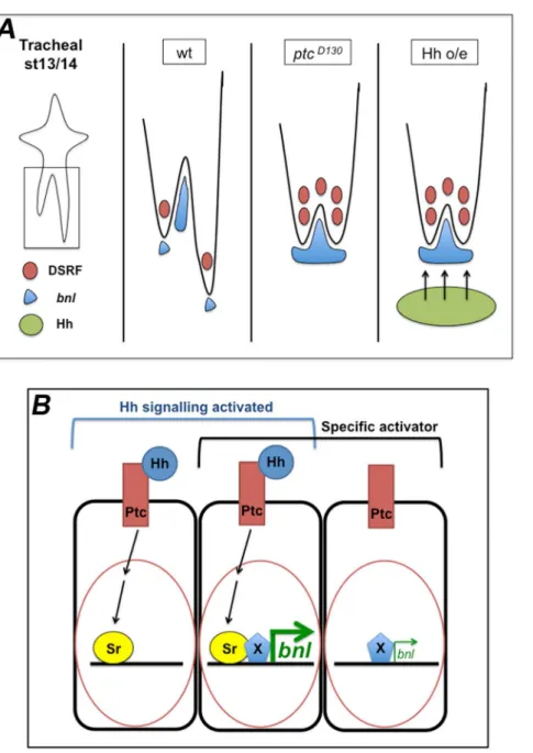
Figure 8. Proposed model of the role of Hh signalling in the regulation ofbnlexpression.(A) Model of changes inbnlexpression dependent on Hh signalling in cells surrounding the tracheal tip-cells. Drawn are the LTa and LTp/GB in the wt,ptcmutant and Hh overexpression scenarios. Both the absence of Ptc activity and overexpression of Hh in cells near the migrating LTp/GB have the same inhibitory effect on GB migration. This is due to higher levels ofbnlexpression induced by the Hh pathway. (B) Proposed model for the activation ofbnlexpression via Sr and a cell/tissue-specific co-activator. When Hh signalling is active, SrB is one of its target genes being expressed inptc-expressing cells. When these cells also express a specificbnlactivator (X),bnlexpression is induced at high levels. In the absence of Sr, X can drivebnlexpression but only at low levels. The activation of the Hh pathway in addition to the expression of specific local activators is responsible for the fast upregulation ofbnlexpression in the tissues surrounding the migrating tracheal cells. According to this model, Sr is involved in the positive regulation ofbnlexpression in a permissive mode.
Methods
D. melanogasterstocks and genetics
The following stocks are described in FlyBase (http://flybase. bio.indiana.edu): Df(2R)ED1742, simGal4, twiGal4, srGAL4, UASsr, 69BGAL4, and UASCD8GFP, hh21, sr155. We also used ptcIIw,ptclacZ,UAS-HhandUAS-Hh-Ptc(from A. Casali, Barcelona, Spain), btlmoeRFP (from M. Affolter, Basel, Switzerland), UASCi (from T. Orenic, Chicago, USA) and Complex2 (from C. Samakovlis, Stockholm, Sweden). Wild-type is yw and ptc refers toptcD130unless otherwise stated. We used the GAL4 system [63] for over or misexpression experiments. inscGal4 (from G.Tear, London, UK) was used as a VNC driver, btlGAL4 (from M. Affolter) as a general tracheal driver,ptcGAL4 (from A. Casali) as a ptc-positive cell driver. Heterozygous embryos were recognized by lacZ and GFP balancer chromosomes. D. melanogasterstocks and crosses were kept under standard conditions at 25uC. All overexpression experiments were conducted at 29uC.
Immunohistochemistry, image acquisition and data analysis
Embryos were staged as described by Campos-Ortega and Hartenstein [64] and stained following standard protocols. For immunostaining, embryos were fixed in 4% formaldehyde for 20-30 minutes. We used antibodies that recognise GFP (Molecular Probes and Roche), RFP (abcam), ßGal (Cappel and Promega), Ptc (from A. Casali), DSRF (Active Motif), Dof (from M. Leptin, Cologne, Germany), Trh (from J. Casanova, Barcelona, Spain), SrB (from T. Volk, Rehovot, Israel) and 2A12 (DSHB). We used HRP, Alexa488, Alexa-555 and Alexa-647, Cy2, Cy3 and Cy5 conjugated secondary antibodies (Jackson ImmunoResearch). For HRP histochemistry, the signal was amplified using the Vectas-tain-ABC kit (Vector Laboratories) when required. In addition, the signal for the DAB reaction was intensified with NiCl2, except for double stainings, where it was omitted from one of the reactions. For tracheal lumen visualisation, we also used fluorescently-conjugated CBP (NEB). In situ hybridisation was performed following standard protocols. ribo-bnl was generated using the whole cDNA as a template and using the Megascript kit (Ambion). Photographs were taken using a Nikon Eclipse 80i microscope. Fluorescent images were obtained with confocal microscopes (Leica TCS-SPE-AOBS and Leica TCS-SP5-AOBS system) and processed using Fiji [65] and Adobe Photoshop (Adobe Systems) softwares. Images are maximum projections of confocal Z-sections. Phenotypes were scored using Nomarski optics on a Nikon Eclipse 80i microscope with a 20x or 40x objective. Cells were counted using Imaris x64 7.6.1 (Bitplane) software.
turer’s instructions. ptc homozygous mutant embryos were separated by means of a GFP balancer. We prepared cDNAs with the RevertAid H Minus First Strand cDNA Synthesis Kit (Fermentas) using random hexamer primers. The nature of the PCR products was confirmed by melting curve analysis. All analyses were performed using the Relative Expression Software Tool (REST) following the user manual and references. The main steps of the automatic REST workflow were as follows: PCR efficiencies were calculated for each pair of primers by generating standard curves at increasing dilutions of cDNA (1:2, 1:4, 1:8, 1:16) and used to correct raw data.rp49was assumed to be equally expressed in wt and mutant/overexpressing embryos and was used as a reference to normalise data. A ratio between the normalised signals of tested genes in mutant and wt embryos was calculated and expressed as fold increase/decrease, and statistically tested by a bootstrap test (10000 randomisations). We used a sample size of 3 repeats per plate, for each embryonic genotype tested, using a set of primers forbnl, and each experiment was repeated twice.
Supporting Information
Figure S1 Different allelic combinations ofptchave the same phenotype and inhibition of apoptosis does not change the GB phenotype. (A–E) Stage 16 wt (A), ptcIIw mutant (B),ptcD130over the deficiency that deletesptc(C),ptcD130 over the deficiency that deletesgrim,reaperandhid(D) andptcD130 heterozygous embryos stained with the tracheal lumen antibody 2A12, using HRP immunohistochemistry for visualization. A B, C, D and E are dorsal views; A9, B9, C9, D9and E9are lateral views and A0, B0, C0, D0 and E0 are ventral views. (F) Sequence comparison between the wild-type andptcD130 mutant; detection of the base difference that leads to an early STOP codon. Represented are nucleotides 1918 to 2067 of theptccDNA. (TIF)
Figure S2 Ptc is expressed in cells surrounding the migrating tracheal branches and inactivation of the Hh pathway does not affect GB migration.(A–F) Different stage embryos expressingptclacZstained with anti-ßgal and anti-Trh. Ptc expression is detected by nuclear bgal presence. Scale bars are 10mm. Panels B, D and F show a single Z-section. (G–I) Ventral views of stage 16 embryos stained with 2A12 to visualize the tracheal lumen.
(TIF)
GB as it migrates towards the VNC. This cell extends long filopodial extensions in response to FGF signalling.
(AVI)
Movie S2 ptc mutant GB migration and generation of filopodial extensions. ptc mutant embryo carrying btlGAL4 and UASCD8GFP constructs to reveal tracheal cell morphology and cytoplasmic extensions. One tip-cell is present at the tip of each GB as it tries to migrate towards the VNC. This cell extends long filopodial extensions in response to FGF signalling, but is not able to advance in the VNC direction.
(AVI)
Tables S1 Supporting tables.Table S1, qRT-PCR Results. Table S2, qRT-PCR Results.
(DOCX)
Acknowledgments
We are very grateful to J. Casanova for his support during this study. Thanks also go to all the members of Casanova’s and Llimargas’ laboratories, M. Llimargas, A. Casali, M. Mila´n, and M. Corominas for many fruitful discussions throughout this study. We thank the Bloomington Stock Center, and M. Affolter, A. Casali, M. Mila´n, T. Orenic, T. Volk and G. Tear for fly stocks and reagents. We thank L. Bardia and A. Llado´ for assistance with confocal microscopy and imaging and Y. Rivera, N. Martin and E. Fuentes for excellent technical support. We are grateful to M. Grillo, G. Lebreton and D. Ricolo for help with some experiments. We thank M. Mila´n, M. Llimargas, M. Furriols, A. Casali, G. Lebreton, M. Corominas and J. Casanova for helpful comments on the manuscript and T. Yates for help with editing.
Author Contributions
Conceived and designed the experiments: SJA. Performed the experiments: EB DM SJA. Analyzed the data: EB SJA. Contributed reagents/materials/ analysis tools: EB SJA. Wrote the paper: SJA.
References
1. Affolter M, Caussinus E (2008) Tracheal branching morphogenesis in Drosophila: new insights into cell behaviour and organ architecture. Develop-ment 135: 2055–2064.
2. Sutherland D, Samakovlis C, Krasnow MA (1996) Branchless encodes a Drosophila FGF homolog that controls tracheal cell migration and the pattern of branching. Cell 87: 1091–1101.
3. Klambt C, Glazer L, Shilo B (1992) FGF receptor homolog, is essential for migration of tracheal and specific midline glial cells. Genes & Development. 4. Lee T, Hacohen N, Krasnow M, Montell DJ (1996) Regulated Breathless
receptor tyrosine kinase activity required to pattern cell migration and branching in the Drosophila tracheal system. Genes Dev 10: 2912–2921.
5. Merabet S, Ebner A, Affolter M (2005) The Drosophila Extradenticle and Homothorax selector proteins control branchless/FGF expression in mesoder-mal bridge-cells. EMBO Rep 6: 762–768.
6. Zhan Y, Maung S, Shao B, Myat MM (2010) The bHLH transcription factor, Hairy, refines the terminal cell fate in the Drosophila embryonic trachea. PLoS ONE 5: e14134.
7. Arau´jo SJ, Casanova J (2011) Sequoia establishes tip-cell number in Drosophila trachea by regulating FGF levels. Journal of Cell Science.
8. Robbins DJ, Fei DL, Riobo NA (2012) The Hedgehog signal transduction network. Science Signaling 5: re6.
9. Ingham PW, Nakano Y, Seger C (2011) Mechanisms and functions of Hedgehog signalling across the metazoa. Nature reviews Genetics.
10. Choy SW, Cheng SH (2012) Hedgehog signaling. Hedgehog Signaling 88: 1–23. 11. Biehs B, Kechris K, Liu S, Kornberg TB (2010) Hedgehog targets in the Drosophila embryo and the mechanisms that generate tissue-specific outputs of Hedgehog signaling. Development 137: 3887–3898.
12. Ruiz i Altaba A (1999) Gli proteins and Hedgehog signaling: development and cancer. Trends Genet 15: 418–425.
13. Lin C, Nozawa YI, Chuang P-T (2012) The path to chemotaxis and transcription is smoothened. Sci Signal 5: pe35.
14. Geisbrecht ER, Sawant K, Su Y, Liu ZC, Silver DL, et al. (2013) Genetic interaction screens identify a role for hedgehog signaling in Drosophila border cell migration. Dev Dyn.
15. Barolo S, Posakony JW (2002) Three habits of highly effective signaling pathways: principles of transcriptional control by developmental cell signaling. Genes & Development 16: 1167–1181.
16. Strutt H, Thomas C, Nakano Y, Stark D, Neave B, et al. (2001) Mutations in the sterol-sensing domain of Patched suggest a role for vesicular trafficking in Smoothened regulation. Curr Biol 11: 608–613.
17. Khaliullina H, Pana´kova´ D, Eugster C, Riedel F, Carvalho M, et al. (2009) Patched regulates Smoothened trafficking using lipoprotein-derived lipids. Development.
18. Briscoe J, Chen Y, Jessell TM, Struhl G (2001) A hedgehog-insensitive form of patched provides evidence for direct long-range morphogen activity of sonic hedgehog in the neural tube. Mol Cell 7: 1279–1291.
19. Caussinus E, Colombelli J, Affolter M (2008) Tip-cell migration controls stalk-cell intercalation during Drosophila tracheal tube elongation. Curr Biol 18: 1727–1734.
20. Samakovlis C, Hacohen N, Manning G, Sutherland DC, Guillemin K, et al. (1996) Development of the Drosophila tracheal system occurs by a series of morphologically distinct but genetically coupled branching events. Development 122: 1395–1407.
21. Duman-Scheel M, Weng L, Xin S, Du W (2002) Hedgehog regulates cell growth and proliferation by inducing Cyclin D and Cyclin E. Nature 417: 299–304. 22. Christiansen AE, Ding T, Fan Y, Graves HK, Herz H-M, et al. (2013) Non-cell
autonomous control of apoptosis by ligand-independent Hedgehog signaling in Drosophila. Cell Death Differ 20: 302–311.
23. Beitel GJ, Krasnow MA (2000) Genetic control of epithelial tube size in the Drosophila tracheal system. Development 127: 3271–3282.
24. Aza-Blanc P, Kornberg TB (1999) Ci: a complex transducer of the hedgehog signal. Trends Genet 15: 458–462.
25. Affolter M, Nellen D, Nussbaumer U, Basler K (1994) Multiple requirements for the receptor serine/threonine kinase thick veins reveal novel functions of TGF beta homologs during Drosophila embryogenesis. Development 120: 3105– 3117.
26. Vincent S, Ruberte E, Grieder NC, Chen CK, Haerry T, et al. (1997) DPP controls tracheal cell migration along the dorsoventral body axis of the Drosophila embryo. Development 124: 2741–2750.
27. Chen CK, Kuhnlein RP, Eulenberg KG, Vincent S, Affolter M, et al. (1998) The transcription factors KNIRPS and KNIRPS RELATED control cell migration and branch morphogenesis during Drosophila tracheal development. Development 125: 4959–4968.
28. Ribeiro C, Ebner A, Affolter M (2002) In vivo imaging reveals different cellular functions for FGF and Dpp signaling in tracheal branching morphogenesis. Dev Cell 2: 677–683.
29. Englund C, Uv AE, Cantera R, Mathies LD, Krasnow MA, et al. (1999) adrift, a novel bnl-induced Drosophila gene, required for tracheal pathfinding into the CNS. Development 126: 1505–1514.
30. Affolter M, Montagne J, Walldorf U, Groppe J, Kloter U, et al. (1994) The Drosophila SRF homolog is expressed in a subset of tracheal cells and maps within a genomic region required for tracheal development. Development 120: 743–753.
31. Guillemin K, Groppe J, Ducker K, Treisman R, Hafen E, et al. (1996) The pruned gene encodes the Drosophila serum response factor and regulates cytoplasmic outgrowth during terminal branching of the tracheal system. Development 122: 1353–1362.
32. Nussbaumer U, Halder G, Groppe J, Affolter M, Montagne J (2000) Expression of the blistered/DSRF gene is controlled by different morphogens during Drosophila trachea and wing development. Mech Dev 96: 27–36.
33. Gervais L, Casanova J (2011) The Drosophila homologue of SRF acts as a boosting mechanism to sustain FGF-induced terminal branching in the tracheal system. Development 138: 1269–1274.
34. Vincent S, Wilson R, Coelho C, Affolter M, Leptin M (1998) The Drosophila protein Dof is specifically required for FGF signaling. Mol Cell 2: 515–525. 35. Imam F, Sutherland D, Huang W, Krasnow M (1999) stumps, a Drosophila
gene required for Fibroblast Growth Factor (FGF)-directed Migrations of Tracheal and Mesodermal Cells. Genetics.
36. Hooper JE, Scott MP (1989) The Drosophila patched gene encodes a putative membrane protein required for segmental patterning. Cell 59: 751–765. 37. Hidalgo A, Ingham P (1990) Cell patterning in the Drosophila segment: spatial
regulation of the segment polarity gene patched. Development 110: 291–301. 38. Torroja C, Gorfinkiel N, Guerrero I (2004) Patched controls the Hedgehog
gradient by endocytosis in a dynamin-dependent manner, but this internaliza-tion does not play a major role in signal transducinternaliza-tion. Development 131: 2395– 2408.
39. Glazer L, Shilo BZ (2001) Hedgehog signaling patterns the tracheal branches. Development 128: 1599–1606.
40. Casali A, Struhl G (2004) Reading the Hedgehog morphogen gradient by measuring the ratio of bound to unbound Patched protein. Nature 431: 76–80. 41. Chen Y, Struhl G (1996) Dual roles for patched in sequestering and transducing
Hedgehog. Cell 87: 553–563.
and patterning of the developing limb bud. Cell 79: 993–1003.
50. Pepicelli CV, Lewis PM, McMahon AP (1998) Sonic hedgehog regulates branching morphogenesis in the mammalian lung. Curr Biol 8: 1083–1086. 51. Zu´n˜iga A HA, McMahon AP, Zeller R (1999) Signal relay by BMP antagonism
controls the SHH/FGF4 feedback loop in vertebrate limb buds. Nature 401: 598–602.
52. Miyake A NY, Konishi M, Itoh N. (2005) Fgf19 regulated by Hh signaling is required for zebrafish forebrain development. Dev Biol 288: 259–275. 53. Blaess S CJ, Joyner AL. (2006) Sonic hedgehog regulates Gli activator and
repressor functions with spatial and temporal precision in the mid/hindbrain region. Development 133: 1799–1809.
54. Barrett A, Krueger S, Datta S (2008) Branchless and Hedgehog operate in a positive feedback loop to regulate the initiation of neuroblast division in the Drosophila larval brain. Developmental Biology 317: 234–245.
62. Fahmy RG, Dass CR, Sun L-Q, Chesterman CN, Khachigian LM (2003) Transcription factor Egr-1 supports FGF-dependent angiogenesis during neovascularization and tumor growth. Nat Med 9: 1026–1032.
63. Brand AH, Perrimon N (1993) Targeted gene expression as a means of altering cell fates and generating dominant phenotypes. Development 118: 401–415. 64. Campos-Ortega AJ, Hartenstein V (1985) The Embryonic Development of
Drosophila Melanogaster. Springer-Verlag New York: 10–84.