Mtp-40 and Alpha Antigen Gene Fragment Amplification for the
Detection of
Mycobacterium tuberculosis
in Colombian
Clinical Specimens
Rosalba Alfonso
+, Rosa Elena Romero, Manuel Elkin Patarroyo, Luis Angel Murillo
Departamento de Biología Molecular, Fundación Instituto de Inmunología de Colombia, Universidad Nacional de Colombia, Carrera 50 # 26-00 Bogotá, D. C., Colombia
In this study, the use of Mtp-40 and alpha antigen polymerase chain reaction (PCR) amplification fragments for the precise tuberculosis (TB) diagnosis was evaluated. One hundred and ninety two different samples were obtained from 113 patients with suspected TB. Mtp-40 and alpha antigen protein genes were amplified by the PCR technique and compared to both the “gold standard” (culture) test, as well as the clinical parameters (including a clinical record and X-ray film exam in 113 patients). Thirty-eight of the 113 patients had a presumptive clinical diagnosis of TB; 74% being detected by PCR technique, 58% by culture and 44% by direct microscopic visualization. We conclude that it is possible to use PCR as a suitable technique for the detection of any mycobacteria by means of the alpha antigen product, or the specific infection of Mycobacterium tuberculosis by means of the mtp-40 gene. This might be a good supporting tool in difficult clinical TB diagnosis and pauci-bacillary cases.
Key words:Mycobacterium tuberculosis - alpha antigen - Mtp-40 - polymerase chain reaction
Despite the advances in the handling of infectious diseases and the availability of an effective short course of chemotherapy (DOTS) and the administration of a BCG vaccine, the tubercle bacillus continues to cause more deaths than any other single infectious agent throughout the world (Snider et al. 1994).
In recent years, there has been an increased incidence of tuberculosis (TB) in both developing and industrial-ized countries, as well as an increase of drug-resistant strains and synergy with the human immunodeficiency virus (HIV). In Colombia the incidence of TB is 19.5 cases for every 100,000 people. A total of 13,000 cases of TB were recorded in 1999 alone (Ministerio de Salud de Co-lombia 1999). The World Health Organization (WHO) estimates that by the year 2020, nearly one billion people will be newly infected, 200 million people will become sick and 70 million will die from TB if existing controls are not strengthened (WHO/OMS 1998).
Even though a presumptive diagnosis of pulmonary TB is usually established on a clinical basis (the patient’s radiological and medical record), a definitive TB case is established by two traditional methods: microscopic ex-amination of acid-fast stained smears and culturing in syn-thetic media. Although Ziehl-Neelsen staining is a rapid and inexpensive method, it lacks sensitivity and can only detect 10,000 organisms/ml or greater in a sample. The “gold standard” diagnosis for TB is the culturing of My-cobacterium tuberculosis. However, it takes a long time to obtain a result in the laboratory, thereby delaying
clini-+Corresponding author. Fax: +57-1-4815269. Ext. 108. E-mail: [email protected]
Received 15 February 2002 Accepted 21 August 2002
cal and therapeutic decisions (De Wit et al. 1990). In past years, several molecular biology-based techniques have been developed to improve the sensitivity, specificity and speed for M. tuberculosis detection. For example, poly-merase chain reaction (PCR) has been used for M. tuber-culosis diagnosis in different tests and has been shown to be as sensitive as or better than the gold standard test (Del Portillo et al. 1991, Manjunath et al. 1991, De Wit et al.
1992). The PCR technique involves amplifying different genetic elements used as templates within mycobacteria, such as DNA, rRNA (Jonas et al. 1993) or single copy genes encoding 65 or 38 kDa structural proteins (Kaneko et al. 1990, Sjobring et al. 1990, Brisson-Noël et al. 1991). The PCR technique is usually used to amplify variable insertion element sequences, for example IS6110 (De Wit et al. 1990, Eisenach et al. 1990, Thierry et al. 1990). Our group decided to focus its PCR approach on using alpha antigen, abundantly secreted into the culture superna-tant of a variety of mycobacterial species (Borremans et al. 1989), and the M. tuberculosis species-specific Mtp-40 fragment. The alpha antigen, also called MPB59 belongs to the 85 complex. This complex is codified by three ho-mologous but distinct structural genes (Ag85A, Ag85B, Ag85C), displaying genetic diversity in species of slow and fast growing mycobacteria (Wiker et al. 1990, Kitaura et al. 1993). The species-specific Mtp-40 gene, identified by our group, has been previously used in several stud-ies as a target element for identification of M. tuberculo-sis (Del Portillo et al. 1991, Herrera & Segovia 1996, Faizal et al. 1996, Del Portillo et al. 1996, Marchetti et al. 1998). This fragment encodes a 14 kDa protein belonging to the phospholipase C family and hybridises only to M. tuber-culosis DNA but not to DNA from other mycobacterial species, including M. tuberculosis complex species (Parra et al. 1991). Recent studies have shown that the Mtp-40 fragment was not always present in all M. tuberculosis
In the present study, we have evaluated the suitability of the PCR technique in different samples from a group of 113 Colombian patients, amplifying both the Mtp-40 and the alpha antigen genes. The second target gene was used for screening patients who presented TB symptoms; the Mtp-40 gene was used exclusively for specific M. tuber-culosis identification. When positive results were found by both Mtp-40 and alpha antigen in the same sample, the presence M. tuberculosis was inferred. A result which was positive for the alpha antigen but negative for Mtp-40, strongly suggests that any mycobacteria, different from
M. tuberculosis, was being detected. Nevertheless, since some rare M. tuberculosis clinical isolates lack the DNA fragment encoding the Mtp-40, the presence of one of these isolates in the sample cannot be conclusively dis-carded.
MATERIALS AND METHODS
Clinical group - The clinical records of a group of 113 patients, ranging from 3 months to 82 years old, with sus-pected TB were studied retrospectively. The group was composed of in-patient and out-patient cases, variously attending Hospital Santa Clara, Clínica Colsubsidio and Hospital San Juan de Dios, all located in Bogotá, Colom-bia, between October 1996 and October 1998. Fifty-three percent (60/113) of the patients presented one or various risk factors for TB disease, such as a previous contact history with a diseased patient (23 cases), an HIV posi-tive test (12 cases), a previous tuberculous disease (8 cases), malnutrition (7 cases), diabetes (5 cases), drug abuse (4 cases) and Addison’s disease (1 patient). The data was double-checked and non-matching laboratory and clinical information was revised and corrected. Con-tingency tables were drawn up in order to evaluate PCR sensitivity and specificity in comparison with the “gold standard” technique for detection of M. tuberculosis. IBM compatible computers, using statistical software (EpiInfo version 6.04, CDC and Stata 5.0 for Windows, 1997), were used for data analysis.
Clinical samples -A total of 192 samples including sputum,bronchoalveolar lavage (BAL), urine, gastric as-pirates, biopsy tissues, pericardic fluid, cerebrospinal fluid (CSF), ascitic fluid and pleural liquid, were obtained from these 113 patients. Samples were taken from patients at the time of a suspected TB diagnosis, with the exception of two patients who were being treated for a period of two months without any clinical improvement. Two contain-ers were used for each sample, one for staining and cul-turing and the other for chromosomal DNA isolation.
Staining and culturing -All samples were previously de-contaminated according to the Petroff technique (1915); sediment was then suspended in 310 ml sterile distilled water. Ziehl-Neelsen (ZN) staining required 10 µl of the suspension and another 100 µl were inoculated into each of three slope tubes (Lowenstein Jensen, Stonebrink-G and Middlebrook 7H10) and incubated at 37°C for 4-8 wks. The sterile samples (i.e. CSF, pericardic fluid) were pro-cessed without the de-contamination step.
Chromosomal DNA isolation - Fluid samples: a 500 µl aliquot was centrifuged for 10 min at 14,000 rpm and the pellet resuspended in 10X TE (1X TE = 10 mM Tris-HCl
pH 8.0, plus 1 mM EDTA pH 8.0). Each sample was incu-bated at 37°C with vigorous shaking for 1.5 h in the pres-ence of lysozyme (2 mg/ml). The bacterial and cell mem-branes were lysed by increasing the temperature to 65°C and adding sodium dodecyl sulphate and proteinase K (Sigma, St. Louis, Mo.) to final concentrations of 1% and 250 µg/ml, respectively. After 1.5 h incubation, the DNA was extracted with chloroform-isoamyl alcohol mix (24:1) and later precipitated with 0.6 volume of isopropanol. The pellet was re-suspended in 1X TE; sputum samples: one volume of sterile distilled water was added to 200 µl ali-quot of sputum and incubated at 95°C for 10 min. The sample was centrifuged at 14,000 rpm for 10 min and the pellet suspended in 10X TE. The DNA was isolated by the method described above; biopsies: each sample was fragmented using a razor-blade, suspended in 10X TE and processed according to the method already described for fluid samples.
PCR primers -Two sets of primers were used for PCR amplification. Generic primers MT1 (5’-TTCCTGA CCAGCGAGCTGCCG-3’) and MT2 (5’- CCCCAGTACT CCCAGCTGTGC-3’) were designed to amplify the alpha antigen present in all reported mycobacteria (Matsuo et al. 1990, 1998, Kitaura et al. 1993, Ohara et al. 1993). Spe-cies-specific primers PT1 (5’-CGGCAACGCGCCG TCGGTGG-3’) and PT2 (5’- CCCCCCACGGCACC GCCGGG-3’), derived from our previously described M. tuberculosis Mtp-40 gene fragment, were also designed (Del Portillo et al. 1991). All primers were synthesised by the solid-phase phosphite triester method on a Gene As-sembler (Pharmacia, Piscataway, New Jersey, USA) and purified in a 20% polyacrylamide gel (Maniatis et al. 1989).
PCR amplification -Ten micro-litres from the sus-pended DNA were used for PCR under standard condi-tions. All reactions were adjusted to a final 50 µl volume, containing: 1X reaction buffer (10 mM Tris-HCl (pH 8.3), 50 mM KCl, 1.5 mM MgCl2 and 0.001% gelatin), 1 U Taq
DNApolymerase, 0.1 mM of each deoxynucleoside triph-osphate and 0.4 µM of each primer. A Perkin Elmer thermocycler (Perkin Elmer Cetus) was programmed for 25 amplification cycles as follows: (i) denaturation at 94°C for 1 min and (ii) annealing and extension at 74°C for 2 min for PT primers or 70°C for 1 min for MT primers. The posi-tive PCR control was 100 ng DNA from M. tuberculosis
H37Rv (ATCC 27294) and sterile distilled water was used as the negative PCR control. One positive and one nega-tive sputum sample were also included as controls for each procedure.
(ICN Pharmaceuticals Inc.) by the Rediprime technique (Amersham International).
Hybridization was done in a solution consisting of 20 ml for each 100 cm2 membrane containing 1X SSC, 5X Denhardt’s (5X: 0.5 g Ficoll Type 400, 0.5 g Polyvinylpyr-rolidone, 0.5 g BSA Pentax Fraction V and H2O to 500 ml), 0.5% SDS and 100 µg/ml salmon sperm DNA at 65°C. Af-ter hybridization, membranes were washed twice in 1X SSC and 0.1% SDS for 15 min at 65°C. They were washed twice again in 0.5 X SSC and 0.1% SDS for 15 min at 62°C. Finally, they were washed twice in 0.1 X SSC and SDS 0.1% for 15 min at 60°C. They were then exposed on Kodak X-Omat film, for 15-18 h at -70°C.
RESULTS
One hundred and thirteen suspected TB patients (hav-ing cough and expectoration, fever, weight-loss, anorexia, adenopathies and other suggestive clinical signs and symptoms according to the clinical criteria of the bian Ministry of Health) (Ministerio de Salud de Colom-bia 1995), were enrolled in the present study. For statisti-cal analysis of TB distribution, and taking into consider-ation that 70% of the Colombian populconsider-ation is already infected by some type of mycobacteria, patients were di-vided into 4 age groups: group 1: 0-15 years (59.3%); group 2: 16-30 years (14.2%); group 3: 31-45 years (14.2%) and group 4: 45-86 years (12.4%). One hundred and ninety two samples (40 sputum, 12 BAL, 7 urine, 102 gastric aspi-rates, 7 biopsy, 2 pericardic fluid, 18 CSF, 1 ascitic fluid and 3 pleural effusions) were analyzed by conventional methods and by PCR. Gastric aspirates corresponded to more than 50% of the total, due to the high frequency of pediatric patients in the present study.
In case of multiple specimens taken from a single pa-tient, the result was considered as being positive if at least one of the specimens was positive. However, there were no discrepancies noted between multiple specimens in terms of their smear, PCR, or culture results. As with any diagnostic test, results should be interpreted within the context of clinical findings.
Having the clinical findings and/or response to therapy, 38 patients were shown to be TB positive by clinical diag-nosis. Of them, 27 (71%) had pulmonary TB disease and 11 (29%) developed extra-pulmonary TB. The distribu-tion of TB by age groups was as follows: 12 cases in group 1; 7 in group 2; 9 in group 3; and 10 in group 4. Also, 22 of 29 patients with pulmonary, milliary and pleu-ral TB presented thoracic X-ray study compatible with a TB disease, according to the radiological parameters re-ported by the Colombian Ministry of Health (Ministerio de Salud de Colombia 1995). PCR amplification and dot-blot hybridization, using both Mtp-40 and alpha antigen genes as targets, were performed on every sample ana-lyzed. All positive PCR experiments showed hybridiza-tion of both Mtp-40 and alpha antigen genes, suggesting that only M. tuberculosis was present in the specimens analyzed. However, M. tuberculosis co-infection with other mycobacteriacannot be discarded from our results; there have only been two reports of such a case in the literature (Massenkeil 1992, Fernandez & Chavez 1994), therefore, this interpretation of our results should not be
taken as the first choice. Among patients with a positive clinical diagnosis of TB, 15 were found positive by PCR, culture and bacilloscopy tests; 7 were found positive us-ing the PCR and culture tests; 2 were bacilloscopy posi-tive with negaposi-tive PCR and culture; 6 were only posiposi-tive by PCR and 8 patients presented negative results by all laboratory methods (Table I). The last group was diag-nosed as being tuberculous by clinical findings and had risk factors predisposing them to the disease (3 had a positive HIV test, 2 had a history of previous tuberculo-sis diagnotuberculo-sis, 2 had a history of TB contact and 1 patient was of advanced age).
The Dot Blot hybridization for some samples from dif-ferent patients and controls, using the radiolabeled Mtp-40 PCR fragment as a probe (Fig. 1). Similar results were obtained for alpha antigen (data not shown). Southern blot analysis for alpha antigen and Mtp40-amplified frag-ments from the same clinical samples (Fig. 2). PCR sensi-tivity, expressed as a percentage (people having the dis-ease detected by PCR/total number of people tested hav-ing the disease), was 100% [95% C.I. (Confidence Inter-val)] and its specificity expressed as a percentage (people without the disease who were negative to the screening test/total number of people tested who did not have the disease) was 93.4% (95% C.I.). Positive test PCR predic-tive value (people having posipredic-tive diagnosis and posipredic-tive test/total people with positive test) was 78.6% and tive test predictive value (people with diagnosis and nega-tive test/total people with neganega-tive test) was 100% (Table II).
Fig. 1: Dot blot hybridizationof Mycobacterium tuberculosis Mtp-40 specie-specific fragment, in some clinical samples and controls. 1-3: samples positive culture, bacilloscopy and PCR; 4-6: samples with culture and polymerase chain reaction (PCR) positives; 7-9: samples only with PCR positive; 10-11: samples with all negative laboratory methods; 12: negative chromosomal DNA extraction and PCR controls; 13: PCR Positive control (M. tuberculosis H37Rv DNA). (Dot blot hybridization for the alpha antigen fragment was similar).
DISCUSSION
dis-ease. Until now, molecular biology techniques have of-fered advantages when compared to traditional methods for TB diagnosis.
In the present study, the distribution of TB cases was slightly similar in the different age groups. The majority of the patients having a final diagnosis of TB presented risk factors, which predisposed them to the disease. It is known that infants and children, who are exposed to adults with pulmonary TB, comprise a group of individuals hav-ing high infection risk. Other high-risk groups include people with human immunodeficiency virus (HIV) infec-tion, drug abusers, low-income populations, those held in correctional facilities and people with specific medical risk factors such as diabetes mellitus, chronic renal failure and immunosuppressive disorders, as described in other
studies (Committee on Infectious Diseases 1994). Eight patients with no TB diagnosis by laboratory test presented risk factors predisposing them to the disease. These patients received anti-mycobacterial treatment and their clinical status improved. Two of the patients (who tested positive by ZN staining and negative for the other detection methods) presented a recurrence of TB after abandoning chemotherapy. It has been reported elsewhere that patients having pulmonary TB may show positive smears with negative sputum cultures at the end of the therapy (WHO 1994-1997, Al-Moamary et al. 1999). Since the introduction of rifampicin, the problem of positive smear with negative culture results has been due to po-tent anti-microbial activity, but the exact mechanism still remains unclear ( Kaneko et al. 1990, Pierre et al. 1991).
TABLE I
Results of three different detection methods for Mycobacterium tuberculosis diagnosis compared with clinical standards No. X-ray Samples ZN Culture PCR TB clinical
study stain diagnosis
1 Nd 1 Urine + + + Urinary
2 Com 1 BAL + + + Milliary
3 Com 3 Sputum, 3 GL + + + Pulmonary
4 Com 2 Sputum + + + Pulmonary
5 Com 1 Sputum, 3 BAL + + + Pulmonary 6 No Com 3 Sputum, 3GL + + + Pulmonary
7 Nd 1 AA + + + Peritoneal
8 No Com 1 RF + + + Pericardic
9 Com 1 RF + + + Pericardic
10 Com 3 GL + + + Milliary
11 Com 1 Sputum + + + Pulmonary
12 Com 1 Sputum + + + Pulmonary
13 Com 1 Sputum + + + Pulmonary
14 Com 1 Sputum + + + Pulmonary
15 Com 1 Sputum + + + Milliary
16 Com 1 PF, 1 PB - + + Pleural
17 Nd 1 CSF - + + Meningitis
18 Com 2 BB - + + Spinal
19 Com 1 Sputum - + + Pulmonary
20 Com 1 Sputum - + + Pulmonary
21 No Com 1 Sputum - + + Pulmonary
22 Com 1 Sputum - + + Pulmonary
23 Com 2 GL - - + Pulmonary
24 Com 3 GL - - + Pulmonary
25 Com 1 Sputum - - + Pulmonary
26 Com 1 Sputum - - + Pulmonary
27 Com 1 Sputum - - + Pleural
28 Com 1 Sputum - - + Pulmonary
29 Com 1 Sputum + - - Pulmonary
30 Com 1 Sputum + - - Milliary
31 No Com 3 GL, 1 BAL - - - Pulmonary
32 No Com 3 GL, 1 BAL - - - Pulmonary
33 RUS com 1 Urine - - - Urinary
34 No Com 3 GL - - - Pulmonary
35 No Com 3 GL, 1 BAL - - - Contact children
36 Nd 1 CSF - - - Meningitis
37 Com 1 CSF - - - Meningitis
38 No Com 1 Sputum - - - Pulmonary
Our aforementioned two patients also presented a negative PCR test for both Mtp-40 and alpha antigen. Although PCR inhibition by anti-tuberculosis drugs has not been reported to date, several inhibitory factors, in-cluding antibiotics, have been described in a standard PCR procedure (Wilson 1997). This fact, in addition to the ongoing rifampicin treatment that these two patients were following suggests that this antibiotic might be respon-sible for the PCR negative results. Consequently, PCR was not a useful tool for TB diagnosis in the case of these two patients who had had various months of chemo-therapy, since false negative results could have been present in the culture and PCR in spite of having had a positive bacilloscopy.
Culturing is considered to be the “gold standard” for confirming a TB diagnosis. However, it is expensive, time-consuming, the results are only available 4-6 weeks later and it is not very sensitive in pauci-bacillary cases. Tak-ing culture as a reference test for confirmTak-ing TB diagno-sis in this study, PCR sensitivity and specificity was 100% and 93.4% respectively. By this method, we were able to detect M. tuberculosis in 74% (28/38) of the cases, being positive to both the mtp-40 and the alpha antigen. This
percentage was significantly higher than the results ob-tained from culture and bacilloscopy: 58% (22/38) and 44% (17/38) respectively.
Among the main problems found in routine PCR use as a diagnostic technique is cross-contamination, this can be overcome with the adequate selection of internal con-trols for both reagents and sample during the set-up pro-cess. In spite of the technical difficulties inherent in the methodology and its relatively high price, the cost-ben-efit balance needs to be addressed, since the improve-ment in pauci-bacillary detection is clearly enhanced. In our study, PCR was able to detect 15.8% (6/28) cases more than the culture; this last group corresponding to patients with a difficult diagnose, from whom it was not possible to take good quality samples and who had low bacillary charge (children and elderly people).
The pulmonary form of TB is usually presented (71% of cases) and constitutes an epidemiological risk in the population, given the greater dissemination of the bacilli. In this study, group 1 (infants and children) was consid-erably more important because it represents more than half of the patients where difficult diagnosis occurs due to pauci-bacillary infections and because most of them have a previous history of TB contacts. Our data corre-lates with previous reports, showing that M. tuberculosis
causes the highest incidence of both pulmonary and ex-tra pulmonary TB cases in Colombia (Ministerio de Salud de Colombia 1999).
Previous studies using Mtp-40 in combination with other target genes have shown that its amplification con-fers a good sensitivity and specificity in the detection of
M. tuberculosis in clinical samples (Herrera et al. 1996b, Wei et al. 1999). Another commonly used target gene tested is the insertion sequence IS6110, present in the M. tuber-culosis complex. This method lacks the M. tuberculosis
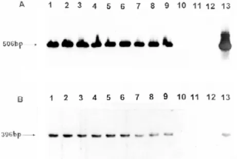
strain-specific detection capacity and, because of its de-Fig 2:Southern blot hybridization patterns for the same samples shown in Fig. 1, using radiolabeled generic alpha antigen gene (A) and Mtp-40 (B) fragments.
TABLE II
Polymerase chain reaction (PCR) sensitivity and specificity Method Culture (+) Culture (-) Total
PCR (+) 22 6 28
PCR (-) 0 85 85
Total 22 91 113
letion in some M. tuberculosis strains, the demonstration of these mycobacteria cannot always be achieved (Yuen et al. 1993, Agasino et al. 1998).
In this study, the utility of two independent amplifica-tions, recognising the presence of M. tuberculosis and related mycobacterial infections in clinical samples, is shown. The combination of these genes increases the TB diagnostic spectrum and confirms that M. tuberculosis is responsible for the infection when both the alpha antigen and Mtp-40 tests are positive. If PCR for alpha antigen is positive and negative for Mtp-40, then the presence of M. tuberculosis cannot be discarded. According to our re-sults, all those clinical isolates, positive for M. tuberculo-sis, analysed in this study contain the Mtp-40 gene.
ACKNOWLEDGMENTS
To Santiago López and Myriam Neira for analyzing the statistics. To Diana Tovar and Martha Silvera for their technical assistance. To Hospital Santa Clara, Clínica Colsubsidio and the Hospital San Juan de Dios for providing the clinical samples from those patients used in this study. To Manuel Alfonso Patarroyo at our Institute for kindly and patiently reviewing this manuscript.
REFERENCES
Agasino CB, Ponce de Leon A, Jasmer RM, Small PM 1998. Epidemiology of Mycobacterium tuberculosis strains in San Francisco that do not contain IS6110. Int J Tuberc Lung Dis2: 518-520.
Al-Moamary MS, Black W, Bessuille E, Elwood RK, Vedal S 1999. The significance of the persistent presence of acid-fast bacilli in sputum smears in pulmonary tuberculosis.
Chest116: 726-731.
Borremans M, De Wit L, Volckaert G, Ooms J, De Bruyn J, Huygen C, Van Vooren JP, Steladre M, Verhofstadt R, Con-tent J 1989. Cloning, sequence determination and expres-sion of a 32-kilodalton-protein gene of Mycobacterium tu-berculosis. Infect Immun57: 3123-3130.
Brisson-Noël A, Aznar C, Chureau C, Nguyen S, Pierre C, Bartoli M, Bonete R, Pialoux G, Gicquel B, Garrigue G 1991. Diagnosis of tuberculosis by DNA amplification in clinical practice evaluation. Lancet 338: 364-366. Committee on Infectious Diseases 1994. Screening for
tubercu-losis in infants and children. Pediatrics 93: 131-134. De Wit D, Maartens G, Steyn L 1992. A comparative study of
the polymerase chain reaction and conventional procedures for the diagnosis of tuberculous pleural effusion. Tuber Lung Dis 73: 262-267.
De Wit D, Steyn L, Shoemaker S, Sogin M 1990. Direct detec-tion of Mycobacterium tuberculosis in clinical specimens by DNA amplification. J Clin Microbiol28: 2437-2441. Del Portillo P, Murillo LA, Patarroyo ME 1991. Amplification
of a species-specific DNA fragment of Mycobacterium tu-berculosis and its possible use in diagnosis. J Clin Microbiol 29: 2163-2168.
Del Portillo P, Thomas MC, Martínez E, Marañon C, Valladares B, Patarroyo ME, López MC 1996. Multiprimer PCR sys-tem for differential identification of Mycobacteria in clini-cal samples J Clin Microbiol34: 324-328.
Eisenach KD, Cave MD, Bates JH, Crawford JT 1990. Poly-merase chain reaction amplification of a repetitive sequence specific for Mycobacterium tuberculosis.J Infect Dis 161: 977-981.
Faizal M, Jimenez G, Burgos C, Del Portillo P, Romero RE, Patarroyo ME 1996. Diagnosis of cutaneous tuberculosis
by polymerase chain reaction using a species-specific gene.
Int J Derm35: 185-188.
Fernandez DF, Chavez F 1994. Mycobacterium scrofulaceum
and Mycobacterium tuberculosis coinfection in an AIDS patient. Med Clin (Barc) 102: 799.
Herrera EA, Segovia M 1996. Evaluation of mtp-40 genomic fragment amplification for specific detection of Mycobacte-rium tuberculosis in clinical specimens. J Clin Microbiol 34: 1108-1113.
Herrera EA, Perez O, Segovia M 1996. Differentiation between
Mycobacterium tuberculosis and Mycobacterium bovis by a multiplex-polymerase chain reaction. J Appl Bacteriol80: 596-604.
Jonas V, Alden MJ, Curry JI, Kamisango K, Knott CA, Lankford R, Wolfe JM, More DF 1993. Detection and identification of Mycobacterium tuberculosis directly from sputum sedi-ments by amplification of rRNA. J Clin Microbiol31: 2410-2416.
Kaneko K, Ondera O, Miyatake T, Tsuji S 1990. Rapid diagno-sis of tuberculous meningitis by polymerase chain reaction.
Neurology40: 1617-1618.
Kitaura H, Ohara N, Matsuo K, Tasaka H, Kobayashi K, Yamada T 1993. Cloning, sequencing and expresion of the gene for the alpha antigen from M. intracellulare and use of PCR for the rapid identification of M. intracellulare.
Biochem Biophys Res Comun169: 1466-1473.
Maniatis T, Fritsch EF, Sambrook J 1989. Molecular Cloning: a Laboratory Manual, 2nd ed., Cold Spring Harbor Labora-tory, Cold Spring Harbor, NY, p. 11.23-11.28.
Manjunath N, Shankar P, Rajan L, Bhargava A, Saluja S, Shriniwas 1991. Evaluation of a polymerase chain reaction for the diagnosis of tuberculosis. Tubercle72: 21-27. Marchetti G, Gori A, Catozzi L, Vago L, Nebuloni M, Rossi
MC, Esposti AD, Ban A, Franzetti F 1998. Evaluation of PCR in detection of Mycobacterium tuberculosis from for-malin-fixed, paraffin-embedded tissues: comparison of four amplification assays. J Clin Microbiol36: 1512-1517. Massenkeil G, Opravil M, Salfinger M, von Graevenitz A,
Luthy R 1992. Disseminated coinfection with Mycobacte-rium avium complex and Mycobacterium kansdasii in a patient with AIDS and liver abscess. Clin Infect Dis 14: 618-619.
Matsuo K, Yamaguchi R, Yamasaki A, Tasaka H, Terasaka K, Yamada T 1990. Cloning and expression of the gene for the cross-reactive alpha antigen of Mycobacterium kansasii. Infect Immun58: 550-556.
Matsuo K, Yamaguchi R, Yamasaki A, Tasaka H, Yamada T 1998. Cloning and expression of the Mycobacterium bovis
BCG gene for extracellular alpha antigen. J Bacteriol170: 3847-3854.
Ministerio de Salud de Colombia 1995. Tuberculosis, Prevención y Control.Manual de Normas, Bogotá, p. 55-56. Ministerio de Salud de Colombia 1999. Epidemiología de la
tuberculosis en Colombia. Bol Epidemiol4: 81-92. Ohara N, Matsuo K, Yamaguchi R, Yamasaki A, Tasaka H,
Yamada T 1993. Cloning and sequencing of the gene for alpha antigen from M. avium and mapping of B cell epitopes.
Infect Immun61: 1173-1179.
Parra CA, Londoño LP, Del Portillo P, Patarroyo ME 1991. Isolation, characterization and molecular cloning of a spe-cific Mycobacterium tuberculosis antigen gene. Identifica-tion of a species-specific sequence. Infect Immun59: 3411-3417.
Petroff SA 1915. A new and rapid method for isolation and cultivation of tubercle bacilli directly from the sputum and feces. J Exp Med21: 38-42.
Hance AJ 1991. Use of reamplification protocol improves the sensitivity of detection of Mycobacterium tuberculosis
in clinical samples by amplification of DNA. J Clin Microbiol 29: 712-717.
Sjobring U, Mecklenburg M, Andersen AB, Miorner H 1990. Polymerase chain reaction for detection of Mycobacterium tuberculosis. J Clin Microbiol28: 2200-2204.
Snider Jr DE, Raviglione M, Kochi AM 1994. Tuberculosis: Pathogenesis, Protection and Control, Bloom BR, Am Soc Microbiol, Washington D.C., p. 2-11.
Thierry D, Brisson-Noel A, Vincent-Levy-Frebault V, Nguyen S, Guesdon JL, Gicquel B 1990. Characterization of a My-cobacterium tuberculosis insertion sequence, IS6110 and its application in diagnosis. J Clin Microbiol28: 2668-2673. Vera-Cabrera L, Howard ST, Laszlo A, Johnson WM 1997. Analysis of genetic polymorphism in the phospholipase region of Mycobacterium tuberculosis. J Clin Microbiol35: 1190-1195.
Wei CY, Lee CN, Chu CH, Hwang JJ, Lee CP 1999. Determina-tion of the sensitivity and specificity of PCR assay using
different target DNAs for the detection of Mycobacterium tuberculosis. Kao Hsiung I Hsueh Ko Hsueh Tsa Chih15: 396-405.
Weil A, Plikaytis BB, Butler WR, Wodley CL, Shinnick TM 1996. The mtp-40 gene is not present in all strains of Myco-bacterium tuberculosis. J Clin Microbiol34: 2309-2311. Wiker HG, Sletten K, Nagai S, Harboe M 1990. Evidence of
three separate genes encoding the proteins of the Mycobac-terial Antigen 85 Complex. Infect Immun 58: 272-274. Wilson IG 1997. Inhibition and facilitation of nucleid acid
am-plification. Appl Environ Microbiol 63: 3741-3751. WHO-Worl Health Organization 1994-1997. Anti-tuberculosis
drug resistance in the world. The WHO/IUATLD Global Projet on Anti-tuberculosis Drug Resistance Surveillance, Global Tuberculosis Programme, Geneva.
WHO/OMS 1998. Tuberculosis. Fact sheet104: 1-5. Yuen LK, Ross BC, Jackson KM, Dwyer B 1993.