Assessment of genetic mutations in the
XRCC2
coding region by high
resolution melting curve analysis and the risk of differentiated thyroid
carcinoma in Iran
Shima Fayaz
1,2*, Pezhman Fard-Esfahani
2,*, Armaghan Fard-Esfahani
3, Ehsan Mostafavi
4, Reza Meshkani
1,
Hossein Mirmiranpour
1and Shahnaz Khaghani
11
Department of Biochemistry, Faculty of Medicine, Tehran University of Medical Sciences, Tehran, Iran.
2Department of Biochemistry, Pasteur Institute of Iran, Tehran, Iran.
3
Research Institute for Nuclear Medicine, Tehran University of Medical Sciences, Tehran, Iran.
4Department of Epidemiology, Pasteur Institute of Iran, Tehran, Iran.
Abstract
Homologous recombination (HR) is the major pathway for repairing double strand breaks (DSBs) in eukaryotes and XRCC2 is an essential component of the HR repair machinery. To evaluate the potential role of mutations in gene re-pair by HR in individuals susceptible to differentiated thyroid carcinoma (DTC) we used high resolution melting (HRM) analysis, a recently introduced method for detecting mutations, to examine the entireXRCC2 coding region in an Iranian population. HRM analysis was used to screen for mutations in threeXRCC2 coding regions in 50 patients and 50 controls. There was no variation in the HRM curves obtained from the analysis of exons 1 and 2 in the case and control groups. In exon 3, an Arg188
His polymorphism (rs3218536) was detected as a new melting curve group (OR: 1.46; 95%CI: 0.432-4.969; p = 0.38) compared with the normal melting curve. We also found a new Ser150
Arg polymorphism in exon 3 of the control group. These findings suggest that genetic variations in theXRCC2 coding re-gion have no potential effects on susceptibility to DTC. However, further studies with larger populations are required to confirm this conclusion.
Key words:DNA repair, gene polymorphism, mutation analysis.
Received: June 12, 2011; Accepted: October 5, 2011.
Introduction
Thyroid cancer is the most common endocrine malig-nancy and its incidence has increased in recent years (Leenhardtet al., 2004; Davies and Welch, 2006; Jemalet al., 2008). Differentiated thyroid carcinoma (DTC) is the most common type of all thyroid carcinomas (accounting for ~90% of all cases) and consists of papillary, follicular and Hürthle cell carcinomas, the latter being a subtype of follicular thyroid carcinoma (Hundahlet al., 1998). Risk factors associated with DTC include exposure to various carcinogenic agents, ethnicity and dietary habits, although exposure to ionizing radiation is still the only recognized risk factor (Ronet al., 1995; Xionget al., 2005; Prestonet al., 2007).
Recent studies have proposed that genetic variation in conserved DNA repair systems may influence susceptibil-ity to cancer (Gatzidouet al., 2010). These systems
nor-mally ensure the genetic intactness of cell populations such that any alteration in the genes related to these systems could lead to a defect in DNA repair pathways and ulti-mately affect the cellular genetic stability and susceptibility to cancer. In severe DNA damage, such as double-strand breaks (DSBs), there are two important recombination sys-tems for cell survival, namely, homologous recombination (HR) and non-homologous end joining (NHEJ) (Paques and Haber, 1999; Peterson and Cote, 2004).
In eukaryotes, HR is the major pathway for DSB re-pair and has an important role in preventing mutations, chromosomal instability and cancer; these functions make HR essential for cell viability and genomic stability (Jack-son, 2002; Thompson and Schild, 2002). The HR repair system functions primarily during the S and G2 phases of the cell cycle. Since HR is an error-free pathway of damage tolerance that allows the replication bypass of lesions dur-ing a template switch it has a distinct advantage over NHEJ (Jackson, 2002; Thompson and Schild, 2002).
TheRAD51gene family is the key component of HR and its impairment can lead to extreme sensitivity to certain DNA damaging agents, intense genomic instability and a
Send correspondence to Shahnaz Khaghani. School of Medicine, Tehran University of Medical Sciences & Health Services, Poursina ST, PO Box 14155-6447 Tehran, Iran. E-mail: [email protected].
risk of cancer (Sonodaet al., 1998; Cuiet al., 1999; Deans et al., 2000, 2003; Takataet al., 2001; Gruveret al., 2005; Sirajet al., 2008). TheRAD51-like gene family in somatic mammalian cells is composed of XRCC2, XRCC3, RAD51L1, RAD51L2 and RAD51L3 (Thacker, 1999). There are indications that XRCC2 has an important role in enhancing the action of RAD51, with a loss of XRCC2 de-laying the early steps of HR, including the integration of RAD51 at the site of DNA damage, nucleoprotein filament formation and strand invasion (Sugawaraet al., 2003; Wol-ner et al., 2003). In XRCC2-deficient cells, RAD51 re-sponses are reduced by approximately five-fold, and this has led toXRCC2being recognized as a repair response-enhancing factor (Tambiniet al., 2010).
XRCC2is a 29,668 kb gene located on human chromo-some 7q36.1 and consists of three exons (1 to 3) that contain 38, 82 and 722 bp, respectively. XRCC2 protein is highly conserved among mammalian species (Tambini et al., 2010). Several studies have examined the association be-tween genetic polymorphisms inXRCC2and different can-cers (Hanet al., 2004; Jiaoet al., 2008; Curtinet al., 2009). In the present report, we describe the first case-control study of an Iranian population to examine the association between mutations in the entire coding region of theXRCC2gene and individual susceptibility to DTC based on high resolution melting (HRM) analysis, a recently introduced method for detecting mutations (Montgomeryet al., 2010).
Materials and Methods
Study subjects
A sample size calculation (Kasiulevicious et al., 2006) indicated that the minimum sample size for this case-control study was ~45 for each group (controls and cases). In addition, the minimum odds ratio (OR) for signif-icance was 2.5 (p < 0.05). The probability of having a mu-tant allele in theXRCC2coding region in control individu-als was estimated as 0.35. Based on these preliminary calculations, our study population consisted of 50 patients with histopathologically confirmed DTC and 50 controls. Informed consent was obtained from all participants before commencement of the study and the study was approved by the ethics committee of Tehran University of Medical Sci-ence. The DTC patients were recruited from the Research Institute for Nuclear Medicine of Shariati Hospital, Tehran, Iran. The control population was matched for age (£50 and > 50 year) and sex, with no previous or concurrent malig-nant disease. The controls were recruited from volunteers at two academic centers in Tehran. Individuals with a history of other cancers, radiation exposure, alcohol consumption or smoking were excluded from the study.
DNA extraction
5 mL of peripheral blood was collected from each subject into tubes containing 1 mL of EDTA (1 g/dL) and
stored at -20 °C until used. Whole blood DNA was ex-tracted by a salting out procedure (Milleret al., 1988).
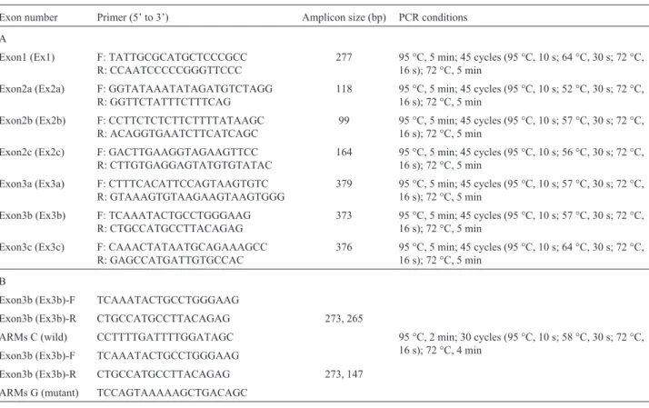
Primer design and assay conditions for PCR-HRM
One primer pair for exon 1 (Ex1), three primer pairs for exon 2 (Ex2) and three primer pairs for exon 3 (Ex3) were used in this work (Table 1). PCR-HRM was done in a Rotor-Gene 6000 real-time rotary analyzer (Corbett Life Sciences). The reactions were prepared in 10mL volumes in 0.1 mL strip tubes of a 72-well rotor. For symmetric PCR-HRM, the amount of DNA in each reaction was ad-justed to 50 ng. Each reaction contained 5mL of 2 x HRM PCR Master Mix (Type-it HRM PCR kit, QIAGEN), 0.7mL (10mM) of primer mix, 1.5mL (50 ng) of DNA and DNA RNase-free water to a final volume of 10mL.
The PCR cycling profile is summarized in Table 1. After amplification, HRM analysis data were collected from 65 °C to 85 °C, with each step raised by 0.05 °C, fol-lowed by a waiting time of 10 s. Samples from the top, mid-dle and bottom of each melting curve group and suspicious melting curves were sequenced. Any new mutation de-tected by PCR-HRM was screened for in the other samples by using the Allele Refractory Mutation System (ARMS). The ARMS primers and fragment lengths are listed in Ta-ble 1.
Statistical analysis
Hardy-Weinberg equilibrium of theXRCC2alleles in the control population was assessed using the Chi square test (c2). The homogeneity of age distribution between the controls and cases was assessed with an independent sam-plet-test and Levene’s test. Allelic and genotypic frequen-cies were compared across groups using the Chi square test. The odds ratio (OR) and the corresponding 95% confidence intervals (CIs) between DTC and a detected polymorphism were calculated using logistic regression. All analyses were done with SPSS v13 software.
Results
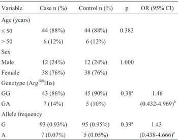
This study included 50 DTC patients and 50 can-cer-free controls. The two groups were matched for sex and age (£50 and > 50 years). The mean age (±SEM) in the cases and controls was 38.4±2.1 and 36.2±1.2 years, re-spectively. Age and sex were not significantly different be-tween the two groups. The general characteristics of both groups are shown in Table 2.
HRM analysis results
a single nucleotide polymorphism (SNP) in the Ex3b frag-ment, the samples in this group were subjected to DNA se-quencing and a previously reported polymorphism (Arg188His, rs3218536) was detected in all of them. No His/His alleles were found in the population. Table 2
sum-marizes the distributions of the allelic and genotypic frequencies in the case and control groups.
Another suspicious melting curve was identified in the Ex3b fragment and DNA sequencing confirmed a novel missense mutation (AGC®AGG, Ser150Arg) in one
sam-Table 1- (A) Primers for theXRCC2coding regions and the PCR conditions used before HRM analysis. (B) Primers used in PCR-ARMS.
Exon number Primer (5’ to 3’) Amplicon size (bp) PCR conditions
A
Exon1 (Ex1) F: TATTGCGCATGCTCCCGCC R: CCAATCCCCCGGGTTCCC
277 95 °C, 5 min; 45 cycles (95 °C, 10 s; 64 °C, 30 s; 72 °C, 16 s); 72 °C, 5 min
Exon2a (Ex2a) F: GGTATAAATATAGATGTCTAGG R: GGTTCTATTTCTTTCAG
118 95 °C, 5 min; 45 cycles (95 °C, 10 s; 52 °C, 30 s; 72 °C, 16 s); 72 °C, 5 min
Exon2b (Ex2b) F: CCTTCTCTCTTCTTTTATAAGC R: ACAGGTGAATCTTCATCAGC
99 95 °C, 5 min; 45 cycles (95 °C, 10 s; 57 °C, 30 s; 72 °C, 16 s); 72 °C, 5 min
Exon2c (Ex2c) F: GACTTGAAGGTAGAAGTTCC R: CTTGTGAGGAGTATGTGTATAC
164 95 °C, 5 min; 45 cycles (95 °C, 10 s; 56 °C, 30 s; 72 °C, 16 s); 72 °C, 5 min
Exon3a (Ex3a) F: CTTTCACATTCCAGTAAGTGTC R: GTAAAGTGTAAGAAGTAAGTGGG
379 95 °C, 5 min; 45 cycles (95 °C, 10 s; 57 °C, 30 s; 72 °C, 16 s); 72 °C, 5 min
Exon3b (Ex3b) F: TCAAATACTGCCTGGGAAG R: CTGCCATGCCTTACAGAG
373 95 °C, 5 min; 45 cycles (95 °C, 10 s; 57 °C, 30 s; 72 °C, 16 s); 72 °C, 5 min
Exon3c (Ex3c) F: CAAACTATAATGCAGAAAGCC R: GAGCCATGATTGTGCCAC
376 95 °C, 5 min; 45 cycles (95 °C, 10 s; 64 °C, 30 s; 72 °C, 16 s); 72 °C, 5 min
B
Exon3b (Ex3b)-F TCAAATACTGCCTGGGAAG
Exon3b (Ex3b)-R CTGCCATGCCTTACAGAG 273, 265
ARMs C (wild) CCTTTTGATTTTGGATAGC 95 °C, 2 min; 30 cycles (95 °C, 10 s; 58 °C, 30 s; 72 °C, 16 s); 72 °C, 4 min
Exon3b (Ex3b)-F TCAAATACTGCCTGGGAAG
Exon3b (Ex3b)-R CTGCCATGCCTTACAGAG 273, 147
ARMs G (mutant) TCCAGTAAAAAGCTGACAGC
F- forward, R- reverse.
Figure 1- (A) HRM curves for exon 3b (Ex3b) of theXRCC2gene in patients and controls. The Arg188His genotype (red curves) was detected as a new melting curve group in a few patients and controls compared with the normal melting curves obtained for the wild type (Arg188) genotype (blue curves).
ple of the control group. No mutation was detected in the other samples screened for this mutation using ARMS.
The difference between the Arg188His and Arg188 genotypic frequencies was not significant and there was no significant association between DTC and the G and A allelic frequencies (Table 2).
Discussion
In this study, we used HRM analysis to screen for ge-netic mutations in the entire coding region of theXRCC2 gene and examined the association between these muta-tions and DTC in a subpopulation from Tehran, Iran. HRM analysis is an innovative technique for genotyping that is based on DNA melting analysis such that any alteration in genotype leads to variation in the HRM curves when com-pared to the wild type (Montgomeryet al., 2010). To our knowledge, this is the first time that the entire coding re-gion of the XRCC2 gene has been analyzed. Two polymorphisms, CGC®CAC (Arg188His, rs3218536) and
AGC®AGG (Ser150Arg), were identified in Ex3b of the XRCC2gene. To detect the Arg188His polymorphism we distinguished between wild homozygote (G::C) and hetero-zygote (A::T, G::C) samples based on a discernible melting transition (Figure 1A).
There was no significant association between DTC and the Arg188His polymorphism and its genotypic fre-quency in the individuals studied (Table 2). Bastoset al. (2009) and Garcia-Quispeset al.(2011) also found no sig-nificant involvement of the XRCC2 Arg188His polymor-phism in thyroid cancer in 109 (OR: 0.8, 95%CI: 0.4-1.6) and 402 (OR: 1.12, 95%CI: 0.80-1.59) patients, respec-tively. There are currently no additional data on the rela-tionship between theXRCC2Arg188His polymorphism and thyroid cancer.
A recent meta-analysis by Yuet al.(2010) reported that there was no evidence of a significant association be-tweenXRCC2Arg188His and the risk of breast cancer in any genetic model. A meta-analysis of three case-control stud-ies in the United Kingdom and a family-based study in the United States found no association between a putatively functional Arg188His SNP and colorectal cancer (Curtinet al., 2009). Similarly, a meta-analysis of 16 studies com-piled by the International Ovarian Cancer Association Con-sortium showed there was also no association between Arg188His and the risk of ovarian cancer (Pearce et al., 2009).However, theXRCC2Arg188His polymorphism may be a genetic modifier for smoking-related pancreatic cancer (Jiaoet al., 2008).
Alignment of the protein sequence of human XRCC2 with that of other species revealed an Arg188His substitution inGallus gallusandPongo abelii(Figure 2). Furthermore, Arg188is not present at the active site of three XRCC2 pro-teins. This finding suggests that the Arg188His polymor-phism generated in homologous recombination repair pathway may not have severe effects on XRCC2 function and may not influence the susceptibility to cancer in hu-mans.
We detected a novel missense mutation (AGC ®
AGG, Ser150Arg) in theXRCC2gene in one sample of the control group. In this G::C conversion, bases switched strands but the GC content was conserved and the melting transitions of heterozygote, wild and variant homozygotes
Table 2- General characteristics of the case and control groups and the allelic and genotypic (Arg188His) frequencies of
XRCC2.
Variable Case n (%) Control n (%) p OR (95% CI)
Age (years)
£50 44 (88%) 44 (88%) 0.383
> 50 6 (12%) 6 (12%)
Sex
Male 12 (24%) 12 (24%) 1.000
Female 38 (76%) 38 (76%)
Genotype (Arg188His)
GG 43 (86%) 45 (90%) 0.38ª 1.46
GA 7 (14%) 5 (10%) (0.432-4.969)b
Allele frequency
G 93 (0.93%) 95 (0.95%) 0.39ª 1.43
A 7 (0.07%) 5 (0.05%) (0.438-4.666)c
ªOne tailed chi-squared analysis comparing genotype distributions and allelic frequencies between cases and controls.
b
OR (95%CI) GA to GG genotype (Arg188His) ofXRCC2for casevs. con-trol groups.
cOR (95%CI) A to G allele (Arg188His) ofXRCC2for casevs.control
groups.
were not clearly distinguishable (Cai et al., 2010). We therefore used PCR-ARMS to detect this SNP in other sam-ples. Ser150, which is involved in the formation of the ATP binding site in XRCC2, is highly conserved among species (Figure 2). Consequently, an Arg substitution at this posi-tion (replacing a neutral amino acid by a positively charged one) would most probably affect the formation of the ATP binding site. However, protein expression studies are needed to confirm this hypothesis.
The major limitation of this study is the sample size, which may be due to lesser probability of detecting any rare mutations in smaller sample size. On the other hand, for de-tected SNP, the larger sample size would make the results more significant. In this regard, the coexistence of other members of theRad51gene family with an impaired func-tion alongside Arg188His should be considered since these proteins are also involved in the HR repair system. For ex-ample, Bastoset al.(2009) showed that the coexistence of three or more variant alleles in theRAD51 and XRCC3 genes was associated with a significantly higher risk of DTC (OR = 2.9, p = 0.036; four variant alleles: adjusted OR = 8.0, p = 0.006), while no associations were observed for polymorphisms inXRCC3alone (Bastoset al., 2009; Garcia-Quispeset al., 2011).
In conclusion, our findings indicate that there is no as-sociation between polymorphisms in theXRCC2coding re-gion and the risk of DTC. However, further studies with a larger population are needed to confirm this conclusion. In addition, it would be prudent for other members of the Rad51gene family also to be screened for SNPs in their coding region. Finally, HRM analysis was found to be very useful for genotyping, although it was unable to distinguish between G::C and A::T conversions because of the low melting curve transition. Other complementary methods are needed to overcome this limitation.
Acknowledgments
This research was supported by Tehran University of Medical Sciences & Health Services (grant no. 89-01-30-10489). We thank the Department of Biochemistry, Pasteur Institute of Iran for contribution in study design and HRM experiments. We also thank Dr. Anis Jafari and Mr. Naser Nejadi for editing the manuscript.
References
Bastos HN, Antao MR, Silva SN, Azevedo AP, Manita I, Teixeira V, Pina JE, Gil OM, Ferreira TC, Limbert E,et al.(2009) Association of polymorphisms in genes of the homologous recombination DNA repair pathway and thyroid cancer risk. Thyroid 19:1067-1075.
Cai Y, Yuan Y, Lin Q and Chan P (2010) Allele-specific exten-sion allows base-pair neutral homozygotes to be discrimi-nated by high-resolution melting of small amplicons. Anal Biochem 406:29-33.
Cui X, Brenneman M, Meyne J, Oshimura M, Goodwin EH and Chen DJ (1999) The XRCC2 and XRCC3 repair genes are required for chromosome stability in mammalian cells. Mutat Res 434:75-88.
Curtin K, Lin WY, George R, Katory M, Shorto J, Cannon-Albright LA, Smith G, Bishop DT, Cox A and Camp NJ (2009) Genetic variants in XRCC2: New insights into colo-rectal cancer tumorigenesis. Cancer Epidemiol Biomarkers Prev 18:2476-2484.
Davies L and Welch HG (2006) Increasing incidence of thyroid cancer in the United States, 1973-2002. JAMA 295:2164-2167.
Deans B, Griffin CS, Maconochie M and Thacker J (2000) Xrcc2 is required for genetic stability, embryonic neurogenesis and viability in mice. EMBO J 19:6675-6685.
Deans B, Griffin CS, O’Regan P, Jasin M and Thacker J (2003) Homologous recombination deficiency leads to profound genetic instability in cells derived from Xrcc2-knockout mice. Cancer Res 63:8181-8187.
Garcia-Quispes WA, Perez-Machado G, Akdi A, Pastor S, Galo-fre P, Biarnes F, Castell J, Velazquez A and Marcos R (2011) Association studies of OGG1, XRCC1, XRCC2 and XRCC3 polymorphisms with differentiated thyroid cancer. Mutat Res 709-710:67-72.
Gatzidou E, Michailidi C, Tseleni-Balafouta S and Theocharis S (2010) An epitome of DNA repair related genes and mecha-nisms in thyroid carcinoma. Cancer Lett 290:139-147. Gruver AM, Miller KA, Rajesh C, Smiraldo PG, Kaliyaperumal
S, Balder R, Stiles KM, Albala JS and Pittman DL (2005) The ATPase motif in RAD51D is required for resistance to DNA interstrand crosslinking agents and interaction with RAD51C. Mutagenesis 20:433-440.
Han J, Hankinson SE, Hunter DJ and De Vivo I (2004) Genetic variations in XRCC2 and XRCC3 are not associated with endometrial cancer risk. Cancer Epidemiol Biomarkers Prev 13:330-331.
Hundahl SA, Fleming ID, Fremgen AM and Menck HR (1998) A National Cancer Data Base report on 53,856 cases of thyroid carcinoma treated in the U.S., 1985-1995. Cancer 83:2638-2648.
Jackson SP (2002) Sensing and repairing DNA double-strand breaks. Carcinogenesis 23:687-696.
Jemal A, Siegel R, Ward E, Hao Y, Xu J, Murray T and Thun MJ (2008) Cancer statistics, 2008. CA Cancer J Clin 58:71-96. Jiao L, Hassan MM, Bondy ML, Wolff RA, Evans DB,
Abbru-zzese JL and Li D (2008) XRCC2 and XRCC3 gene poly-morphism and risk of pancreatic cancer. Am J Gastroenterol 103:360-367.
Kasiulevicious V, Sapoka V and Filipaviciute R (2006) Sample size calculation in epidemiological studies. Gerontologija 7:225-231.
Leenhardt L, Grosclaude P and Cherie-Challine L (2004) In-creased incidence of thyroid carcinoma in France: A true ep-idemic or thyroid nodule management effects? Report from the French Thyroid Cancer Committee. Thyroid 14:1056-1060.
Montgomery JL, Sanford LN and Wittwer CT (2010) High-resolution DNA melting analysis in clinical research and di-agnostics. Expert Rev Mol Diagn 10:219-240.
Paques F and Haber JE (1999) Multiple pathways of recombina-tion induced by double-strand breaks in Saccharomyces cerevisiae. Microbiol Mol Biol Rev 63:349-404.
Pearce CL, Near AM, Van Den Berg DJ, Ramus SJ, Gentry-Maharaj A, Menon U, Gayther SA, Anderson AR, Edlund CK, Wu AH,et al.(2009) Validating genetic risk associa-tions for ovarian cancer through the International Ovarian Cancer Association Consortium. Br J Cancer 100:412-420.
Peterson CL and Cote J (2004) Cellular machineries for chromo-somal DNA repair. Genes Dev 18:602-616.
Preston DL, Ron E, Tokuoka S, Funamoto S, Nishi N, Soda M, Mabuchi K and Kodama K (2007) Solid cancer incidence in atomic bomb survivors: 1958-1998. Radiat Res 168:1-64.
Ron E, Lubin JH, Shore RE, Mabuchi K, Modan B, Pottern LM, Schneider AB, Tucker MA and Boice Jr. JD (1995) Thyroid cancer after exposure to external radiation: A pooled analy-sis of seven studies. Radiat Res 141:259-277.
Siraj AK, Al-Rasheed M, Ibrahim M, Siddiqui K, Al-Dayel F, Al-Sanea O, Uddin S and Al-Kuraya K (2008) RAD52 polymorphisms contribute to the development of papillary thyroid cancer susceptibility in Middle Eastern population. J Endocrinol Invest 31:893-899.
Sonoda E, Sasaki MS, Buerstedde JM, Bezzubova O, Shinohara A, Ogawa H, Takata M, Yamaguchi-Iwai Y and Takeda S (1998) Rad51-deficient vertebrate cells accumulate chromo-somal breaks prior to cell death. EMBO J 17:598-608.
Sugawara N, Wang X and Haber JE (2003)In vivoroles of Rad52, Rad54, and Rad55 proteins in Rad51-mediated recombina-tion. Mol Cell 12:209-219.
Takata M, Sasaki MS, Tachiiri S, Fukushima T, Sonoda E, Schild D, Thompson LH and Takeda S (2001) Chromosome insta-bility and defective recombinational repair in knockout mu-tants of the five Rad51 paralogs. Mol Cell Biol 21:2858-2866.
Tambini CE, Spink KG, Ross CJ, Hill MA and Thacker J (2010) The importance of XRCC2 in RAD51-related DNA damage repair. DNA Repair (Amst) 9:517-525.
Thacker J (1999) A surfeit of RAD51-like genes? Trends Genet 15:166-168.
Thompson LH and Schild D (2002) Recombinational DNA repair and human disease. Mutat Res 509:49-78.
Wolner B, van Komen S, Sung P and Peterson CL (2003) Recruit-ment of the recombinational repair machinery to a DNA double-strand break in yeast. Mol Cell 12:221-232. Xiong P, Zheng R, Wang LE, Bondy ML, Shen H, Borer MM,
Wei Q and Sturgis EM (2005) A pilot case-control study of gamma-radiation sensitivity and risk of papillary thyroid cancer. Thyroid 15:94-99.
Yu KD, Chen AX, Qiu LX, Fan L, Yang C and Shao ZM (2010) XRCC2 Arg188His polymorphism is not directly associated with breast cancer risk: Evidence from 37,369 subjects. Breast Cancer Res Treat 123:219-225.
Associate Editor: Jeremy A. Squire