Supported in part by Consejo de Desarrollo Científico y Humanístico. Universidad de los Andes. Mérida, Venezuela. Grant M-1011-11-07-B. (1) Instituto de Investigaciones Cardiovasculares.
(2) Instituto de Inmunologia Clinica. Universidad de Los Andes. Mérida, Venezuela.
MUSCARINIC ANTIBODIES AND HEART RATE RESPONSES TO DYNAMIC EXERCISE AND TO THE
VALSALVA MANEUVER IN CHRONIC CHAGASIC PATIENTS
Barbara C. DAS NEVES(1), Mey Lyn BACILIO(1), Lisbeth BERRUETA(2), Siham SALMEN(2), Darrell L. PETERSON(3), Jose H. DONIS(1), Tulio J. NUÑEZ(1) & Diego F. DAVILA(1)
SUMMARY
We have studied the cardiac chronotropic responses to the Valsalva maneuver and to dynamic exercise of twenty chronic chagasic patients with normal left ventricular function and no segmental wall abnormalities by two-dimensional echocardiogram. The absolute
increase in heart rate of the patients (∆ = 21.5 ± 10 bpm, M±SD) during the maneuver was significantly diminished when compared to
controls (∆ = 31.30 ± 70, M±SD, p = 0.03). The minimum heart rate (58.24 ± 8.90 vs. 62.80 ± 10, p = 0.68) and the absolute decrease in heart rate at the end of the maneuver (∆ = 38.30 ± 13 vs. ∆ = 31.47 ± 17, p = 0.10) were not different from controls. The initial heart rate acceleration during dynamic exercise (∆ = 12 ± 7.55 vs. ∆ = 19 ± 7.27, M±SD, p = 0.01) was also diminished, but the heart rate
recovery during the first ten seconds was more prominent in the sero-positive patients (Median: 14, Interquartile range: (9.75-17.50
vs. 5(0-8.75, p = 0.001). The serum levels of muscarinic cardiac auto-antibodies were significantly higher in the chagasic patients
(Median: 34.58, Interquartile Range: 17-46.5, Optical Density) than in controls (Median: 0, Interquartile Range: 0-22.25, p = 0.001)
and correlated significantly and directly (r = 0.68, p = 0.002) with early heart rate recovery during dynamic exercise. The results of this investigation indirectly suggest that, the cardiac muscarinic auto-antibodies may have positive agonist effects on parasympathetic heart rate control of chagasic patients.
KEYWORDS: Chagas disease; Muscarinic receptors; Cardiac muscarinic auto-antibodies; Parasympathetic; Dynamic exercise; Valsalva Maneuver.
INTRODUCTION
The natural history of Chagas’ disease is characterized by an acute and chronic phase. The latter includes an indeterminate and a cardiac form31.
Although the responsible mechanisms are yet to be determined, Chagas heart disease appears to evolve from localized myocardial damage to a clinical form of congestive cardiomyopathy with extensive myocardial
damage10,13,39. A cardiac autoimmune response,aimed at the cardiac
muscarinic receptors, and several other hypotheses have been postulated in order to explain the progression of myocardial damage4,28,29,33,34,36.
A cardiac autoimmune response arises in chagasic patients because of antigenic mimicry between the parasite and cardiac muscarinic receptors32.
The second extracellular (o2) and the third intracellular loops (i3), of these receptors, are considered as autoimmune epitopes in patients with
chronic Chagas’ disease22,40. The autoimmune response occurs early on
the natural history of the disease and it is considered to be responsible for the abnormalities of parasympathetic control of heart rate14,21,23,41-44. These
investigations have demonstrated that, the chronotropic responses to cardiac
autonomic tests are apparently impaired in the indeterminate form of the disease. However, we7-9,20,21,37 and other investigators16 have found that
chagasic patients who are in different stages of natural history of the disease
may have normal, abnormal2 or even enhanced responses to conventional
cardiac autonomic tests15,27. Moreover, the frequency and time domain
indexes of parasympathetic modulation may be suppressed in the supine position, but become similar to controls in the standing position and while performing isometric exercise6,24,27,45. Since, muscarinic auto-antibodies
may behave as positive allosteric modulators of parasympathetic activity25;
a diminished high frequency component of heart rate variability41 and the
presence of a slow heart rate44 could be an indirect expression of the agonist
effect. Thus, an alternative explanation for the “impaired” chronotropic response to the cardiac autonomic tests would be over stimulation with saturation of the parasympathetic system3,11,12,42. Indeed, as stated by
chronotropic responses to the Valsalva maneuver and to dynamic exercise and correlated them with the serum levels of cardiac muscarinic auto-antibodies, of chagasic patients who were in the indeterminate form of the disease.
METHODS
We studied twenty asymptomatic patients with at least two positive serologic tests for Chagas’ disease. The tests used for the serologic diagnosis of Chagas disease were: 1. Test ELISA Chagas III. Grupobios S.A. (Santiago de Chile). 2. CruziElisa. Diagnostico y Genetica (Diagen), (Mérida, Venezuela) 3. Chagas AB Rapid. Standard Diagnostics, Inc. (Yongin-si, Korea). All patients had clinical examinations, surface EKG’s, Chest x-rays and two-dimensional echocardiograms. Patients came from western Venezuela (States of Mérida, Táchira and Zulia). They had no other co-morbidities (i.e., Cardiac, Pulmonary or Metabolic diseases) and none of them were receiving other medication. Eighteen sero-negative subjects from the same geographical area and matched by age and EKG abnormalities, were included as a control group. On the day of the study, patients were in the post absorptive state and had not ingested coffee or smoked cigarettes for the last 12-24 hours. All procedures and blood tests were performed in the same environment (room temperature 20-24 °C) and at the same time of the day. The Valsalva maneuver and the dynamic exercise test were performed on separate days. Patients and sero-negative individuals were informed about the purpose of the investigation and signed an informed consent. This protocol was approved by the Commission for Clinical Investigations of the Instituto de Investigaciones Cardiovasculares of the University of Los Andes in Mérida, Venezuela.
Valsalva maneuver protocol: All patients practiced the maneuver in the sitting position two or three times in order to become familiar with it. Following a normal intake of breath, patients were instructed to blow into a handmade mouth piece connected to a mercury manometer and maintain a pressure of 40 mm Hg for 20 seconds. Lead II of the surface EKG was used to continuously record the heart rate changes prior to, during and after recovery from the maneuver. The following criteria were used to assess the validity of the maneuver: facial plethora and jugular vein distension. The resting heart rate was obtained by averaging the R-R intervals during three minutes prior to the definitive maneuver. The maximum tachycardia (shortest R-R interval while performing the maneuver) and maximum bradycardia (longest R-R interval at the end of the maneuver) were used to calculate the Valsalva index. The absolute increase in heart rate was obtained by subtracting the shortest R-R interval during the maneuver, from the longest R-R interval prior to the maneuver. The absolute decrease in heart rate was obtained by subtracting the shortest R-R interval during the maneuver, from the longest R-R following the maneuver19,26. The data were stored in a hard
disk and played back for analysis. Data are expressed as beats per minute.
Dynamic exercise protocol: The heart rate changes during the early
stages30,35 and recovery phases of dynamic exercise were assessed by
performing a maximal standard stress test5. Heart rate prior to, during and
after exercise was stored in a hard disk and played back for analysis. The resting heart rate was obtained, in the standing position, by averaging the R-R intervals during one minute prior to exercise. The initial heart rate acceleration (first 10 seconds) was obtained by subtracting the shortest R-R interval, at this moment of exercise, from the longest R-R recorded prior to exercise. The heart recovery at 10 seconds, and at one and two minutes following exercise, was obtained by subtracting the shortest R-R interval
at peak exercise (maximum heart rate) from the longest R-R intervals at 10 seconds, first minute and second minute following cessation of exercise18.
Cloning and expression of loop2 M2 Muscarinic receptor: The loop2 fragment was obtained by PCR from human cDNA M2 muscarinic receptor (MAR 020000020, www.cdna.org.). The following primers were used: Forward [5’-GGCCATGGTTCATTGTAGGGGTGAGAACTGTG-3’] engineered to contain the NcoI restrictionsite while retaining the start codon, and a reverse primer [5’-CCAAGCTTAAATAGCCGTACCAA
AGGTGACAGC-3’], that was designedto contain a stop codon and the
HindIII restriction site. Following amplification of the fragment using
Pfu Turbo DNA polymerase (Stratagene), the amplified fragment was
digested with NcoI and HindIII and ligated to pET30a. Transformed
E. coli (BL21) were selected on kanamycin (25 µg/mL) and screened for insert and sequenced to verify the correct clone. Both pET30a expression
vector (Novagen, Inc., Madison, Wis.), which containedan N-terminal
hexahistidine tag, plus the thrombin sequence followed by loop2 sequence, and vector alone (only with thrombin sequence), were purified
and used to transform E. coli BL21-CodonPlus-RIPL, then expanded
and induced for protein synthesis with 0.5 mM of IPTG over the course of three hours, afterwards the proteins were purified using Sepharose Ni/nitrilotriacetic acid (NTA) column (BioRad). The presence of 6xHis-thrombin-loop2 and 6xH-thrombin was monitored by electrophoresis and Western blot.
Enzyme-linked immunosorbent assay (ELISA): ELISA was performed according to standard protocols. Briefly, 96 flat well bottomed polystyrene plates were coated overnight with recombinant proteins (thrombin-loop2 and thrombin) previously resuspended at 10 mg/mL in carbonate buffer pH 9.6. To avoid background interference, antigen-coated plates were blocked with PBS containing 1% BSA and 0.1% Tween 20. Sera were obtained from 20 chagasic patients and 18 healthy individuals. Triplicates from each sample were used in 1:1000 dilution in order to improve specificity of the antigen-antibody reaction, and were added to the antigen-bound surface and incubated at room temperature for one hour. After extensive washing, the plates were subsequently incubated for one hour with anti-human IgG horseradish peroxidase-conjugated antibodies (1:5,000) (Santa Cruz, Biotechnology, CA) and thoroughly washed. The read out of the reaction was achieved by color development obtained by incubation with the substrate o-phenylenediamine (Sigma-Aldrich, St. Louis, MO) and measured at 490 nm in a microplate reader (BioTek ELx800; Cole-Parmer). In order to further improve specificity, optical densities read out generated from thrombin alone were subtracted from the optical density signal generated by His-trom-loop. Absorbance values were expressed in milliunits of optical density (mU OD) to
multiply by 1000 times the absorbance values17. A ROC curve analysis
will be elaborated using lectures of optical densities for antibodies against muscarinic receptors obtained from all analyzed samples vs. serological tests for Chagas disease per each patient.
Statistical methods: Data are expressed as absolute numbers and percentages. The one sample Kolmogorov-Smirnov and the Shapiro-Wilk tests were used to analyze for an normal or abnormal distribution of the data. Continuous normally distributed variables are expressed as mean ± standard deviation and abnormally distributed variables as median and interquartile range (IQRs) (25th and 75th percentiles). Categorical variables
variables and the Mann-Whitney test for abnormally distributed variables. Correlation analysis of the heart rate responses and muscarinic cardiac auto-antibodies of chagasic was performed by Spearman test. Statistical significance was considered as p < 0.05.
RESULTS
Clinical and echocardiographic characteristics: We have studied twenty sero-positive patients who were in the indeterminate (n = 13) and cardiac (n = 7) stages of Chagas’ disease. Mean age was 46 ± 13.29. Males predominated (62.50%). Chest x-rays and surface electrocardiograms were normal except for the presence of right bundle branch block, in 35% of patients and in 38% of sero-negative controls. Left ventricular function and wall motion, as assessed by two dimensional echocardiograms, were normal in all sero-positive patients. The serum levels of muscarinic cardiac auto-antibodies were significantly higher in the chagasic patients (Median: 34.58, IQR 17- 46.50 (mU OD)) as compared to controls
(Median: 0, IQR 0-22.25 p = 0.001).
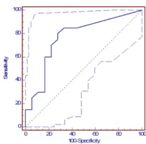
A cut-off value of 11 (mU OD) was determined from the Receptor-Operator-Curve (Serology for Chagas’ disease vs. Serum levels of muscarinic auto-antibodies). The area under the curve was 0.768 with a sensitivity of 80% (Confidence interval 56-94)) and a specificity of 72.20% (Confidence intervals 46-90). Thus, five sero-negative subjects (27.80%) had serum values above the cut-off value and four sero-positive patients (20%) had serum values below the cut-off point (Fig. 1).
Heart rate responses to the Valsalva maneuver: The Valsalva maneuver was performed successfully on only seventeen of the twenty patients with positive serology. The maximum heart rate achieved during the maneuver (89.71 ± 18.40 beats per minute, bpm) and the absolute increase in heart rate (∆ = 21.50 ± 10 bpm) were significantly diminished in the chagasic patients in comparison to controls (101.10 ± 12.90 bpm,
∆ = 31.30 ± 70 bpm) (Table 2). However, the minimum heart rate achieved following the maneuver (58.24 ± 8.90, bpm) and the absolute decrease in heart rate (∆ = 38.30 ± 13 bpm) of the sero-positive patients were similar
to controls (62.80 ± 10 bpm, ∆ = 31.47 ± 17) (Table 2). The Valsalva
Index was similar in both groups.
Heart rate responses to dynamic exercise: Basal heart rates of the chagasic patients were not different from those of the controls. The initial heart rate acceleration of the chagasic patients (∆ = 12 ± 7.55 bpm) was significantly diminished in comparison to controls (19 ± 7.27 bpm,
Fig. 1 - Receptor operator curve: Serology for Chagas disease vs serum levels of muscarinic auto-antibodies. A cut-off value of 11 milliunits of optical density (mU OD) was determined. The area under the curve was 0.768 with a sensitivity of 80% (Confidence interval 56-94) and a specificity of 72.20% (Blue dotted lines = confidence intervals 46-90).
Table 1
Clinical and echocardiographic characteristics
Variables
Negative Serology
(n = 18)
Positive Serology (n = 20)
p value
Age (years)) 42 ± 9.75 46 ± 13.29 NS
Male (%) 50 62.50 NS
Weight (Kg) 71.56 ± 8.85 71.50 ± 9.70 NS
Height (cm) 164.44 ± 9.18 162.88 ± 8.89 NS
Chest-X-Ray
CTI < 0.50 (%) 18 20 NS
CTI > 0.50 (%) 0 0 NS
EKG
Normal (%) 62.00 65.00 NS
RBBB (%) 38.00 35.00 NS
Echocardiogram
LV mass Index g/m2 71.70 ± 16.21 83.50 ± 23.4 NS
LV relative wall thickness 0.41 ± 0.08 0.39 ± 0.03 NS
Ejection fraction 60.69 ± 4.09 58.56 ± 5.22 NS
Segmental wall motion
Normal 18 20 NS
CTI: cardiothoracic index. RBBB: right bundle branch block. LV = Left ventricle. NS = No significance.
Table 2
Heart rate responses during the Valsalva’s maneuver
Variable Negative
Serology (n = 17)
Positive Serology (n = 15)
p value
Basal heart rate (bpm) 68.8 ± 9.90 63.65 ± 11.10 NS
Maximum heart rate 101.10 ± 12.90 89.71 ± 18.40 0.05
∆acceleration 31.30 ± 7.00 21.50 ± 10 0.03
Minimun heart rate (bpm) 62.80 ± 10 58.24 ± 8.90 0.68
∆recovery 31.47 ± 17 38.30 ± 13 0.10
Valsalva index 1.63 ± 10.28 1.55 ± 0.32 0.17
p = 0.01). Heart rate recovery during the first ten seconds of the exercise
was more prominent in the sero-positive patients (∆10R= 14 (9.75 - 17.50 bpm) vs. 5 (0-8.75 bpm, p = 0.001). Heart rate recovery during the first and second minutes following exercise as well as duration of exercise was similar in both groups (Table 3).
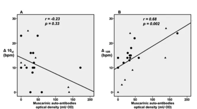
Correlation analysis between the heart rate responses and serum levels of cardiac auto-antibodies: Correlation analysis, of the heart rate responses and the serum levels of muscarinic auto-antibodies, of sero-positive patients showed: 1. that the heart rate responses to the Valsalva maneuver did not correlate with the serum levels of muscarinic auto-antibodies;2. thatthe initial heart rate acceleration during the first 10 seconds of dynamic exercise did not correlate with the muscarinic auto-antibodies (Table 4, Fig. 2A); 3. that the absolute decrease in heart rate, following cessation of exercise (first ten seconds), correlated directly and significantly with the serum levels of muscarinic auto-antibodies (Table 4, Fig. 2B).
DISCUSSION
Heart rate changes, during the early and late phases of the Valsalva maneuver and of dynamic exercise, indirectly reflect modulation of the autonomic nervous system on the sinus node. Thus, parasympathetic withdrawal is mostly responsible for the heart rate acceleration observed during the early phase of the Valsalva maneuver and the first seconds of dynamic exercise. Reactivation of parasympathetic activity, on the other hand, determines the magnitude of heart rate deceleration following termination of the maneuver and immediately after cessation of dynamic exercise. These two tests assess the responsiveness of the cardiac autonomic nervous system, particularly the negative chronotropic
effects of the parasympathetic system on heart rate5,19,26,30,35.
Parasympathetic effects on heart rate, of asymptomatic chagasic patients who are in the indeterminate and cardiac stages of the disease, have been studied under stressful2,20,27, and under steady-state
conditions6,24,45. The findings of these clinical investigations have been
conflicting and controversial. Nonetheless, parasympathetic effects on heart rate are currently considered, by most investigators, as being
impaired by cardiac muscarinic auto-antibodies14,21-23,42,43. We have
postulated, on the contrary, that parasympathetic activity is enhanced by the allosteric effects of the muscarinic cardiac auto-antibodies11,12.
The present clinical investigation included asymptomatic chagasic patients, who were in the indeterminate and cardiac forms of the disease and had normal two-dimensional echocardiograms. Heart rate acceleration, during the early phases of the Valsalva maneuver and of dynamic exercise, was significantly diminished in the chagasic patients. However, the heart rate changes, during the late phases of the Valsalva maneuver and of dynamic exercise, revealed a normal and enhanced response respectively. These apparently “discordant” results would indirectly suggest that: 1. the diminished initial heart rate acceleration during the early phases of both of these tests is indicative of reduced or impaired resting parasympathetic activity; 2. the normal or augmented heart rate recovery at the end of these two same tests would indicate, on the other hand, that parasympathetic reactivation is normal and even enhanced. Thus, chronotropic responses to parasympathetic withdrawal would be apparently impaired, but the chronotropic responses to parasympathetic reactivation are normal or even accentuated. How can one reconcile these apparently “discordant” results, with current knowledge on cardiac parasympathetic function in chronic chagas heart disease?
Muscarinic receptors are known to be up-regulated by Trypanosoma
cruzi infection and muscarinic cardiac auto-antibodies potentiate the chronotropic effects of acetylcholine on the cardiac muscarinic receptors of the sinus node25,38. Therefore, the postsynaptic muscarinic receptors,
which mediate the negative chronotropic effects of parasympathetic Table 3
Heart rate responses during dynamic exercise
Variable Negative
Serology (n = 18)
Positive Serology (n = 20)
p value
Basal heart rate (bpm)
78 ± 11.60 69 ± 7.4 0.31
∆ 10sI (bpm) 19 ± 7.27 12 ± 7.55 0.01
Maximum heart rate (bpm)
165.50 (155-179) * 159.50 (150-176) * 0.26
Heart rate recovery
∆10R 5(0 - 8.75)* 14 (9.75-17.50) * 0.001
∆1Rmin 30 ± 13.08 32 ± 11.26 0.27
∆2Rmin 43 ± 12.12 43 ± 13.5 0.76
Exercise time (minutes)
8.2 ± 2.35 7.64 ± 2.32 0.83
Data are expressed as Mean ± Standard deviation (M ± SD). *Data are expressed as Median and interquartile range (25th and 75th percentiles). *Mann-Whitney test was used for intergroup comparisons. ∆ 10sI = Magnitud of initial heart accelera-tion during the first 10 seconds. bpm = beats per minute. ∆10R = Magnitud of heart rate recovery during the first 10 seconds post-exercise. ∆1Rmin = Magnitud of heart rate recovery during the first minute post-exercise. ∆2Rmin = Magnitud of heart rate recovery during the second minute post-exercise.
Table 4
Correlation analysis between the heart rate responses and serum levels of muscarinic auto-antibodies
Variable Negative Serology Positive Serology
r p value r p value
Valsalva’s maneuver
∆acceleration 0.42 0.11 - 0.09 0.69
∆recovery 0.36 0.51 0.18 0.47
Dynamic exercise
∆10sI 0.23 0.31 0.23 0.33
∆10R -0.11 0.62 0.68 0.002
activity, are numerically increased and positively influenced by
Trypanosoma cruzi infection. In this particular context, we have found that the serum levels of the cardiac muscarinic auto-antibodies correlated directly with the magnitude of cardiac deceleration, following cessation of exercise. Therefore, the more prominent heart rate recovery of the chagasic patients could be an expression of a positive allosteric effect of the muscarinic auto-antibodies on the membrane muscarinic receptor. Alternatively, these results could be due to a direct agonist effect of the auto-antibodies on the muscarinic receptor and thereby potentiate early heart recovery. Consequently, the results of this investigation, although not conclusively demonstrating the presence of saturation of the cardiac parasympathetic system, indirectly suggest that the cardiac muscarinic auto- antibodies may have a positive agonist action on parasympathetically-mediated heart rate recovery, at the cessation of dynamic exercise.
Limitations of the study: The Valsalva maneuver was not successfully performed by all the chagasic patients included in this investigation. During the maneuver, an aspect such as the maximum depth of inspiration before straining is not uniform in all subjects. A smaller sample size and methodological aspects of the maneuver may, in part, explain the absence of a significant correlation between the serum levels of cardiac muscarinic auto-antibodies and the heart rate responses to the maneuver. Moreover, the auto-antibodies did not correlate with the heart rate acceleration of the early stages of both autonomic tests. These limitations definitively argue against our hypothesis of a saturation of the cardiac parasympathetic system versus parasympathetic dysautonomia. Nonetheless, the results of this investigation clearly show that, concerning parasympathetic withdrawal or reactivation-mediated heart rate responses, the former can be diminished, but the latter are normal or even enhanced. Further studies are needed to clarify the true nature of these apparently “discordant” heart rate responses.
In summary, we have studied the heart rate responses of sero-positive patients to the Valsalva maneuver and to dynamic exercise. These heart rate responses were apparently depressed during the early phases of both autonomic tests, but preserved in the late phases. Particularly, the magnitude of early heart rate recovery at the cessation of exercise was more prominent in the chagasic patients and correlated directly and significantly with the serum levels of the cardiac muscarinic auto-antibodies. These results indirectly suggest that the cardiac muscarinic auto-antibodies may have positive agonists effects on parasympathetically-mediated heart rate recovery of chronic chagasic patients.
RESUMO
Anticorpos muscarínicos e resposta da frequência cardíaca ao exercício dinâmico e a manobra de Valsalva na doença de Chagas
crônica
Foram estudadas as respostas cronotrópicas cardíacas à manobra de Valsalva e ao exercício dinâmico de vinte pacientes chagásicos com função ventricular esquerda normal e sem alterações da contractilidade segmentar por ecocardiografia bidimensional. O aumento absoluto
da frequência cardíaca dos pacientes (∆ = 21,5 ± 10 bpm, M ± DP)
durante a manobra de Valsalva foi significativamente menor quando se
comparava ao grupo controle (∆ = 31,30 ± 70, p = 0,03). A frequência
cardíaca mínima (58,24 ± 8,90 vs 62,80 ± 10, p = 0,68) e a diminuição
da frequência cardíaca absoluta no final da manobra (∆ = 38,30 ± 13
vs ∆ = 31,47 ± 17, p = 0,10) não foram diferentes em comparação com
o grupo controle. A aceleração inicial da frequência cardíaca durante o exercício dinâmico (∆ = 12 ± 7,55 vs ∆ = 19 ± 7,27, p = 0,01) também foi
menor, mas a recuperação da frequência cardíaca, durante os primeiros dez segundos, foi maior no grupo sero-positivos [mediana:14 (intervalo
interquartil: 9,75-17,50) vs 5 (0 - 8,75), p = 0,001]. Os níveis séricos
de auto-anticorpos muscarínicos cardíacos foram significativamente maiores nos pacientes chagásicos do que no grupo controle [(mediana: 34,58 densidade óptica (intervalo interquartil 17 - 46,5) vs (mediana: 0, intervalo interquartil 0 - 22,25) p = 0,001] e a correlação é significativa
e direta (r = 0,68, p = 0,002) com o início da recuperação da frequência cardíaca durante o exercício dinâmico. Os resultados desta investigação sugerem que indiretamente, os auto-anticorpos muscarínicos cardíacos, podem ter ação agonista positiva sobre o controle parassimpático da frequência cardíaca dos pacientes chagásicos.
REFERENCES
1. Acquatella H. Echocardiography in Chagas heart disease. Circulation. 2007;115:1124-31.
2. Amorim DS, Manco JC, Gallo L Jr, Marin-Neto JA. Chagas` heart disease as an experimental model for studies of cardiac autonomic function in man. Mayo Clin Proc. 1982;57(Suppl):48-60.
3. Benchimol-Barbosa PR. Comments on the letter by Ribeiro et al.: Impairment of parasympathetic-mediated autonomic modulation of the heart in Chagas cardiomyopathy: parasympathetic modulation vs tonus. Int J Cardiol. 2009;133:126-7. 4. Bonney KM, Engman DM. Chagas heart disease pathogenesis: one mechanism or
many? Curr Mol Med. 2008;8:510-8.
5. Cole CR, Blackstone EH, Pashkow FJ, Snader CE, Lauer MS. Heart-rate recovery immediately after exercise as a predictor of mortality. N Engl J Med. 1999;341:1351-7. 6. Correia D, Junqueira LF Jr, Molina RJ, Prata A. Cardiac autonomic modulation
evaluated by heart interval variability is unaltered but subtly widespread in the indeterminate Chagas’ disease. Pacing Clin Electrophysiol. 2007;30:772-80. 7. Davila, DF, Donis JH, Navas M. Fuenmayor AJ, Torres A, Gottberg C. Response
of heart-rate to atropine and left ventricular function in Chagas heart disease. Int J Cardiol.1988;21:143-52.
8. Davila DF, Rossell RO, Donis JH. Cardiac parasympathetic abnormalities: cause or consequence of Chagas heart disease? Parasitol Today. 1989;5:327-9.
9. Dávila DF, Inglessis G, Mazzei de Dávila CA. Chagas› heart disease and the autonomic nervous system. Int J Cardiol. 1998;66:123-7.
10. Davila DF, Donis JH, Torres A, Ferrer JA. A modified and unifying neurogenic hypothesis can explain the natural history of chronic Chagas heart disease. Int J Cardiol. 2004;96:191-5.
11. Dávila DF, Santiago JJ, Odreman W. Vagal dysfunction and the pathogenesis of chronic Chagas disease. Int J Cardiol. 2005;100:337-9.
12. Dávila DF, Donis JH, Dávila LA, Odreman WA, de Bellbarba GA, Villarroel V. Anti-muscarinic autoantibodies and vagal modulation in Chagas disease: positive allosteric modulators vs desensitization and downregulation of M2 cardiac acetylcholine receptors. Int J Cardiol. 2008;123:328-9.
13. Davila-Spinetti DF, Colmenares-Mendoza HJ, Lobo-Vielma L. Mecanismos causantes de la progresión del daño miocárdico en la enfermedad de Chagas crónica. Rev Esp Cardiol. 2005;58:1007-9.
14. de Oliveira SF, Pedrosa RC, Nascimento JH, Campos de Carvalho AC, Masuda MO. Sera from chronic chagasic patients with complex cardiac arrhythmias depress electrogenesis and conduction in isolated rabbit hearts. Circulation. 1997;96:2031-7. 15. Decourt LV, Sosa EA, Pileggi F. Electrophysiological studies in the indeterminate
form of Chagas´disease. Arq Bras Cardiol. 1981;36:227-34.
16. de Resende LA, Molina RJ, Ferreira BD, Carneiro AC, Ferreira LA, da Silva VJ, et al. Cardiac autonomic function in chagasic elderly patients in an endemic area: a time and frequency domain approach. Auton Neurosci. 2007;131:94-101. 17. Sehr P, Zumbach K, Pawlita M. A generic capture ELISA for recombinant proteins
fused to glutathione S-transferase: validation for HPV serology. J Immunol Methods. 2001;253:153-62.
18. Fletcher GF, Balady G, Froelicher VF, Hartley LH, Haskell WL, Pollock ML. Exercise standards. A statement for healthcare professionals from the American Heart Association. Writing Group. Circulation. 1995;91:580-615.
19. Freeman R. Assessment of cardiovascular autonomic function. Clin Neurophysiol. 2006;117:716-30.
20. Fuenmayor AJ, Rodríguez L, Torres A, Donis J, Navas M, Fuenmayor AM, et al. Valsalva maneuver: a test of the functional state of cardiac innervation in chagasic myocarditis. Int J Cardiol. 1988;18:351-6.
21. Goin JC, Borda E, Leiros CP, Storino R, Sterin-Borda L. Identification of antibodies with muscarinic cholinergic activity in human Chagas’ disease: pathological implications. J Auton Nerv Syst. 1994;47:45-52.
22. Goin JC, Peres-Leiros C, Borda E, Sterin-Borda E. Interaction of human chagasic IgG with the second extracellular loop of the human heart muscarinic acetylcholine receptor: functional and pathological implications. FASEB J. 1997;11:77-83. 23. Goin JC, Borda ES, Auger S, Storino R, Sterin-Borda L. Cardiac M2 muscarinic
cholinoceptor activation by human chagasic autoantibodies: association with bradycardia. Heart. 1999;82:273-8.
24. Guzzetti S, Iosa D, Pecis M, Bonura L, Prosdocimi M, Malliani A. Impaired heart rate variability in patients with chronic Chagas’ disease. Am Heart J. 1991;121:1727-34. 25. Hernandez CC, Nascimento JH, Chaves EA, Costa PC, Masuda MO, Kurtenbach E, et
al. Autoantibodies enhance agonist action and binding to cardiac muscarinic receptors in chronic Chagas’ disease. J Recept Signal Transduct Res. 2008;28:375-401. 26. Junqueira LF Jr. Teaching cardiac autonomic function dynamics employing the
Valsalva (Valsalva-Weber) maneuver. Adv Physiol Educ. 2008; 32:10010-6. 27. Junqueira LF Jr, Soares JD. Impaired autonomic control of heart interval changes to
Valsalva manoeuvre in Chagas’ disease without overt manifestation. Auton Neurosci. 2002;97:59-67.
28. Kierszenbaum F. Mechanisms of pathogenesis in Chagas disease. Acta Parasitol. 2007;52:1-12.
29. Lescure FX, Le Loup G, Freilij H, Develoux M, Paris L, Brutus L, et al. Chagas disease: changes in knowledge and management. Lancet Infect Dis. 2010;10:556-70. 30. Maciel BC, Gallo L Jr, Marin Neto JA, Lima Filho EC, Martins LE. Autonomic nervous control of the heart rate during dynamic exercise in normal man. Clin Sci (Lond). 1986;71:457-60.
31. Marin-Neto JA, Cunha-Neto E, Maciel BC, Simões MV. Pathogenesis of chronic Chagas heart disease. Circulation. 2007;115:1109-23.
32. McCormick TS, Rowland EC. Trypanosoma cruzi: cross-reactive anti-heart autoantibodies produced during infection in mice. Exp Parasitol. 1989;69:393-401. 33. Medei EH, Matheus Nascimento JH, Coury Pedrosa R, Campos de Carvalho AC. Role of autoantibodies in the physiopathology of Chagas´disease. Arq Bras Cardiol. 2008;91:257-62.
35. Nobrega AC, Castro CI, Araujo CG. Relative roles of the sympathetic and parasympathetic systems in the 4-s exercise test. Braz J Med Biol Res. 1990;23:1259-62.
36. Nussinovitch U, Shoenfeld Y. The diagnostic and clinical significance of anti-muscarinic receptor autoantibodies. Clin Rev Allergy Immunol. 2012;42:298-308. 37. Odreman R, Dávila DF, Donis JH, Torres A, Ferrer JA, Inglesis I. Valsalva maneuver in
chagasic patients with documented past medical history of acute chagasic myocarditis. Int J Cardiol. 2004;93:163-7.
38. Peraza-Cruces K, Gutiérrez-Guédez L, Castañeda Perozo D, Lankford CR, Rodríguez-Bonfante C, Rodríguez-Bonfante-Cabarcas R. Trypanosoma cruzi infection induces up-regulation of cardiac muscarinic acetylcholine receptors in vivo and in vitro. Braz J Med Biol Res. 2008;41:796-803.
39. Rassi A Jr., Rassi A, Rasi SG. Predictors of mortality in chronic Chagas disease. A systematic review of observational studies. Circulation. 2007;115:1101-8. 40. Retondaro FC, Dos Santos Costa PC, Pedrosa RC, Kurtenbach E. Presence of
antibodies against the third intracellular loop of the m2 muscarinic receptor in the sera of chronic chagasic patients. FASEB J. 1999;13:2015-20.
41. Ribeiro AL, Giménez LE, Hernandez CC, de Carvalho AC, Teixeira MM, Guedes VC, et al. Early occurrence of anti-muscarinic autoantibodies an abnormal vagal modulation in Chagas disease. Int J Cardiol. 2007;117:59-63.
42. Ribeiro AL, Carvalho AC, Texeira MM, Lombardi F, Rocha MO. Chagas disease: impaired vagal modulation has been demonstrated, enhanced parasympathetic activity remains to be proved. Int J Cardiol. 2008;123;330-2.
43. Sterin-Borda L, Borda E. Role of neurotransmitter autoantibodies in the pathogenesis of chagasic peripheral dysautonomia. Ann N Y Acad Sci. 2000;917:273-80. 44. Talvani A, Rocha MO, Ribeiro AL, Borda E, Sterin-Borda L, Teixeira MM. Levels of
anti-M2 and anti-β1 autoantibodies do not correlate with the degree of heart dysfunction in Chagas´heart disease. Microbes Infect. 2006;8:2459-64.
45. Vasconcelos DF, Junqueira LF Jr. Distinctive impaired cardiac autonomic modulation of heart rate variability in chronic Chagas’ indeterminate and heart diseases. J Electrocardiol. 2009;42:281-9.