(1) Universidade Federal de São Paulo - São Paulo/SP Brasil
Conlict of interest: non-existent
Semicircular superior canal dehiscence: cases reports
Síndrome da deiscência do canal semicircular superior:
relato de dois casos
Carolina Calsolari Figueiredo de Godoy(1)
Kelle Cristine Erhrdt Wiggers Ávila(1)
Adriana Neves de Andrade(1)
Daniela Gil(1)
Received on: September 21, 2016 Accepted on: January 17, 2017
Mailing address:
Carolina Calsolari Figueiredo de Godoy Rua Werner Goldberg, 77 - Torre Sabiá Apto 26 - Jd Tupanci - Barueri/SP CEP: 06414-025
E-mail: [email protected]
ABSTRACT
The Superior Semicircular Canal Dehiscence Syndrome (SSCDS) is characterized by bone wear layer overlying the superior semicircular canal. Common symptoms of SSCDS the presence of vertigo asso -ciated with nystagmus induced by intense sound stimuli or changes in intracranial pressure or middle ear. The aim of this study is to describe the audiological and vestibular indings of two patients diagnosed with Superior Semicircular Canal Deiscence Syndrome, with conirmed diagnosis by computed tomo -graphy. Meatoscopy, anamnesis, pure tone audiometry and vocal followed by the acoustic impedance measurements, audiometric Weber, research Tulio phenomenon and Valsalva maneuver, performed by the same researcher in one session were held. Air-bone gap were observed, type A tympanometric curve and acoustic relex. The air-bone gap is presented with greater amplitude at low frequencies. Hearing complaints were not reported by patients as the irst symptoms. Weber showed lateralization in both cases, conirming the presence of gap. The Thulium phenomenon is positive for vertigo in both cases. The Valsalva maneuver showed a change in only one case.
Keywords: Hearing; Audiometry; Semicircular Canals; Nystagmus, Pathologic; Dizziness
RESUMO
A Síndrome da Deiscência do Canal Semicircular Superior (SDCSS) é caracterizada pelo desgaste da camada óssea que recobre o canal semicircular superior. São sintomas comuns da SDCSS a presença de vertigem associada à nistagmos induzidos por estímulos sonoros intensos ou por modiicações das pressões intracraniana ou da orelha média. O objetivo deste trabalho é descrever os achados audiólogi -cos e vestibulares de dois pacientes com diagnóstico de Síndrome da Deiscência do Canal Semicircular Superior, com diagnóstico conirmado por meio de tomograia computadorizada. Foram realizadas mea -toscopia, anamnese, audiometria tonal e vocal seguida das medidas de imitância acústica, Weber audio -métrico, pesquisa do fenômeno de Túlio e manobra de Valsalva, realizados pela mesma pesquisadora em uma única sessão. Foram observados gap aéreo-ósseo, curva timpanométrica tipo A e relexos acústi -cos presentes. O gap aéreo-ósseo apresenta-se com maior amplitude nas frequências baixas. As queixas auditivas não foram relatadas pelas pacientes como os primeiros sintomas. O Weber mostrou lateraliza -ção, nos dois casos, conirmando a presença de gap. O fenômeno de Túlio apresentou-se positivo para vertigem em ambos os casos. A manobra de Valsalva apresentou alteração em apenas um caso.
Descritores: Audição; Audiometria; Canais Semicirculares; Nistagmo Patológico; Tontura
In tympanometry, patients presented a type A curve
and present acoustic relexes9,10. Acoustic relexes are
present in patients with SSCDS, contrary to what was
expected for individuals with hearing impairment due to alteration of the middle ear 7.
Tullio phenomenon and the Valsava maneuver in patients with SSCDS may show dizziness or
nystagmus in the presence of high intensity sounds and pressure variation in the external acoustic canal,
respectively9-12.
The Symptoms for Superior Semicircular Canal
Dehiscence Syndrome may be similar to the symptoms
of other diseases such as: otosclerosis and tubal dysfunction, Ménierè’s disease, temporomandibular dysfunction8, which may delay the diagnosis. It is important to establish a differential evaluation protocol when the suspicion of Superior Semicircular Canal
Dehiscence Syndrome is present.
Based on the previous considerations, the purpose
of this study is to describe the audiological and vestibular indings of two patients diagnosed with
Superior Semicircular Canal Dehiscence Syndrome.
PRESENTATION OF CASES
This case report project was analyzed and approved
by the Federal University of São Paulo research
ethics committee under No. 1717/08. The study was
conducted at the Audiological Outpatient Clinic of the Discipline of Auditory Disorders of the Department of Speech-Language Pathology and Audiology, from the Federal University of São Paulo.
The eligibility criteria were: both sexes, Superior Semicircular Channel Dehiscence Syndrome conirmed
by computed tomography.
Subjects were informed about the procedures performed and signed a consent form before partici -pating in the study.
Meatoscopy, anamnesis, tonal and vocal audiometry followed by acoustic immitance measure -ments, audiometric Weber, Tullio phenomenon
research and Valsalva maneuver were performed by
the same researcher in a single session.
Pure tone audiometry (air and bone conduction),
speech audiometry, audiometric Weber and Tullio
phenomenon were performed with the Interacoustics
MA-41 audiometer. For acoustic immitance measure -ments, the Interacoustics AZ7 impedance meter was used.
INTRODUCTION
Superior Semicircular Canal Dehiscence (SSCDS)
was irst described as the wear of the bone layer that
covers the superior semi-circular canal, causing an
abnormal exposure of the vestibular membranous labyrinth in the cranial middle fossa 1.
This bone dehiscence results in a third movable window, allowing pressure to dissipate as the membranous labyrinth projects inwardly and the
endolymph lows away from the ampulla. Following this pathophysiological scenario, some symptoms of
Superior Semicircular Canal Dehiscence Syndrome (SSCDS) may appear, such as elevated air conduction
thresholds and maintenance of bone conduction
thresholds, as well as vestibular symptoms induced by
intense sonorous stimuli and by modiications of intra -cranial pressure or by the medium ear2.
A microscopic study aiming at determining the
prevalence of SSDCS in the general population
analysed 1,000 temporal bones obtained through
autopsies, and found that the superior semi-circular canal dehiscence occurred in approximately 0.7% of the individuals studied, reafirming the low incidence of disease. The authors of this study afirmed,
however, that not all patients with SSCDS present the
symptoms of the Syndrome, and that the percentage of symptomatic ones among them is not yet known3. Other studies indicating the incidence of SSCDS in its clinical form were not found.
The etiology of SSCDS is still unclear and it has
been much debated in order to determine whether it is
congenital, acquired or a mixture of both. Some authors have postulated that it is a developmental imperfection that becomes clinically relevant in adulthood after a trauma or as a cause of an increased intracranial pressure which leads to a rupture of the bone 4.
The presence of vertigo associated with nystagmus
induced by intense sonorous stimuli or by changes in intracranial or middle ear pressures are common
symptoms of SSCDS5.
Some patients diagnosed with SSCDS may present autophony and conductive hearing loss, although
these characteristics are less frequent than for patients
with vestibular symptoms.
Patients with SSCDS present a more signiicant air-bone gap for the low frequencies, caused by the presence of the third window that dissipates acoustic
energy 6,7 It is known that the bone pathway threshold
Both cases were submitted to the following
procedures: anamnesis, air and bone pathway tonal audiometry, logoaudiometry, Imitanciometry
(tympanometry and research of contralateral stapedial relexes), audiometric Weber’s test, tullio’s
phenomenon and Valsalva maneuver.
RESULTS
Case 1
RPS was admitted in the Otorhinolaryngology Clinic of the institution of origin, with complaints already
reported in the case presentation. An otolaryngological
evaluation was performed, which revealed normal
otoscopy and no other complaints were reported.
The following two cases are presented:
• Case 1- RPS, female, 42 years of age. She was admitted in the Otorhinolaryngology outpatient
clinic of the institution of origin with a complaint of dizziness. The patient reported rotatory and non-rotatory dizziness, lasting approximately 40 minutes, as well as auricular fullness in vehicles.
• Case 2 - NVSS, female, 52 years of age. In 2010,
she presented a complaint of intense vertigo in short-term crises 20 years before, accompanied by neurovegetative manifestations, spatial disorien -tation and panic to go out without a company. She reported sporadic acute pitch tinnitus and high
discomfort for loud sounds.
Table 1. Results for the procedures performed in case 1
Date Complaint Tonal
Audiometry
Vocal
Audiometry Timpanometry
Acoustic
Relexes Audiometric Weber
Tullio Phenomenon
Valsalva Manouver
2005
Rotating and non-rotating dizziness;
Auricular fullness.
Normal Normal Type A bilaterally bilaterally present Not made Not made Not made
2006 2007
Tinnitus; Rotating dizziness;
Auricular fullness; Crises start with temperature
changes.
Normal Normal Type A bilaterally bilaterally present Not made Not made Not made
2009
Dizziness; Nausea; Discomfort with intense sounds; Bilateral intense tinnitus.
Left Ear: GAP at 250, 500, 1000, 3000 and 4000Hz frequencies, and hearing loss at 6000Hz;
Right Ear: Normal
TA Compatible Type A bilaterally Bilaterally present
1000 and 2000Hz lateralized to the
right; 500 and 4000Hz
lateralized to the left.
Vertigo +
Nystagmus - Nystagmus -Vertigo +
TA: Tonal audiometry; HZ: Hertz.
Audiometry with normal thresholds in both ears was
performed. In the tympanometry, type A tympanometric curves and acoustic relexes were observed in both ears. The patient underwent a vestibular examination,
which diagnosed irritative peripheral bilateral vestibular syndrome.
In August 2006, the patient returned to the outpa-tient clinic complaining that the crisis began with the change in temperature. The crisis began with tinnitus,
In April 2009, RPS returned to the outpatient clinic complaining of dizziness and nausea in evolution for 5
years, as well as intense bilateral tinnitus and
uncom-fortable for intense sounds, without diminishing the
auditory sensitivity.
In the audiological evaluation performed in 2009 for this study, hearing loss was observed only at the 6000 Hz frequency on the left side, with air-bone gap in the frequencies of 250, 500, 1000, 3000 and 4000 Hz
and logoaudiometry compatible with the audiometric
results. Good mobility of the tympano-ossicular system
was observed, with type “A” curve and contralateral
stapedial relexes present in both ears at adequate levels of intensity.
frontal pulsatile irradiation with nausea, aversion to sound and light. A new audiometry was performed,
which results again revealed auditory thresholds within
the limits of normality.
When the patient returned in June 2007, she reported
that she presented three crisis of rotatory dizziness with
nausea and scotomas, and it was necessary to go to the emergency room. She also reported mild dizziness that
lasted 10 minutes whenever she got up from a chair, auricular fullness when on the subway or on the bus in
addition to tinnitus and hearing loss. In August 2007, a
temporal bone CT was performed, which indicated total bone dehiscence in the left superior semicircular canal.
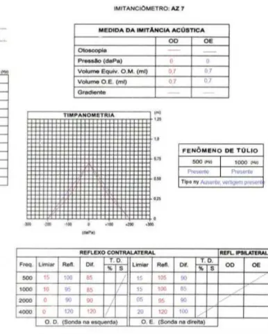
Figure 1. Results of the audiological evaluation of case 1
The audiometric Weber test lateralized to the right
at 1000 and 2000 Hz and to the left at 500 and 4000 Hz. The Tullio phenomenon was performed at 500 and 1000 Hz at 100 dB with a positive result for vertigo, but
no nystagmus was observed. In the Valsalva maneuver,
there was vertigo without the presence of nystagmus.
Case 2
NVSS was admitted in the Otorhinolaryngology
outpatient clinic of the institution of origin in 2010, with
the complaints already reported in the case
presen-tation. An otolaryngological evaluation was performed,
degree of hearing loss bilaterally . Good mobility of the
ossicle tympanic system was observed, with type “A”
curve and contralateral stapedial relexes present in both ears at adequate levels of intensity.
In the audiometric evaluation, it was observed
the presence of mild conductive hearing loss and ascending coniguration, with a signiicant air-bone gap in the frequencies of 250, 500 and 4000 Hz. The
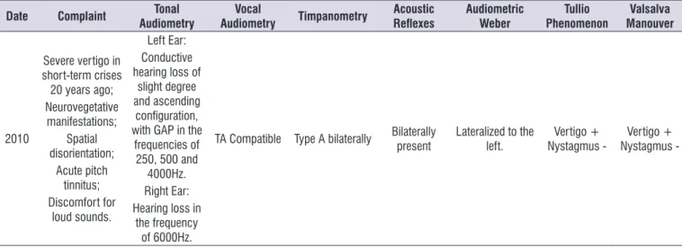
logoaudiometry was compatible with the type and Table 2. Results for the procedures performed in case 2
Date Complaint Tonal
Audiometry
Vocal
Audiometry Timpanometry
Acoustic
Relexes Audiometric Weber
Tullio Phenomenon
Valsalva Manouver
2010
Severe vertigo in short-term crises 20 years ago; Neurovegetative
manifestations; Spatial disorientation;
Acute pitch tinnitus; Discomfort for
loud sounds.
Left Ear: Conductive hearing loss of
slight degree and ascending
coniguration, with GAP in the
frequencies of 250, 500 and
4000Hz. Right Ear: Hearing loss in
the frequency of 6000Hz.
TA Compatible Type A bilaterally Bilaterally present Lateralized to the left. Nystagmus -Vertigo + Nystagmus -Vertigo +
TA: Tonal audiometry; HZ: Hertz.
The Tullio phenomenon was positive for vertigo in
both cases, in agreement with the results described in the literature 9,10,12.
The Valsalva maneuver also presented alteration, in
case 1 for vertigo, alterations were also present in other
studies described in the literature. However, in case report 2, the Valsalva maneuver was negative.
For this study, the data collection was performed for more than one year, and only two cases of SSCDS were diagnosed, reafirming the rare incidence of this
pathology 3,15.
Since it is an uncommon alteration, audiological and
otoneurological evaluations are essential for diagnosis. The differential diagnosis of the SSCDS should be
sought whenever there is an apparent incompatibility between tonal audiometry, tympanometry and acoustic
relex research, which is the starting point of the
diagnostic process
In cases with suspicion of SSCDS, it is important
that all individuals undergo an evaluation protocol with
the tests studied in order to conirm the diagnosis. It is
suggested to carry out new studies with this population in order to elaborate a protocol to be used whenever
there is suspicion of SSCDS.
FINAL CONSIDERATIONS
The audiological and vestibular evaluation of
two patients with superior semicircular canal
dehis-cence syndrome revealed: Pure tone audiometry with conductive loss; Air-bone gap in the frequencies of 250, 500, 1000, 3000 and 4000 Hz in case 1 and in the frequencies of 250, 500 and 4000 Hz in case 2; Greater air-bone gap in the frequency of 250 Hz; Acoustic
immitance measurements with tympanometric curves
type A and present acoustic relexes; Weber lateralized to the side of the gap; Túlio phenomenon with presence of vertigo and absence of nystagmus; Valsalva maneuver with presence of vertigo and absence of
nystagmus, only in case 1.
REFERENCE
1. Minor LB, Solomon D, Zinreich JS, Zee DS. Sound
and/or pressure induced vertigo due to bone
dehiscence of the superior semicircular canal. Arch Otolaryngol Head Neck Surg. 1998;124(3):249-58. 2. Hirvonen TP, Carey JP, Liang CJ, Minor LB.
Superior canal dehiscence: mechanisms of
pressure sensitivity in a chinchilla model. Arch
The Weber test presented lateralization to the left in the low frequencies, conirming the presence of gap. The Tullio phenomenon was performed at 500 and 1,000 Hz at 100 dB with a positive result for vertigo, but
no nystagmus was observed. The Valsava maneuver
proved to be negative, that is, there was no presence of
nystagmus or vertigo.
DISCUSSION
SSCDS is characterized by vestibular symptoms
associated with the presence of nystagmus induced by
intense sonorous stimuli or by changes in intracranial
or middle ear pressures resulting from wear of the bone
layer that covers the superior semicircular canal1. This is a disease with few studies conducted in Brazil, most of which are literature reviews of studies performed abroad11.
In case 1, the patient presented dizziness at 38
years of age and was diagnosed with SSCDS at age 40. In case 2, the patient was 52 years old when she presented the crises and received the diagnosis of
SSCDS. As it can be observed, case 2 presents similar results to the study that observed a decrease in bone
coverage of the superior semicircular canal after the age of 45, but in case 1 the pathology was diagnosed with a lower age than the one found by the author13.
In both cases, the patients presented air-bone gap,
type A tympanometric curves and acoustic relexes, similarly to other studies that found air-bone gap, type A tympanometric curve and acoustic relexes in their
patients6,7,9,10.
The air-bone gap was higher in the low frequencies, in these cases the largest difference occurred in the frequency of 250 Hz, corroborating with a survey that found a more pronounced gap in the low frequencies. This inding emphasizes the importance of including bone threshold test in the frequency of 250Hz in cases of suspicion of SSCDS.
Auditory complaints were not reported by the
patients of the present study as being the irst
symptoms, however in the literature there are studies
in which the patients reported that the irst symptoms
were the auditory alterations14.
Otoneurological symptoms were reported in both cases presented, either in dizziness or in vertigo.
Such indings are important for the audiological and
otoneurological practice, indicating the need to
15. Tsunoda A, Terasaki O. Dehiscence of the bony roof of the superior semicircular canal in the middle cranial fossa. J Laryngol Otol Suppl. 2002;116(7):514-8.
3. Carey JP, Minor LB, Nager GT. Dehiscence or thinning of bone overlying the superior semicircular
canal in a temporal bone survey. Arch Otolaryngol
Head Neck Surg. 2000;126(2):137-47.
4. Minor LB. Clinical Manifestations of Superior
Semicircular Canal Dehiscence. Laryngoscope. 2005;115(10):1717-27.
5. Cremer PD, Minor LB, Carey JP, Della Santina CC.
Eye movements in patients with superior canal dehiscence syndrome align with the abnormal
canal. Neurology. 2000;55(12):1833-41.
6. Minor LB, Carey JP, Cremer PD, Lustig LR, Streubel SO. Dehiscence of bone overlying the superior canal as a cause of apparent conductive hearing loss. Otol Neurotol. 2003;24(2):270-8.
7. Mikulec AA, McKenna MJ, Ramsey MJ, Rosowski JJ, Herrmann BS, Rauch SD, Curtin HD, Merchant
SN. Superior semicircular canal dehiscence presenting as conductive hearing loss without
vertigo. Otol Neurotol. 2004;25(2):121-9.
8. Zhou G, Gopen Q, Poe DS. Clinical and Diagnostic Characterization of Canal Dehiscence Syndrome: A Great Otologic Mimicker. Otol Neurotol.
2007;28(7):920-6.
9. Halmagyi GM, Swee T, McGarvie LA, Todd MJ,
Bradshaw A, Yavor RA et al. Superior semicircular canal dehiscence simulating otosclerosis. J Laryngol Otol Suppl. 2003;117(7):553-7 .
10. Rajenderkumar D, Farrel KL, Alles RM, Savy L. Multiple dehiscence of semicircular canals. J
Laryngol Otol Suppl. 2006;121(1):80-2.
11. de Lima MT. Síndrome de Deiscência de Canal
Semicircular Superior. Rev. Bras. Otorrinolaringol.
2006;72(3):414-8.
12. Woodeson JM, Heitzmann T, Morales LR.
Dehiscencia del conducto semicircular superior:
presentación de un caso y revisión bibliográica. Acta Otorrinolaringol Esp. 2005;55(04):171-5. 13. Crovetto MA, Whyte J, Rodriguez OM, Lecumberri
I, Martinez C, Fernandez C et al. Inluence of Aging and Menopause in the Origin of the Superior
Semicircular Canal Dehiscence. Otol Neurotol.
2012;33(4):681-4.
14. Lee GS, Zhou G, Poe D, Kenna M, Amin M, Ohlms L et al. Clinical experience in diagnosis and management of superior semicircular