331 331 331 331 331 Mem Inst Oswaldo Cruz, Rio de Janeiro, Vol. 100(3): 331-337, M ay 2005
Lymphocyte subset analyses in healthy adults vaccinated with
yellow fever 17D D virus
Ana Paula dos Santos/+ +, Álvaro Luiz Bertho* , D aniela Capuzzo D ias/+ +, Jaciara Ramos Santos* , Rugimar M arcovistz/+
Laboratório de Tecnologia Imunológica, Bio-Manguinhos-Fiocruz *Laboratório de Imunoparasitologia, Departamento de Imunologia, IOC-Fiocruz, Av. Brasil 4365, 21045-900 Rio de Janeiro, RJ, Brasil
In this study the kinetics of humoral and cellular immune responses in first-time vaccinees and re-vaccinees with the yellow fever 17DD vaccine virus was analyzed. Flow cytometric analyses were used to determine percentual values of T and B cells in parallel to the yellow fever neutralizing antibody production. All lymphocyte subsets analyzed were augmented around the 30th post vaccination day, both for first-time vaccinees and re-vaccinees. CD3+ T cells increased from 30.8% (SE ± 4%) to 61.15% (SE ± 4.2%), CD4+ T cells from 22.4% (SE ± 3.6%) to 39.17% (SE ± 2%) with 43% of these cells corresponding to CD4+CD45RO+ T cells, CD8+ T cells from 15.2% (SE ± 2.9%) to 27% (SE ± 3%) with 70% corresponding to CD8+CD45RO+ T cells in first-time vaccinees. In re-vaccinees, the CD3+ T cells increased from 50.7% (SE ± 3%) to 80% (SE ± 2.3%), CD4+ T cells from 24.9% (SE ± 1.4%) to 40% (SE ± 3%) presenting a percentage of 95% CD4+CD45RO+ T cells, CD8+ T cells from 19.7% (SE ± 1.8%) to 25% (SE ± 2%). Among CD8+CD38+ T cells there could be observed an increase from 15 to 41.6% in first-time vaccinees and 20.7 to 62.6% in re-vaccinees. Regarding neutralizing antibodies, the re-vaccinees presented high titers even before re-vaccination. The levels of neutralizing antibodies of first-time vaccinees were similar to those presented by re-vaccinees at day 30 after vaccination, indicating the success of primary vaccination.Our data provide a basis for further studies on immunological behavior of the YF 17DD vaccine.
Key words: yellow fever - vaccine - flow cytometry - neutralizing antibodies - lymphocyte subsets
Yellow fever (YF) is a viral illness transmittedby in-fected mosquitoes (Aedes and Haemagogus genus) with yellow fever virus (YFV), which belongs to the Flavivirus
genus, Flaviviridae family (Monath & Heinz 1996). It re-mains a serious health problem in endemic areas of tropi-cal and subtropitropi-cal Africa and South America (Vasconcelos 2003). Since the 19th century, studies about YF immuniza-tion have been accomplished (WHO 2001a). The YF 17D vaccine virus strain is one of the most effective and safe vaccines available. Immunization with the 17D vaccine strain induces a long-term neutralizing antibody response and provides excellent protection against infection with the virulent YFV (Wisseman & Sweet 1962). In 1937, the production of the YF 17DD vaccine virus substrain was started in Brazil, and has been in continuous use for 60 years (Post et al. 2001). The YFV 17DD vaccine is highly immunogenic and induces neutralizing antibody persis-tent at least 10 years and in some individuals up to 30 years or more (WHO 2001b).
Although the YF 17D virus strain and the 17DD substrain are the most successful vaccines developed to date, recently, rare cases of postvaccinal neurological dis-orders have been recorded (Chan et al. 2001, Martin et al. 2001, Vasconcelos et al. 2001). The virus isolates from two fatal cases after 17DD vaccination demonstrated genetic
+Corresponding author. E-mail: rugimar@bio.fiocruz.br ++CNPq fellowships
Received 28 July 2004 Accepted 15 April 2005
stability and attenuated phenotype, suggesting that some peculiarities of health status of the host might have been responsible for such adverse events (Galler et al. 2001).
Despite the large literature on the humoral immune response to the YF vaccine virus, few works of the evalu-ation of cellular immune response, in particular T cell re-sponses have been published (Reinhardt et al. 1998, Co et al. 2002, van der Most et al. 2002). The capacity of vac-cines to activate the cellular immune system and induce T cell memory is an important mechanism of protection against wild-type viral infection. T cells often recognize more conserved epitopes that do not change due to anti-body mediated selection pressure (Whitton & Oldstone 1996, Reinahrdt et al. 1998). Co and collaborators (2002) studying the T cell responses to YFV 17D in four volun-teers could observed proliferation and cytolytic responses in all subjects. Their results present CD8 T cell responses directed against at least four different HLA-B35 restricted YFV epitopes.
In order to monitor the immunological behavior of first-time YF 17DD virus vaccinated and re-vaccinated volun-teers, T and B lymphocyte subsets were analyzed by flow cytometry, which is an efficient tool for definition and quantification of lymphocytes employing monoclonal antibodies against cell surface proteins, to determine the importance of knowing the cellular immune response acti-vation related to the YF 17DD vaccine.
MATERIALS AND METHODS
3 3 2 3 3 2 3 3 2 3 3 2
3 3 2 T cell subsets in YF 17D D vaccination • Ana Paula dos Santos et al.
Oswaldo Cruz Foundation, Brazil. Eight individuals, des-ignated first-time vaccinees, had not been vaccinated with the YF 17DD vaccine, and had had no previous infection or contact with the YF wild-type virus. Nine volunteers, designated re-vaccinees, had been vaccinated once or more with the YF 17DD vaccine, 10 or more years before. All volunteers gave their written consent after the study was explained. Each volunteer was injected subcutane-ously with 0.5 ml of vaccine. Blood was collected before vaccination (day 0) and 2, 4, 7, 10, 15, 30, and 60 days post vaccination. The volunteers were advised to report all clinical symptoms and side effects after vaccination. The procedures followed were in accordance with the ethical standards of the responsible committee on human experi-mentation (CEP/Fiocruz 145/01).
Separation of peripheral blood mononuclear cells
(PBMC)-PBMC were obtained by Histopaque (Sigma-H8889, St Louis, MS, US) gradient sedimentation, as de-scribed elsewhere (Noble et al. 1968, Boyle & Chow 1969).
Flow cytometric analyses - Freshly isolated PBMC were adjusted to 106 cells/ml and dual-staining labeled monoclonal antibodies (CD3(IgG1)-FITC/B4-RD1; CD4-PC5/ CD45RO-PE; CD8-CD4-PC5/CD45RO-PE; CD8-PE/CD38-FITC) and IgG1-FITC/IgG1-PE isotype antibody (nega-tive control) (Immunotech - Beckmann Coulter, Marseille, France) were used. After incubation for 20 min at 4ºC, the mixture was washed twice with PBS-azide (PBS contain-ing 0.1% sodium azide and 2% fetal bovine serum), and the cells were fixed in 1% paraformaldehyde (PFA). Stained cells were run in an EPICS ALTRA flow cytometer (Beckmann Coulter, Hialeah, FL, US) equipped with an argon ion laser. Ten thousand events were acquired and analyzed using the Expo32 software (Beckman Coulter). Their forward scatter and side scatter profiles distinguish lymphocytes and monocytes, an electronic gate was cre-ated around lymphocytes.
Plaque reduction neutralization test (PRNT)- PRNT were carried out in 96-well plates as described elsewhere (Stefano et al. 1999). In brief, dilutions of the sera ranging from 1:4 to 1:512 were mixed with 1:1200 of the 17DD virus preparation (7.8 log10 PFU/ml). After 1 h of incubation at 37oC, 100 µl of Vero cells (1.6 x 10⁶ cells/ml) were added. A 3% CMC/199 solution supplemented with 5% FCS was overlaid after 3 h of incubation. The cultures were main-tained for 7 days at 37oC and then the cells were fixed with formaldehyde and stained by violet crystal.
Viremia- For determination of viremia were used the plaque assay (PA) (Wheelock & Sibley 1965, Reinhardt et al. 1998, Marchevsky et al. 2003) and the reverse tran-scriptase-polymerase chain reaction (RT-PCR) (Reinhardt et al. 1998) methods, in serum samples before vaccination and on days 2, 4, and 7 after vaccination. RT-PCR prod-ucts were obtained in the presence of pairs of flavivirus type-specific primers (RG65-antisense, RG36-sense) (Galler et al. 2001), which were kindly provided by Dr Ricardo Galler (Oswaldo Cruz Institute - Fiocruz). Primer pairs were deduced from a RNA sequence from the envelope coding region of the yellow fever virus. The RT-PCR techniques were performed using the Geneamp kit (Applied
Biosystems RNA PCR Core Kit, Roche, Branchburg, New Jersey, US) according to the manufacturer’s instructions.
Statistical analyses - Fisher’s least significant differ-ence (LSD) procedure was used to evaluate statistically significant differences between means. With this method, there is a 5% risk of calling each pair of means signifi-cantly different when the actual difference equals 0. Dif-ferences were considered as statistically significant if p
value were equal or below 0.05.
RESULTS
The 17DD vaccine was shown to be very efficient and safe for all volunteers, activating their humoral and cellu-lar immune response, although viremia was not detected through of the methods used (RT-PCR and PA). During the kinetics blood collection in this study, the blood cell count and biochemical tests, including transaminases, did not present alterations after vaccination for any of the volunteers.
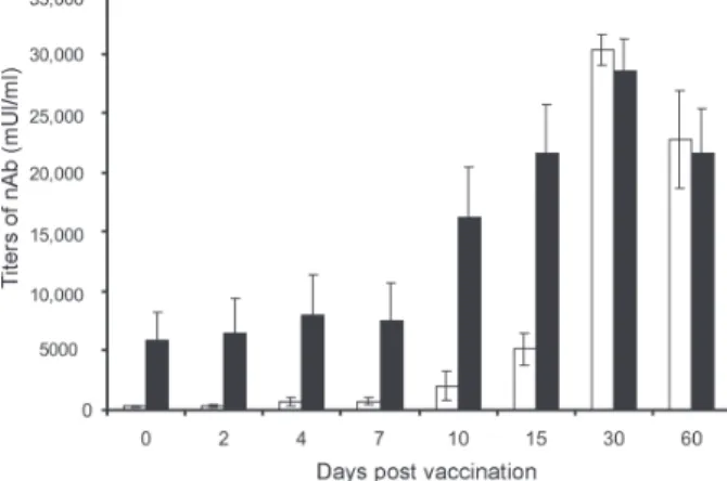
None of the first-time vaccinees showed pre-existing neutralizing antibodies (nAb) against the YF 17DD virus substrain (< 256 mUI/ml, limit of antibody detection for our test). Four days after vaccination, low levels of nAb were detected in 3 of the 8 first-time vaccinees (mean 698 mUI/ml, SE ± 301), however, 15 days after vaccination, the nAb were detected in all of them. High levels of nAb were reached 30 days after vaccination in this group of volun-teers with a slight decrease 30 days later, 30,321 mUI/ml (SE ± 1300) and 22,756 mUI/ml (SE ± 4126), respectively.
All re-vaccinees had persistent nAb before re-vacci-nation, with a mean titer of 5840 mUI/ml (SE ± 2431), de-spite the interval of, at least, 10 years since the last vacci-nation. The high standard error achieved was due to the high nAb titer of one volunteer, above 14,000 mUI/ml, before revaccination. After re-vaccination, nAb levels in-creased to a mean titer of 28,526 mUI/ml (SE ± 2691) and 21,598 mUI/ml (SE ± 3821) 30 and 60 days later, respec-tively. Only one volunteer showed no changes in nAb levels.
The induction of nAb was significantly different be-tween both groups studied among day 0 and day 15 after vaccination (p < 0.05) (Fig. 1).
3 3 3 3 3 3 3 3 3 3 3 3 3 3 3 Mem Inst Oswaldo Cruz, Rio de Janeiro, Vol. 100(3), M ay 2005
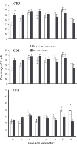
Figs 2, 3, and 4 show the T cell subsets from volun-teers vaccinated with the YF 17DD virus. In all volunvolun-teers an increase in CD3+ T cells was observed. In first-time vaccinees, the level of circulating CD3+ T cells has sig-nificantly increased, from 30.8% (SE ± 4.0%) of the mono-nuclear cells before vaccination to 61.15% (SE ± 4.2%) on the 30th day post vaccination (p = 0.0002) and in re-vaccinees from 50.7% (SE ± 3.0%) to 66.5% (SE ± 2.6%) on the 7th day for 6 of the 9 volunteers (p = 0.0001) and 80% (SE ± 2.3%) on the 30th day after vaccination in the other volunteers (n = 3) (p = 0.0464).
In first-time vaccinees a significant increase in the lev-els of circulating CD8+ T cells was observed. The initial
mean in first-time vaccinees increased from 15.2% (SE ± 2.9) to 27% (SE ± 3.0%) on the 30th day post vaccination (p = 0.0276), with 70% of the cells being identified as
memory cells, according to the analysis of the CD8+ CD45RO+ T cell subset. Among re-vaccinees, the initial concentration of these cells was 19% (SE ± 1.8%) before vaccination and 22.5% (SE ± 2.0%) in 5 volunteers be-tween the 4th and the 10th day (p = 0.2499) and 25% (SE ± 2.0%) on the 30th day post vaccination in the four other volunteers (p = 0.0083) with identification of 90% of the cells as being CD8+CD45RO+ T cells in all volunteers, at day 30. Among the CD8+CD38+ T cells, a marker for acti-vated T cells, a significant increase could be observed, from 15 to 41.6% in first-time vaccinees (p = 0.0207) and from 20.7 to 62.6% in re-vaccinees (p = 0.0017).
Analyzing the levels of CD4+ T cells, it could be ob-served that a significant increase in first-time vaccinees occurred, from 22.4% (SE ± 3.6) to 39.17% (SE ± 2.0%) on the 30th day post vaccination (p = 0.0026), with 43% of these cells being identified as CD4+CD45RO+ T cells. The re-vaccinees have presented levels of circulating CD4+ T cells from 24.9% (SE ± 1.4%) to 35.24% (SE ± 1.2%) be-tween the 4th and the 10th day (n = 5) (p = 0.1613), and to 40% (SE ± 3.0%) on the 30thday post vaccination in the four other volunteers (p = 0.0051), presenting a percent-age of 90 and 95% of CD4+CD45RO+ T cells, respectively. The CD4/CD8 ratios were above one (varying from 1.3 to 2.1) with no difference between the two groups studied (Table).
DISCUSSION
The YF 17DD vaccine, produced in Bio-Manguinhos-Fiocruz, Brazil, is known to be a safe and efficient vaccine, leading to suitable results with regards to nAb produc-tion (Stefano et al. 1999, WHO 2001b, Marchevsky et al. 2003). However, the cellular immune response profile of the YF 17DD vaccine has not been known as yet, although some rare adverse events after vaccination have been suggested as a failure in the cellular immune response of these patients (Galler et al. 2001, Mariannaeu et al. 2001, Vasconcelos et al. 2001).
In this work, we have analyzed some parameters of humoral and cellular immune responses after YF 17DD vaccination.
The volunteers of this study have well tolerated the vaccination. No significant changes were detected in red and white blood cell and platelet counts or in the tran-saminase levels after vaccination.
The methods used to monitor the viremia, PA and RT-PCR, were not able to detect viruses or viral RNA frag-ments in the sera of the first-time vaccinees or re-vaccinees, despite of these methods were used with success by other authors (Reinhardt et al. 1998). A hypothesis for this lack of detection of viremia could be the high titers of neutral-izing antibodies, which would be able to suppress the presence of circulating virus (Wisseman Jr & Sweet 1962) and the decrease of viral circulation that is prompted by YF 17DD virus, unlike the wild type virus (Monath 1998). In a previous study, the RT-PCR was shown to be more sensitive than PA, it was seen that the RT-PCR detects stretches of virus-specific RNA that may represent only RNA fragments or defective viral particles, as a positive assay does not necessarily reflect the presence of viable virus (Reinhardt et al. 1998).
3 3 4 3 3 4 3 3 4 3 3 4
3 3 4 T cell subsets in YF 17D D vaccination • Ana Paula dos Santos et al.
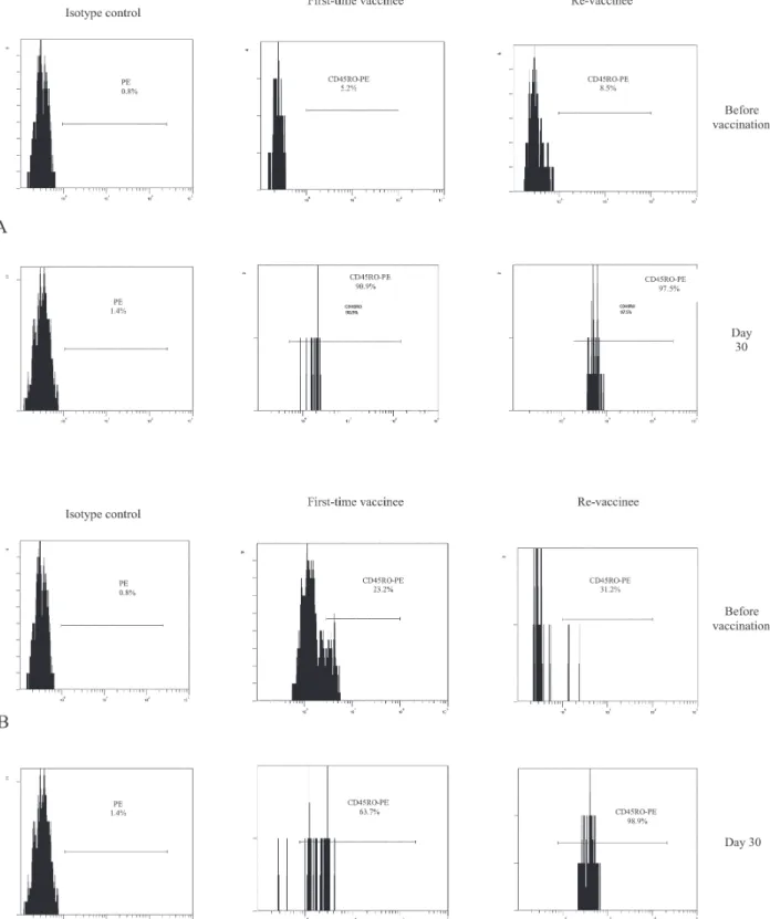
Fig. 3: human peripheral blood mononuclear cell-derived lymphocytes, from volunteers before vaccination and 30th day post immuniza-tion with yellow fever 17DD virus, were dual-stained with CD8-PC5/CD45RO-PE and CD4-PC5/CD45RO-PE. The histograms represent the percentage of CD45RO+ cells gated on CD8+ (A) or CD4+ (B) T cells on day 0 (before vaccination) and 30th day after vaccination in first-time vaccinees and re-vaccinees. Isotype antibodies were used as negative control.
All re-vaccinees have presented high titers of persis-tent neutralizing antibodies before re-vaccination, although they have not been in yellow fever endemic areas within ten years of re-vaccination. These results strengthen the idea of viral presence in lymphatic tissues suggested by
3 3 5 3 3 5 3 3 5 3 3 5 3 3 5 Mem Inst Oswaldo Cruz, Rio de Janeiro, Vol. 100(3), M ay 2005
group of volunteers were similar to those shown for the re-vaccinees.
The CD3+ T cell levels were analyzed to evaluate the profile of cellular immune responses in all vaccinated sub-jects. All volunteers have presented a significant increase in his T cell subset, suggesting an activation of the cellu-lar immune response. With respect to CD8+ T cell counts, it was observed a significant augmentation in all first-time vaccinees and in four re-vaccinees volunteers. This in-crease of CD8+ T cells was expected due to the impor-tance of these cells in the mechanism of the immune re-sponse for intracellular attenuated pathogens vaccinees described by other authors (van der Most et al. 2000, 2002, Co et al. 2002). In our study, the vaccination induced the activation of CD8+ T cells, as could be observed on the significant increase of the CD8+CD38+ T cells in all volun-teers.
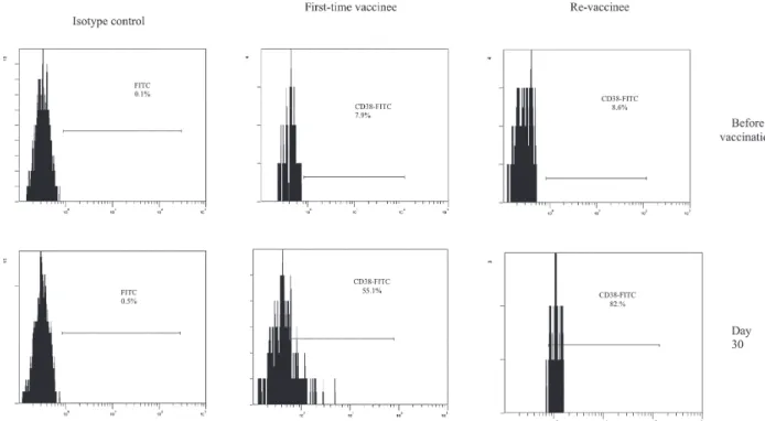
Fig. 4: human peripheral blood mononuclear cell-derived lymphocytes, from volunteers before vaccination and 30th day post immuniza-tion with yellow fever 17DD virus, were dual-stained with CD8-PE/CD38-FITC. The histograms represent the percentage of CD38+ cells gated on CD8+ T cells, in first-time vaccinees and re-vaccinees, day 0 and 30th day after vaccination.
TABLE
Percentage values and ratio of CD4 and CD8 T cell subsets
Days post First-time vaccinees Revaccinees
vaccination %CD4 %CD8 CD4/CD8 %CD4 %CD8 CD4/CD8
0 22.5 15.3 1.5 24.9 19.7 1.3
2 22.8 18.2 1.3 28.4 20.5 1.4
4 35.2 20.7 1.7 31.3 21.3 1.5
7 22.7 17.7 1.3 29.7 17.4 1.7
10 32.0 22.5 1.4 27.0 20.3 1.3
15 29.3 18.2 1.6 24.7 14.3 1.7
30 39.1 24.2 1.6 29.4 20.7 1.4
60 38.8 18.0 2.1 22.7 15.4 1.5
Average of percentage values of CD4+ and CD8+ T cell subsets from 8 first-time vaccinees and 9 re-vaccinees with yellow fever 17DD virus. For standard error see Fig 2.
CD4+ T cells are involved in several steps in the me-chanics of immune response, as antibodies and cytokine production, among other events during the type-2 immune response, through B cells activation and posterior plasma cell differentiation (Mills et al. 2000, Liu & Chambers 2001). As was observed in this study, all first-time vaccinees have presented a significant increase in their numbers of B cells (data not shown) and in the CD4+ T cell subsets. In the group of the re-vaccinees, only four volunteers have presented a significant increase in the latter lym-phocyte subset. It is important to note that these volun-teers also had their CD8+ T cells augmented significantly, as discussed above.
3 3 6 3 3 6 3 3 6 3 3 6
3 3 6 T cell subsets in YF 17D D vaccination • Ana Paula dos Santos et al.
opposite of the infection with flavivirus which presents CD4/CD8 ratio inversion (Lei et al. 2001, Liu et al. 2002).
Not all re-vaccinees have presented significant in-crease in the levels of CD4+ and CD8+ T cells, but the percentage of their activated cell subsets (CD4+CD45RO+ and CD8+CD45RO+ cell markers) was more highly aug-mented than those of the first-time vaccinees. This result has suggested that the re-vaccinees were able to answer to the YF 17DD virus without need for a co-stimulation.
The importance of the CD4+ T cell and CD8+ T cell subsets in nAb production could be observed through analysis of the results from one of the volunteers with leucopoenia, anemia and thrombocytopenia before vacci-nation. In this volunteer, the increase of the cell subsets analyzed did not occur and the nAb induction was ob-served later than in the other volunteers.
Despite of several studies carried out analyzing the levels of lymphocyte subset standards in healthy humans, it is known that such values are not constant, some fac-tors can affect lymphocyte subset counts, such as stress, physical exercises, smoking and alcohol (Goff et al. 1985, Uppal et al. 2003).
The data presented herein provide a basis for further studies on immunological response to the YF 17DD vac-cine in human beings. Therefore, a study of the T cell immune response activation associated to lymphocyte proliferation and cytokine production in YF vaccination is underway.
ACKNOWLEDGMENTS
To Centro de Pesquisa Hospital Evandro Chagas-Fiocruz for measuring blood cells counts and serum biochemical assays and the technical assistance of Adriana de Oliveira Honorato, Bio-Manguinhos-Fiocruz. To Dr Vera Bongertz from De-partamento de Imunologia, Instituto Oswaldo Cruz-Fiocruz for reading the manuscript.
REFERENCES
Boyle W, Chow A 1969. Isolation of human lymphocytes by a Ficoll barrier method. Transfusion 9:151 - 155.
Chan RC, Penney DJ, Little D, Carter IW, Roberts JA, Rawlinson 2001. Hepatitis and death following vaccina-tion with 17D-204 yellow fever vaccine. The Lancet 358: 121-126.
Co MDT, Terajima M, Cruz J, Ennis FA, Rothman AL 2002. Human cytotoxic T lymphocyte responses to live attenu-ated 17D Yellow Fever vaccine: Identification of HLA-B35-restricted CTL epitopes on nonstructural proteins NS1, NS2b, NS3, and the structural protein E. Virology 293:151-163.
Galler R, Pugachev KV , Santos CLS, Ocran SW , Jabor AV, Rodrigues SG, Marchevsky RS, Freire MS, Almeida LFC, Cruz ACR, Yamamura AMY, Rocco IM, Travassos da Rosa ES, Souza LTM, Vasconcelos PFC, Guirakhoo F, Monath TP 2001. Phenotypic and molecular analyses of yellow fever 17DD vaccine viruses associated with serious ad-verse events in Brazil. Virology 290: 309-319.
Goff LK, Habeshaw JA, Rose ML, Gracie JA, Gregory W 1985. Normal values for the different classes os venous blood mononuclear cells defined by monoclonalantibodies. J Clin Pathol 38: 54-59.
Lei HY, Yeh TM, Liu HS, Lin YS, Chen SH, Liu CC 2001. Immunopathogenesis of dengue virus infection. J Biomed Sci 8:377-388.
Liu CC, Huang KJ, Lin YS, Yeh TM, Liu HS, Lei HY 2002. Transient CD4/CD8 ratio inversion and aberrant immune activation during dengue virus infection. J Med Virol 68: 241-252.
Liu T, Chambers TJ 2001. Yellow fever virus encephalitis: Properties of the brain-associated T-cell response during virus clearance in normal and gamma interferon-deficient mice and requirement for CD4+ lymphocytes. J Virol 75: 2107-2118.
Marchevsky RS, Freire MS, Coutinho ESF, Galler R 2003. Neurovirulence of yellow fever 17DD vaccine vírus to rhesus monkeys. Virology 316: 55-63.
Marianneau P, Georges-Courbot MC, Deubel V 2001. Rarity of adverse effects after 17D yellow-fever vaccination. The Lancet 358:84-85.
Martin M, Tsai TF, Cropp B, Chang G-JJ, Holmes DA, Tseng J, Shieh W-J, Zaki SR, Al-Sanouri I , Cutrona AF , Ray G, Weld LH , Cetron MS 2001. Fever and multisystem organ failure associated with 17D-204 yellow fever vaccination a report of four cases. The Lancet 358:98-104.
Mills CD, Kincaid K, Alt JM, Heilman MJ, Hill AM 2000. .M-1/M-2 Macrophages and the Th1/Th2 Paradigm. J Immunol 164:6166-6173.
Monath TP 1998. Yellow fever In S Plotkin, EA Montmer Junior (eds), Vaccine, 3rd ed., WB Saunders, Philadelphia, p. 815-879.
Monath TP, Heinz FX 1996. Flaviviruses. In BN Fields, DM Knipe, PM Howley (eds), Fields Virology, 3rd ed., Lippincott, Raven Publishers, Philadelphia, New York, p. 1009-1016.
Noble PB, Cutts JH, Carroll KK 1968. Ficoll flotation for the separation of blood leukocyte types. Blood 31:66-73.
Oehen S, Waldner H, Kiinding TM, Hengartner H, Zinkernagel RM 1992. Antivirally protective cytotoxic T cell memory to lymphocytic choriomeningitis virus is governed by per-sisting antigen. J Exp Med 176: 1273-1281.
Post PR, Carvalho R, Freire MS, Galler R 2001. The early use of yellow fever vírus strain 17D for vaccine production in Brazil – A review. Mem Inst Oswaldo Cruz 96:849-857.
Reeves WC, Huston GA, Bellamy RE, Scrivany RP 1958. Chronic latent infections of birds with western equine encephalomyelitis virus. Proc Soc Exp Biol Med 97: 733-736.
Reinhardt B, Jaspert R, Niedrig M, Kostner C, L’age-Stelvr J 1998. Development of viremia and humoral and cellular parameters of immune activation after vaccination with yellow fever virus strain 17D: a model of human flavivirus infection. J Med Virol 56:159-167.
Stefano I, Sato HK, Pannuti CS, Omoto TM, Mann G, Freire MS, Yamammura AMY, Vasconcelos PFC, Oselka GW, Weckx LW, Salgado MF, Noale LFO, Souza VAUF 1999. Recent immunization against measles does not interfere with the sero-response to yellow fever vaccine. Vaccine 17: 1042-1046.
3 3 7 3 3 7 3 3 7 3 3 7 3 3 7 Mem Inst Oswaldo Cruz, Rio de Janeiro, Vol. 100(3), M ay 2005
CD8 lymphocyte subsets in healthy Indian adults and the effects of sex, age, ethnicity, and smoking. Cytometry Part B (Clinical Cytometry) 52B:32-36.
van der Most RG, Harrington LE, Giuggio V, Mahar PL, Ahmed R 2002. Yellow fever virus 17D envelope and NS3 proteins are major targets of the antiviral T cell response in mice. Virology 296:117-124.
van der Most RG, Murali-Krishna K, Ahmed R, Strauss JH 2000. Chimeric yellow fever/dengue virus as a candidate dengue vaccine: quantitation of the dengue virus-specific CD8 T-cell response. J Virol 74:8094-8101.
Vasconcelos PFC 2003. Febre amarela. Rev Soc Bras Med Trop 36:1-29.
Vasconcelos PFC, Luna EJ, Galler R, Silva LJ, Coimbra TL, Barros VLRS, Monath TP, Rodrigues SG, Laval C, Costa ZG, Vilela MFG, Santos CLS, Papaiordanou CMO, Alves VAF, Andrade LD, Sato HK, Rosa EST, Froguas GB, Laçava E, Almeida LMR, Cruz ACR, Rocco IM,
Santos RTM, Oliva OFP, Brazilian Yellow Fever Vaccine Evaluation Group 2001. Serious adverse events associ-ated with yellow fever 17DD vaccine in Brazil: a report of two cases. The Lancet 358:91-97.
Wheelock EF, Sibley WA 1965. Circulating virus, interferon and antibody after vaccination with the 17D strain of yel-low fever virus. New England J Med 273:194-198.
Whitton JL, Oldstone MBA 1996. Immune response to viruses In BN Fields, DM Knipe, PM Howley (eds), Fields Virol-ogy, 3rd ed., Lippincott, Raven Publishers, Philadelphia, New York, p. 345-374.
Wisseman Jr CL, Sweet BH 1962. Immunological studies with group B arthropod-borne viruses, III. Response of human subjects to revaccination with 17D strain yellow fever vaccine. Am J Trop Med Hyg 11: 570-575.
WHO 2001a. http://www.who.int/emc/diseases/yellowfever/