ISSN 1546-9239
© 2012 Science Publications
Corresponding Authors: Jintanaporn Wattanathorn, Department of Physiology, Faculty of Medicine, Khon Kaen University, Khon Kaen 40002, Thailand
600
Neuroprotective Effect against
Cerebral Ischemia of Passiflora foetida
1,2
Jintanaporn Wattanathorn,
3Natcha Sattroopinat,
1,2
Terdthai Tong-Un,
1,2Supaporn Muchmapura
1,2
Panakporn Wannanond and
4Panee Sirisa-Ard
1
Department of Physiology,
2
Integrative Complimentary Alternative Medicine Research and Development Group,
3
Department of Physiology and Graduate School (Neuroscience Program),
Faculty of Medicine, Khon Kaen University, Khon Kaen 40002, Thailand
4
Department of Pharmacognosy and Pharmaceutical Technology, Faculty of Pharmacy,
Chiangmai University, Chiang Mai, 50200 Thailand
Abstract: Problem statement: Although cerebral ischemia induced by stroke has been regarded as the important problem worldwide, the therapeutic efficacy is still inadequate. Since the free radicals are implicated in the pathophysiology of cerebral ischemia, the prophylactic protection against stroke with neuroprotective agent possessing antioxidant effect has gained much attention. Therefore, this study was designed to determine whether the alcoholic extract of Passiflora foeida, a plant possessing antioxidant activity, could protect against brain damage and impairment in the cerebral ischemia induced by the occlusion of Middle Cerebral Artery Occlusion (MCAO). Approach: Male Wistar rats, weighing 300-350 g, were orally given the extract once daily at doses of 25, 100 and 400 mg kg−1 BW at a period of 2 weeks before and 3 weeks after the occlusion of right Middle Cerebral Artery (MCAO). The animals were assessed the cerebral infarction volume at 24 h after occlusion while the neurological score and % of foot withdrawal reflex in respond to mechanical stimuli were performed after single dose and every 7 days throughout the experimental period. Results: Rats subjected to
P.foetida at dose of 25 mg kg−1 BW significantly decreased brain infarct volume both in cortical and sub cortical structures. The increasing doses further to 100 and 400 mg kg−1 BW could produce the significant reduction only in cerebral cortex. In addition, it was found that the plant extract could enhance neurological score and improved sensory response to both mechanical and temperature stimuli. Conclusion: The current study clearly demonstrates the neuroprotective effect of P.foetida. Therefore P.foetida may provide the advantage as functional food to protect against cerebral ischemia induced by stroke. However, further researches about possible active ingredient and the precise underlying mechanism are still necessary.
Key words: Passiflora foeida, neuroprotective, certebral ischemia
INTRODUCTION
Cerebral ischemia induced by stroke is recognized as the important problem worldwide because it is one of the major causes of death and disability. Despite the advances of pharmacotherapy nowadays, clinical therapy of the deliberating disorder is still inadequate. Therefore, it has attracted more and more attention for developing novel strategies aim at preventing and reducing impairment induced by stroke. Recently, it has been suggested that prophylactic protection against
stroke with neuroprotective agent may offer useful approach and improve the outcome. However, the agent to be used prophylactic ally should be efficacious, safe, orally available and affordable (Gupta et al., 2010).
against cerebral ischemia (Simonyi et al., 2005) and have a relative higher therapeutic window, lesser side effects and lesser cost consuming, they have gained a lot of acceptance in the recent years and can be potential candidates for prophylactic treatment in stroke.
Passiflora foetida Linn, a plant in a family of Passifloraceae, has been used in traditional folklore for a long time including detoxification, wound healing, antipyretic and analgesic effect. Recent study showed that P.foetida exerted antidepression like effect via dopaminergic and serotonergic system. Moreover, it also possesses antioxidant activity (Osman et al., 2009). Therefore, we hypothesized that P.foetida extract could protect against cerebral ischemia. To date, less scientific about its neuroprotective effect is available, therefore, the current study is set up to determine the neuroprotective effect of P.foetida in experimental model of focal stroke induced by right Middle Cerebral Artery Occlusion (MCAO).
MATERIALS AND METHODS
Experimental animals: Healthy male Wistar rats (300-350gm) were obtained from National Animal Center, Salaya, Nakorn Pathom. They were randomly housed 5 per cage and maintained in 10:14 light: dark cycle and given access to food and water ad labium. The experiments were performed to minimize animals suffering and the experiment protocols were approved by the Institutional Animal Care and Unit Committee Khon Kaen University, Thailand.
Plant material and preparation: The aerial parts of
P.foetidawas collected and authenticated by Associate Professor Panee Sirisa-ard, Department of Pharmacognosy and Pharmaceutical Technology, Faculty of Pharmaceutical Sciences, Chiangmai University, Thailand and the voucher specimen was also kept there. P.foetida was prepared using Soxhlet extraction with ethanol 95%. The percent yield of the final product was 22.67.
The experimental design: Rats were randomly divided in to various groups as described following: (1) Vehicle + MCAO; (2) Aricept + MCAO (positive control); (3) Piracetam + MCAO (positive control); (4) Vitamin C + MCAO (positive control); (5) P.foetida extract (25mg kg−1 BW) + MCAO and (6) P.foetida extract (100mg kg−1 BW.) + MCAO and (7) P.foetida extract (400mg kg−1 BW) + MCAO. All animals were treated with
vehicle or positive control or P.foetida extract at a period of 2 weeks before and 3 weeks after right Middle Cerebral Artery Occlusion (MCAO).
Surgical procedure: Rats were anesthetized by thiopental sodium at dose of 50 mg kg−1 BW. The right common carotid artery and the right external carotid artery were exposed through a ventral midline neck incision and were legated proximally. A silicone coated nylon monofilament (4-0) suture (USS DGTM sutures; Tyco Healthcare group LP, Connecticut, USA) with its tip rounded by heating near a flame was inserted through an arteriectomy in the common carotid artery just below the carotid bifurcation and then advanced into the internal carotid artery approximately 17-18 mm distal to the carotid bifurcation until a mild resistance was felt. Occlusion of the origins of the anterior cerebral artery, the middle cerebral artery and the posterior communicating artery was thereby achieved. Then, the wound was sutured, the rats were returned to their cages with free access to food and water. The incision sites were infiltrated with 10% Providence- Iodine Solution for anti-septic postoperative care.
Infarct volume measurement: The infarct volume was assessed with 2% 2, 3, 5-Triphenyl Tetrazolium Chloride (TTC) solution in saline for 20 min at 37°C. Images of stained sections were digitized and infarction volumes were determined using Olympus light microscope model BH-2(made in Japan) and then they were quantified by an image analysis system
Assessment of neurological deficit: All animals were subjected to neurological evaluation by using 6-points postural reflex test. The deficit was graded from 0-5 as follow: Grade 0: no spontaneous activity; Grade 1: spontaneous circling; Grade 2: circling if pulled by tail; Grade 3: lowered resistance to lateral push without circling; Grade 4: contralateral forelimb flexion; Grade 5: no apparent deficit.
Determination of foot withdrawal reflex time via hot plate test: In the hot plate test, the withdrawal response latency was measured by stopwatch to a maximum cutoff time of 12 s at 56° Celsius and 15 s at 52° Celsius. After the cutoff time, if the rat’s hindpaw still remained on the hot plate, the hindpaw was removed from it to prevent heat injury (i.e., blistering). At each time point, the Withdrwal Reflex Latency (WRL) was measured in triplicate and the mean of these three measurements was considered to be the WRL for that time point.
Statistical analysis: All data were presented as mean Standard Error of Mean (S.E.M). One way Analysis Of Variance (ANOVA) and (Post-Hoc Test) were performed to determine the statistical different. Statistical different was accepted when p-value less than 0.05.
RESULTS
It was found that both Piracetam and Vitamin C, the positive control, used in this study could decrease the infarct volume in cerebral area while no significant change was observed in subcortical area as shown in Fig. 1. Rats subjected to P.foetida extract at dose of 25 mg kg−1 BW significantly decreased the brain infarct volume both in cerebral cortex and subcortical structure (p-value<0.01 and 0.05 respectively; compared to vehicle + MCAO). The increasing dose further to 100 and 400 mg kg−1 BW significantly produced the reduction of brain infarction volume only in cerebral cortex (p-value<0.01 all; compared to vehicle + MCAO).
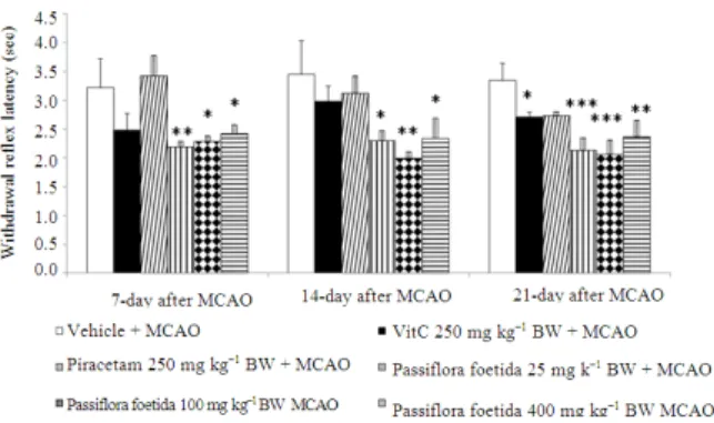
Since cortical and subcortical brain damages could disturb the function of affected areas, we also determined the neurological score and the response of foot withdrawal reflex in respond to both mechanical and temperature stimuli. Figure 2 showed that rats which received Piracetam increased rate of response to mechanical stimuli at 1 g intensity at 14 and 21 days after MCAO while rats which received Vitamin C showed the increased response rate only at 21 days after MCAO. Rats subjected to P.foetida extract at dose of 25 mg kg−1 BW significantly increased the response rates both at 14 and 21 days after MCAO (p-value<0.05 all; compared to vehicle + MCAO). However, the extract at doses of 100 and 400 mg kg−1 BW produced significant increases in response rate only at 14 days after MCAO (p-value<0.05 all; compared to vehicle + MCAO).
Fig. 1: Effect of P.foetida extract on brain infarction induced by right middle cerebral artery occlusion (MCAO). Rats received vehicle or the extract of P.foetida at doses of 25, 100 and 400 mg kg−1 BW once daily at 14 days before and 21 days after MCAO. Then, they were determined brain infarction volume ia TTC staining 24 h after MCAO. (n = 6/group) Results are expressed as mean ± S.E.M. *,**p-value<0.05 and 0.01 respectively; compared to vehicle + MCAO)
Fig. 3: Effect of P.foetida extract on response rate of foot withdrawal reflex in respond to temperature stimuli of rats subjected to right Middle Cerebral Artery Occlusion (MCAO). Rats received vehicle or the extract of P.foetida at doses of 25, 100 and 400 mg kg−1 BW once daily at 14 days before and 21 days after MCAO. Then, they were determined foot withdrawal reflex latency in respond to temperature stimuli. (n = 6/group) Results are expressed as mean ± S.E.M.*,**,*** p-value<0.05, .01and .001 respectively; compared to vehicle + MCAO)
Fig. 4: Effect of P.foetida extract on neurological score of rats subjected to right Middle Cerebral Artery Occlusion (MCAO). Rats received vehicle or the extract of P.foetida at doses of 25, 100 and 400 mg kg−1 BW once daily at 14 days before and 21 days after MCAO. Then, they were determined neurological score. (n = 6/group) Results are expressed as mean ± S.E.M
In addition, it was found that Vitamin C treated rats showed the significant withdrawal reflex latency (WRL) (p-value<0.05; compared to vehicle + MCAO) while no significant change was observed in Piracetam treated group. Rats subjected to the extract at low dose significantly decreased WRL at 7, 14 and 21 days after MCAO (p-value<0.01, .05, .001respectively; compared to vehicle + MCAO). The increasing doses further to 100 and 400 mg kg−1 BW also produced significant reduction of WRL throughout the 21-day experimental
period (p-value<0.05 all; 0.01 and 0.05 respectively; 0.001 and 0.01 respectively; compared to vehicle + MCAO) as shown n Fig. 3.
It was also found that at 7 days after MCAO, only rats subjected to Piracetam treatment significantly increased neurological score (p-value<0.01; compared to vehicle + MCAO). The significant change was also observed at 14 and 21 days after MCAO (p-value<0.001 all; compared to vehicle + MCAO). At 14days after MCAO, only rats which received the extract at dose of 100 mg kg−1 BW showed the significant enhanced neurological score although all doses used in this study increased the neurological score (p-value<0.05; compared to vehicle + MCAO). When the treatment duration was further increased to 21 days, the increased neurological scores were still observed in rats subjected to the extract treatment at all doses used in this study. However, the significant changes were observed only at doses of 25 and 400 mg kg−1 BW (p-value<0.01 and 0.05 respectively; compared to vehicle + MCAO) as shown in Fig. 4.
DISCUSSION
The present study clearly demonstrates that the extract of aerial part of P.foetida decreases the brain infarction and improves brain impairment.
MCAO has been reported to be one of the validated models suitable for testing the neuroprotective agents because it produces a significant ischemic penumbra early after MCA occlusion (Khan et al., 2009; Yousuf et al., 2009). Moreover, this model has been reported to produce impairments that well mimic the clinical characteristic of stroke especially the sensory-motor deficit. It has been proposed that oxidative stress plays a critical role in the development of pathogenesis of cerebral ischemia and the elevation of oxidative stress has been observed in the MCAO model (Loh et al., 2010). Therefore, MCAO has been used as model in this study.
(Gutierrez-Merino et al., 2011) leading to the neuroprotection against cerebral ischemia (Durukan and Tatlisumak, 2007). Therefore, the neuroprotective effect of P.foetida extract might be related to flavonoids content.
In this study, no dose dependent effect of P.foetida
was observed. The possible explanation might be associated with the masking effect of various ingredients in the extract which could possibly mitigate the effect of active ingredient. In addition, numerous mechanisms were playing crucial roles on the neuroprotection as mentioned earlier. Therefore, the relationship between the concentration of extract and the neuroprotective effect was not simple so it failed to show simple relationship.
CONCLUSION
To the best of our knowledge, this is the first demonstration of neuroprotective potential of P.foetida
on brain injury induced by cerebral ischemia. At the present time, the precise protective mechanism and active ingredient are yet to be verified. This study was just concentrated on general effect of P.foetida on neuroprotection against cerebral ischemia induced by stroke. Further researches focusing on the precise underlying mechanism are still essential.
ACKNOWLEDGEMENT
This study was supported by North-Eastern Stroke Research Group and Integrative Complimentary Alternative Research and Development Group, Khon Kaen University, Khon Kaen, Thailand 40002.
REFERENCES
Bei, W., L. Zang, J. Guo, W. Peng and A. Xu et al., 2009. Neuroprotective effects of a standardized flavonoid extract from Diospyros kaki leaves. J. Ethnopharmacol., 126: 134-142. DOI: 10.1016/j.jep.2009.07.034
Durukan, A. and T. Tatlisumak, 2007. Acute ischemic stroke: Overview of major experimental rodent models, pathophysiology and therapy of focal cerebral ischemia. Pharm. Biochem. Beh., 87: 179-197. PMID: 17521716
Gupta, Y.K., S. Breeyal and A. Gulati, 2010. Therapeutic potential of herbal drugs in cerebral ischemia. Indian J. Pharmacol. Phys., 54: 99-122. PMID: 21090528
Gutierrez-Merino, C., C. Lopez-Sanchez, R. Lagoa, A.K. Samhan-Arias and C. Bueno et al., 2011. Neuroprotective actions of flavonoids. Curr. Med. Chem., 18: 1195-212. PMID: 21291366
Khan, M.M., A. Ahmad, T. Ishrat, G. Khuwaja and P. Srivastawa et al., 2009. Rutin protects the neural damage induced by transient focal ischemia in rats. Brain Res., 1292: 123-135. PMID: 19631195 Loh, K.P., J. Qi, B.K. Tan, X.H. Liu and B.G., Wei et
al., 2010. Leonurine protects middle cerebral artery occluded rats through antioxidant effect and regulation of mitochondrial function. Stroke, 41: 2661-2668. PMID: 20947850
Osman, H., A.A. Rahim, N.M. Isa and N.M. Bakhir, 2009. Antioxidant activity and Phenolic content of Paederia foetida and Syzygium aqueum. Molecules, 14: 970-978. PMID: 19305354
Simonyi, A., Q. Wang, R.L. Miller, M. Yusof and P.B. Shelat et al., 2005. Polyphenols in cerebral ischemia: Novel targets for neuroprotection. Mol Neurobiol, 31: 135-147. PMID: 15953817
Ulubelen, A., S. Oksuz, T.J. Mabry, G. Dellamonica and J. Chopin, 1982. C-glycosyl flavonoids from Passiflora Pittieri, P. Alata, P., Ambigua and Adenia Mannii. J. Nat Prod., 45: 783-783. DOI: 10.1021/np50024a030