ARTICLE
Pre, intra and post-ischemic hypothermic
neuroprotection in temporary focal cerebral
ischemia in rats: morphometric analysis
Neuroproteção hipotérmica pré, intra e pós-isquêmica na isquemia cerebral focal
temporária em ratos: análise morfométrica
Roberto Alexandre Dezena1, Benedicto Oscar Colli2, Carlos Gilberto Carlotti Junior3, Luís Fernando Tirapelli4
Experimental Neurosurgery Laboratory, Department of Surgery and Anatomy, Ribeirão Preto Medical School, Universidade de São Paulo SP, Brazil.
1M.D., Ph.D.; Neurosurgeon, Division of Neurosurgery, Department of Surgery and Anatomy, Ribeirão Preto Medical School, Universidade de São Paulo (USP),
Ribeirão Preto SP, Brazil; Division of Neurosurgery, Universidade Federal doTriângulo Mineiro (UFTM), Uberaba MG, Brazil;
2M.D., Ph.D.; Professor of Neurosurgery, Head and Chairman, Division of Neurosurgery, Department of Surgery and Anatomy, Ribeirão Preto Medical School,
USP, São Paulo SP, Brazil;
3M.D., Ph.D.; Professor of Neurosurgery, Division of Neurosurgery, Department of Surgery and Anatomy, Ribeirão Preto Medical School, USP, São Paulo SP, Brazil; 4M.Sc., Ph.D.; Professor of Anatomy, Division of Anatomy, Department of Surgery and Anatomy, Ribeirão Preto Medical School, USP, São Paulo SP, Brazil.
Correspondence: Roberto Alexandre Dezena; Rua Veríssimo 1.264/202-2; 38022-090 Uberaba MG - Brasil; E-mail: [email protected]
Support: Ribeirão Preto Medical School, Universidade de São Paulo (FMRP-USP); Fundação de Amparo à Pesquisa do Estado de São Paulo (FAPESP); Conselho Nacional de Desenvolvimento Cientíico e Tecnológico (CNPq).
Conflict of interest: There is no conlict of interest to declare.
Received 04 March 2012; Received in inal form 11 April 2012; Accepted 18 April 2012
ABSTRACT
Objective: To evaluate the neuroprotection of mild hypothermia, applied in different moments, in temporary focal cerebral ischemia in rats. Methods: Rats was divided into Control (C), Sham (S), Ischemic-control(IC), Pre-ischemic Hypothermia (IH1), Intra-ischemic Hypothermia (IH2), and Post-ischemic Hypothermia (IH3) groups. Morphometry was performed using the KS400 software (Carl Zeiss®) in coronal sections
stained by Luxol Fast Blue. Ischemic areas and volumes were obtained. Results: Statistically, blue areas showed difference for C vs. IC, IC vs. IH1 and IC vs. IH2 (p=0.0001; p=0.01; p=0.03), and no difference between C vs. S, IC vs. IH3 and IH vs. IH2 (p=0.39; p=0.85; p=0.63). Red areas showed difference between C vs. IC, IC vs. IH1 and IC vs. IH2 (p=0.0001; p=0.009; p=0.03), and no difference between C vs. S, IC vs. IH3 and IH1 vs. IH2 (p=0.48; p=0.27; p=0.68). Average ischemic areas and ischemic volumes showed difference between IC vs. IH1 and IC vs. IH2 (p=0.0001 and p=0.0011), and no difference between IC vs. IH3 and IH1 vs. IH2 (p=0.57; p=0.79). Conclusion: Pre-ischemic and intra-ischemic hypother-mia were shown to be similarly neuroprotective, but this was not true for post-ischemic hypotherhypother-mia.
Key words: brain ischemia, mild hypothermia, morphometry, neuroprotection, reperfusion.
RESUMO
Objetivo: Avaliar a neuroproteção da hipotermia leve, aplicada em diferentes momentos, durante isquemia cerebral focal temporária em ratos .Métodos: Ratos foram divididos em grupos: Controle (C), Sham (S), Controle-isquêmico (IC), Hipotermia Pré-isquêmica (IH1), Hipo-termia Intra-isquêmica (IH2) e HipoHipo-termia Pós-isquêmica (IH3). A morfometria foi realizada em secções coronais coradas por Luxol Fast Blue através do programa KS400 (Carl Zeiss®). Foram calculados áreas e volumes isquêmicos. Resultados: Estatisticamente, áreas azuis
demonstraram diferença entre os grupos C vs. IC, IC vs. IH1 e IC vs. IH2 (p=0,0001; p=0,01; p=0,03), e nenhuma diferença entre C vs. S, IC vs. IH3 e IH vs. IH2 (p=0,39; p=0,85; p=0,63). Áreas vermelhas demonstraram diferença entre C vs. IC, IC vs. IH1 e IC vs. IH2 (p=0,0001; p=0,009; p=0,03), e nenhuma diferença entre C vs. S, IC vs. IH3 e IH1 vs. IH2 (p=0,48; p=0,27; p=0,68). Áreas isquêmicas médias e volumes isquêmicos demonstraram diferença entre os grupos IC vs. IH1 e IC vs. IH2 (p=0,0001 and p=0,0011), e nenhuma diferença entre IC vs. IH3 and IH1 vs. IH2 (p=0,57; p=0,79). Conclusão: Hipotermias pré-isquêmica e intra-isquêmica demonstraram neuroproteção em grau semelhante, o que não ocorreu com hipotermia pós-isquêmica.
Cerebral ischemia is a very common disease, with unpre-dictable clinical outcome, and limited prophylaxis and treat-ment. Vasospasm after subarachnoid hemorrhage and tempo-rary vascular occlusion on vascular microneurosurgery are the main situations of focal cerebral ischemia for the neurosurgeon. Marked decrease of cerebral blood low triggers a series of bio-chemical events, with histopathological consequences which, if not blocked, leads to neuronal death. hree concentric areas are histopathologically deined in focal ischemia: the central ischemic zone, the ischemic penumbra zone and the external zone of normal tissue1. In ischemic penumbra zone initially
the cells remain morphologically normal, but lose their elec-trical function, featuring an isoelectric electroencephalogram and, depending on the time of ischemia and on the anasto-motic supply, cell death will occur. Moreover, this area is the most frequent site of apoptosis1. On the other hand, this area
is likely to save when neuroprotective strategies are adopted. Several animals have been used for experimental studies on cerebral ischemia, and among them, the rat is the most used2.
he neuroprotective efect of hypothermia in experimental ce-rebral ischemia is described since 1950’s3. Neuroprotection
af-forded by hypothermia is classically attributed to reduction of metabolic demands. In addition, other mechanisms have been proposed1,4,5. Hypothermia can be applied at diferent times of
ischemia: before (pre-ischemic), during (intra-ischemic), and after ischemia (post-ischemic). he intra-ischemic hypother-mia is the most studied, and it is the most efective4. his
mo-dality of hypothermia signiicantly reduces the total volume of experimental infarction in the basal ganglia and cortex6,7, and
also improves clinical evaluation8. In the post-ischemic mode,
hypothermia acts mainly by inhibiting the “reperfusion injury”9.
Although less efective than intra-ischemic hypothermia, it re-duces experimental neuronal loss in hippocampus induced by ischemia10, reduce the volume of cerebral infarction6,11 and
typi-cally reduces cortical infarct volume12. Classically, to obtain the
maximum neuroprotective efect with this mode of hypother-mia in experimental temporary ischehypother-mia, it must be institut-ed within 15 minutes after reperfusion13. However, recent
stud-ies demonstrate a viable window of tolerance up three to four hours to get maximum of neuroprotection14,15. his window can
vary according the grade and duration of hypothermia4. As far
as our knowledge, there are no reports in the literature about the efect of pre-ischemic hypothermia. his study aimed to compare the neuroprotective efect of mild hypothermia (32– 34ºC) at diferent times of ischemia: (pre, intra and post-isch-emia) in rats submitted to focal cerebral ischemia by temporary occlusion of the middle cerebral artery (MCA).
METHODS
his study was approved by the Ethics Committee on Animal Research, Ribeirão Preto Medical School, Universidade de São Paulo (Protocol nº108/2009).he experiments were performed
according to the standards proposed by the Brazilian College of Animal Experimentation and the International Council for Laboratory Animal Science. We used adult male Wistar rats, weighing 280‒310 g. Before the surgical procedure, the animals were randomly distributed, by ballot, in six groups. All groups, except the Control and Sham were submitted to 60 minutes of ischemia, and 24 hours of reperfusion, using or not diferent patterns of hypothermia. hey were divided in Control group (C): ten animals in normothermic conditions euthanized after anesthesia and stabilization of the homeostasis parameters, without the surgical procedure; Sham group (S): ten animals in normothermic conditions euthanized after simulation of the surgical procedure, with the introduction of the obstructer, but without occlusion of the MCA; Ischemic Control group (IC): 10 animals submitted to 60 minutes of ischemia and 24 hours of reperfusion; Pre-Ischemic Hypothermia group (IH1):10 ani-mals submitted to 30 minutes of pre-ischemic hypothermia ex-tended into the ischemic period itself (plus 60 minutes), and 24 hours of reperfusion; Intra-Ischemic Hypothermia group (IH2): 10 animals submitted to 60 minutes of intra-ischemic hy-pothermia, returning to normothermia at the beginning of 24 hours of reperfusion; Post-Ischemic Hypothermia group (IH3): 10 animals submitted to 60 minutes of ischemia and 6 hours of post-ischemic hypothermia, initiated at the time of reperfu-sion. Anesthesia was induced through inhalation of halothane in a glass campanula and kept with a vaporizer linked to a me-chanical respirator after endotraqueal intubation. Mean inva-sive blood pressure was monitored through a PE 50 catheter inserted in the tails’ ventral artery, and arterial blood samples were collected after 15 minutes the mechanical ventilation was started and during the inal 15 minutes of ischemia, to mea-sure parameters of homeostasis (PaCO2, hemoglobin,
hemato-crit, blood glucose). hese parameters were kept according to previous standartization2, and blood pressure and PaCO
2 were
kept adjusting the parameters of the respirator. Body temper-ature was intermittently recorded using a rectal thermistor and kept between 37 and 38°C using a 220V lamp next to the animal, for the groups performed using normothermia. Mild (32–34°C) pre- and intra-ischemic hypothermia were obtained moisten the anesthetized animals with cooled saline solution and reducing the laboratory temperature with air conditioning equipment. Post-ischemic hypothermia was obtained by plac-ing the animals in a container containplac-ing ice, immediately af-ter extubation. he animals were kept sedated with gauze im-mersed in halothane placed on the nose every hour, until the end of six hours. he surgery was performed, according previ-ously described technique16. he left cervical vessels were
euthanized with massive inhalation of halothane, and under-went thoraco-phreno-laparotomy, the abdominal aorta was clamped and a manual transcardiac perfusion with bufered sa-line solution (0.1 M phosphate bufer, pH 7.3 to 4° C) was per-formed, followed by infusion of ixative solution of 4% parafor-maldehyde in bufer 0.1 M phosphate at pH 7.3). he brains were ixed in 10% bufered formalin, dehydrated cleared and embed-ded in parain. Sequential 10 µm thick coronal sections were performed throughout the ischemic area resulting in about a thousand sections for each brain. One in each sequential 15 was selected for coloring, resulting in a total of 60 coronal sections stained for each brain. he sections were stained by Luxol Fast Blue to emphasize the neuronal bodies and non-myelinated ibers in pink-purple, and myelin ibers in blue-green17. Digital
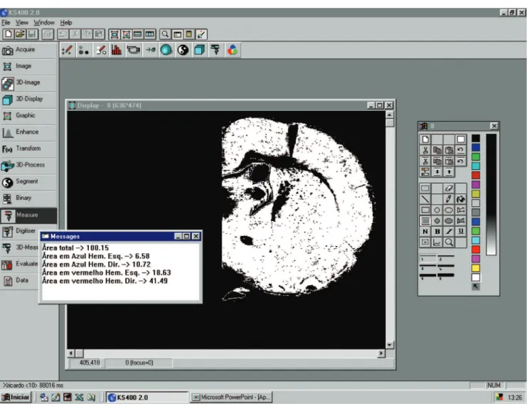
images of stained sections were captured using analogical-digi-tal camera model JVC TK1270, with 55 mm wide angle lens, set at a vertical holder. A ruler was used for calibration for subse-quent conversion of digital measures (pixels) in area measures (mm2). he measurement areas of each section were performed
using the image analysis program KS400, Carl Zeiss, version 2.0, in a microcomputer IBM-PC and video capture card FG1, Carl Zeiss. he total area of the coronal section, the blue areas (my-elin ibers) and red areas (neuron bodies and non-my(my-elinated
areas), of both hemispheres were measured successively by the program (Fig 1), and the respective averages of these areas were calculated for each animal. Ratios (percentage) of left (isch-emic)/right (normal) hemispheres, for both blue and red areas, were calculated using the average areas18. he ischemic area of
each section was obtained by the diference between the total area and the sum of all measures in blue and red areas, of both hemispheres. he partial ischemic volume, for 60 stained sec-tions, was calculated of each animal using measurements of ischemic areas, the thickness of each section and the number of sections using Bartustechnique19 and corrected when
nec-essary according to Chang technique20. he approximate
isch-emic volume was obtained by the partial volume x 1,000/60. Statistical analysis was performed using the GraphPad InStat program for Windows, version 3.06 (GraphPad Software Inc., San Diego, USA). Analyses of the homeostasis parameters were performed using the analysis of variance (ANOVA) test. For group to group comparison of the percentage of the left hemi-sphere over the right hemihemi-sphere of each animal, of the stained areas, and of the average ischemic areas (mm2), and
approxi-mated ischemic volumes (mm3), was used the nonparametric
test of Mann-Whitney-Wilcoxon. A signiicance level of p<0.05 was considered signiicant for the two-tailed tests.
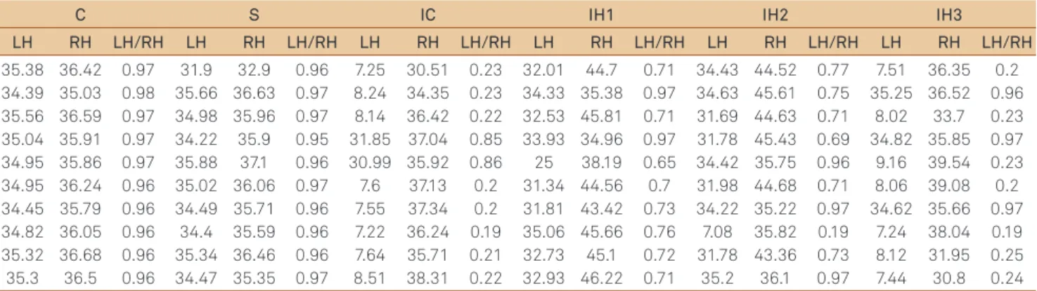
Table 1. Averages of blue areas (mm2), of both hemispheres, and respective ratios for animals of all experimental groups.
C S IC IH1 IH2 IH3
LH RH LH/RH LH RH LH/RH LH RH LH/RH LH RH LH/RH LH RH LH/RH LH RH LH/RH
6.31 4.71 6.36 4.97 5.96 5.54 5.9 5.27 5.79 5.2 6.69 4.96 6.8 5.66 6.65 6.26 6.38 5.92 6.45 5.77 0.94 0.94 0.93 0.87 0.89 0.88 0.92 0.89 0.89 0.9 6.97 6.28 5.34 4.79 5.79 5.01 5.25 5.55 6.28 5.86 7.62 6.54 5.9 5.25 6.46 5.46 5.64 6.18 6.57 6.52 0.91 0.96 0.9 0.91 0.89 0.91 0.93 0.89 0.95 0.89 2.94 2.99 2.99 5.84 4.86 2.75 2.37 2.58 2.72 2.9 6.13 6.27 6.33 6.76 5.69 5.84 5.04 5.29 5.72 6.2 0.47 0.47 0.47 0.86 0.85 0.47 0.47 0.48 0.47 0.46 4.28 5.57 4.19 6.15 4.62 4.06 3.87 4.64 4.52 4.41 6.39 6.52 6.21 6.71 7.06 6.22 5.69 6.78 6.52 6.52 0.66 0.85 0.67 0.91 0.65 0.65 0.68 0.68 0.69 0.67 4.24 4.58 4.09 4.45 5.39 4.27 5.59 2.37 3.85 5.41 5.89 6.99 5.92 6.62 5.88 6.35 6.2 5.2 5.67 5.72 0.71 0.65 0.69 0.67 0.91 0.67 0.9 0.45 0.67 0.94 2.78 6.22 2.73 6.81 2.91 2.99 6.48 2.43 3.37 3.07 5.86 6.88 5.76 7.48 6.43 6.45 6.84 5.19 8.28 6.24 0.47 0.9 0.47 0.91 0.45 0.46 0.94 0.46 0.4 0.49
C: Control group; S: Sham group; IC: Ischemic-control group; IH1: Pre-ischemic hypothermia group; IH2: Intra-ischemic hypothermia group; IH3: Post-ischemic hypothermia group; LH: left brain hemisphere; RH: right brain hemisphere.
There was signiicant difference between C vs. IC, IC vs. IH1, and IC vs. IH2 groups (respectively, p=0.0001; p=0.01; and p=0.03 – Mann-Whitney-Wilcoxon).There was no signiicant difference between C vs. S, IC vs. IH3, and IH1 vs. IH2 groups (respectively, p=0.39; p=0.85; and p=0.63 – Mann-Whitney-Wilcoxon).
RESULTS
Seventy four male Wistar rats were operated. Of these, six animals were excluded during surgery by inadequate homeo-stasis parameters. here were eight deaths in this phase ( four animals in IH1 group, two animals in IH3 group, and one animal in IC and IH2 group). All rejected and dead animals were replaced totalizing ten animals per experimental group. he laboratory parameters during surgical procedure were
within the limits established for this type of experiment2.
here was no signiicant diference among all groups in rela-tion to initial and inal values of PaCO2 (respectively, p=0.47
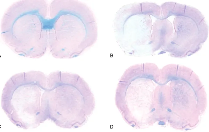
and p=0.4, ANOVA), hemoglobin (respectively, p=0.84 and p=0.49, ANOVA), hematocrit (respectively, p=0.57 and p=0.56, ANOVA), and blood glucose (p=0.34 and p=0.09, ANOVA). Fig 2 presents coronal sections of brains of animals of control and ischemic groups and Tables 1 and 2 present the average of respectively blue and red areas for the animals of all groups.
Fig 2. Coronal sections of the brain of animals from Control and ischemic groups stained with Luxol Fast Blue (myelinated structures stained in blue-green, and neuronal bodies and non-myelinated structures in pink-purple). (A) Control group. Animals of the Sham group showed similar aspect. (B) Ischemic-control group. Note the left hemisphere ischemic area (clear), extending from basal ganglia to the cortical region. (C) Pre-ischemic hypothermia group. Smaller left hemisphere ischemic area when compared to Ischemic-control group (B). (D) Intra-ischemic hypothermia group. Smaller left
hemisphere ischemic area, when compared to Ischemic-control group (B) and similar to Intra-ischemic hypothermia group (C).
C
D
Table 2. Averages of red areas (mm2), of both hemispheres, and respective ratios for animals of all experimental groups.
C S IC IH1 IH2 IH3
LH RH LH/RH LH RH LH/RH LH RH LH/RH LH RH LH/RH LH RH LH/RH LH RH LH/RH
35.38 34.39 35.56 35.04 34.95 34.95 34.45 34.82 35.32 35.3 36.42 35.03 36.59 35.91 35.86 36.24 35.79 36.05 36.68 36.5 0.97 0.98 0.97 0.97 0.97 0.96 0.96 0.96 0.96 0.96 31.9 35.66 34.98 34.22 35.88 35.02 34.49 34.4 35.34 34.47 32.9 36.63 35.96 35.9 37.1 36.06 35.71 35.59 36.46 35.35 0.96 0.97 0.97 0.95 0.96 0.97 0.96 0.96 0.96 0.97 7.25 8.24 8.14 31.85 30.99 7.6 7.55 7.22 7.64 8.51 30.51 34.35 36.42 37.04 35.92 37.13 37.34 36.24 35.71 38.31 0.23 0.23 0.22 0.85 0.86 0.2 0.2 0.19 0.21 0.22 32.01 34.33 32.53 33.93 25 31.34 31.81 35.06 32.73 32.93 44.7 35.38 45.81 34.96 38.19 44.56 43.42 45.66 45.1 46.22 0.71 0.97 0.71 0.97 0.65 0.7 0.73 0.76 0.72 0.71 34.43 34.63 31.69 31.78 34.42 31.98 34.22 7.08 31.78 35.2 44.52 45.61 44.63 45.43 35.75 44.68 35.22 35.82 43.36 36.1 0.77 0.75 0.71 0.69 0.96 0.71 0.97 0.19 0.73 0.97 7.51 35.25 8.02 34.82 9.16 8.06 34.62 7.24 8.12 7.44 36.35 36.52 33.7 35.85 39.54 39.08 35.66 38.04 31.95 30.8 0.2 0.96 0.23 0.97 0.23 0.2 0.97 0.19 0.25 0.24
C: Control group; S: Sham group; IC: Ischemic-control group; IH1: Pre-ischemic hypothermia group; IH2: Intra-ischemic hypothermia group; IH3: Post-ischemic hypothermia group; LH: left brain hemisphere; RH: right brain hemisphere.
There was signiicant difference between C vs. IC, IC vs. IH1, and IC vs. IH2 groups (respectively, p=0.0001; p=0.009; and p=0.03 – Mann-Whitney-Wilcoxon). There was no signiicant difference between C vs. S, IC vs. IH3, and IH1 vs. IH2 groups (respectively, p=0.48; p=0.27; and p=0.68 – Mann-Whitney-Wilcoxon).
Table 3. Average ischemic areas (mm2) for animals of the ischemic groups.
IC IH1 IH2 IH3
23.36 33.8 35.8 9.51 11.7 37.71 41.72 40.12 37.53 38.14 9.07 7.18 7.43 5.58 13.74 9.25 10.43 4.31 9.13 6.43 8.23 4.65 8.93 8.45 6.4 9.19 4.81 39.53 10.55 4.75 38.23 3.73 32.21 3.13 38.73 38.64 4.98 43.05 24.95 22.7
IC: Ischemic-control group; IH1: Pre-ischemic hypothermia group; IH2: Intra-ischemic hypothermia group; IH3: Post-Intra-ischemic hypothermia group; LH: left brain hemisphere; RH: right brain hemisphere.
There was signiicant difference between IC vs. IH1 and IC vs. IH2 groups (respectively, p=0.0001 and p=0.0011 – Mann-Whitney-Wilcoxon). There was no signiicant difference between IC vs. IH3 and IH1 vs. IH2 groups (respectively, p=0.57 and p=0.79 – Mann-Whitney-Wilcoxon).
Table 4. Approximate ischemic volumes (mm3) for animals of the ischemic groups.
CI IH1 IH2 IH3
239.7 344.76 361.76 96.9 119.34 384.54 425.51 409.19 382.67 388.96 92.48 73.1 75.65 56.78 140.08 94.35 106.25 43.86 92.99 65.45 83.81 47.43 90.95 86.19 65.28 93.67 48.96 403.07 107.61 48.45 389.81 37.91 328.44 31.79 394.91 394.06 50.66 439.11 254.49 231.54
IC: Ischemic-control group; IH1: Pre-ischemic hypothermia group; IH2: Intra-ischemic hypothermia group; IH3: Post-Intra-ischemic hypothermia group; LH: left brain hemisphere; RH: right brain hemisphere.
There was signiicant difference between IC vs. IH1 and IC vs. IH2 groups (respectively, p=0.0001 and p=0.0011 – Mann-Whitney-Wilcoxon). There was no signiicant difference between IC vs. IH3 and IH1 vs. IH2 groups (respectively, p=0.57 and p=0.79 – Mann-Whitney-Wilcoxon).
Concerning ratios of averages of blue areas (myelinated i-bers) there was signiicant diference between groups C vs.
IC, IC vs. IH1 and IC vs. IH2 (respectively, p=0.0001, p=0.01,
and p=0.03, Mann-Whitney-Wilcoxon), and there was no sig-niicant diference between groups C vs. S, IC vs. IH3, and
IH1 vs. IH2 (respectively, p=0.39, p=0.85, and p=0.63,
Mann-Whitney-Wilcoxon). Concerning ratios of averages of red ar-eas (neuronal bodies and non-myelinated ibers), there was signiicant diference between groups C vs. IC, IC vs. IH1, and
IC vs. IH2 (respectively, p=0.0001, p=0.009, and p=0.03,
Mann-Whitney-Wilcoxon), and there was no signiicant diference between groups C vs. S, IC vs. IH3, and IH1 vs. IH2
(respective-ly, p=0.48, p=0.27, and p=0.68, Mann-Whitney-Wilcoxon). he analysis for average ischemic areas Table 3 and approximate ischemic volumes Table 4 showed signiicant diference be-tween IC vs. IH1 and IC vs. IH2 groups (respectively, p=0.0001
and p=0.0011, Mann-Whitney-Wilcoxon), and showed no sig-niicant diference between IC vs. IH3 and IH1 vs. IH2 groups
(p=0.57 and p=0.79, Mann-Whitney-Wilcoxon). Fig 3 presents the approximate ischemic volumes of the ischemic groups.
Isc hemic v olumes (mm 3) Experimental groups IH3
IC IH1 IH2
450 400 350 300 250 150 50 0 100 200
DISCUSSION
he strain of Wistar rats has the advantage of not sufer a so intense ischemia, and consequently edema, as occurs in spontaneous hypertensive rat (SHR) strain that has little col-lateral circulation, nor sufer a limited ischemia, as occurs in the Long Evans rat’s strain, especially when there is reperfu-sion, because it has abundant collateral circulation14. Male
animals were chosen because there is evidence that females have neuroprotection, due to sex speciic hormones1. Adult
animals were used, because younger rats, usually weighing less than 300 g, have more developed collateral vessels, pro-viding greater resistance to infarction21. he procedure used
to induce ischemia2,16 was efective, and also at viable cost. Its
great advantage was the possibility of reversion of ischemia, allowing reperfusion when intraluminal obstructer suture is removed, diferently from others which use craniotomy with coagulation and/or ligation of the MCA22. his method allows
until 70% of positivity for ischemia, and the fact that some animals do not demonstrate ischemia, in ischemic groups, is possibly due individual anatomical circulation variations of each animal21. Deaths occurred mainly in the hypothermic
groups, probably due to exposure to low temperature for a long time, and the rate got in this study did not difer from other current studies of the same nature18,23,24. he anesthetic
agent used for all experimental groups was the halothane, be-cause it does not produce signiicant changes on intracranial pressure, as well as because of its accessibility and low cost-efective25. A neuroprotective efect attributed to the
halo-thane is questioned in previous studies18,26. In our study this
possible efect probably had not an important role because it was used in the same way for all experimental groups. he parameters of the homeostasis (PaCO2, hemoglobin,
hemato-crit and blood glucose) that could somehow change the out-come of ischemic results were always kept within accepted values for this type of experiment2, to avoid interference in
the ischemic insult. Morphometric analysis of area and vol-ume of focal cerebral ischemia represents an objective way to estimate the extent of ischemic injury and is commonly used for measuring the eicacy of neuroprotective agents7,18,27. In
this study, we used a new method of semi-automated mor-phometric analysis using the KS400 computer program, (Carl Zeiss®), described by Santana18. his technique uses coronal
sections of the ischemic brain stained with Luxol Fast Blue17.
his stain allows diferentiation between neuronal bodies and myelinated ibers, and the execution of morphometry di-rect from the lamina, without additional procedures. Moreover, it permits the exclusion of natural empty space, as ventricles and cisterns, or artifactual spaces from histological processing. he method was efective for detecting tempo-rary brain ischemia of 90 minutes, with 30 days18. he
pro-gram provided values for areas stained in blue (myelinated ibers) and red (neuronal bodies), of each hemisphere
separately, for each coronal section, and allows to calculate the means for all these values. Because volumes were ob-tained by multiplying the average ischemic areas by the thickness of the cuts, the average ischemic areas and the ap-proximate ischemic volumes showed similar statistical be-havior in the ischemic groups. Hypothermia has been consid-ered the most efective resource, including drugs, to reduce brain injury caused by ischemia in experimental studies25. It
can change many of the harmful efects consequent to isch-emia by suppressing molecular pathways of cell death, as ob-served in experimental models1. Since the irst description of
the neuroprotective efect of mild hypothermia in intra-isch-emic temporary ischemia in rats25, numerous experimental
studies has demonstrated its beneicial efect. he intra-isch-emic modality highly reduces brain infarct size in most ex-perimental models6,7. Most studies show that intra-ischemic
mild hypothermia in temporary focal ischemia is able to greatly reduce the cerebral infarction14. In a meta-analysis of
experimental models for the study of cerebral ischemia it was concluded that the intra-ischemic mild hypothermia can re-duce infarct volume by 44%28. In rats under the post-ischemic
hypothermia, the neuroprotective efect is usually minor10,
and in experimental studies, there is a window of up to 60 minutes of permanent cerebral ischemia and up to 180 min-utes for temporary ischemia14,15, for what recovery of the
ischemic insult can occurs. Increasing the time of the hypo-thermia may increase its efectiveness14,23. he efect of
pre-ischemic hypothermia is unclear. In this study, we used mild hypothermia, which is used in most experimental models be-cause fewer side efects28, and it was applied before, during
and after focal temporary brain ischemia (pre, intra and post-ischemia). Our results are in agreement with these data. We also noted that neuroprotection occurred for both neuronal bodies and myelinated ibers. Furthermore, we demonstrate for experimental temporary focal ischemia, with 24 hours of reperfusion, that pre-ischemic mild hypothermia, started 30 minutes before ischemia, ofers no additional protection compared to the intra-ischemic hypothermia. However, we do not rule out the possibility that pre-ischemic cooling used was insuicient to increase the protection. he neuroprotec-tor efect of post-ischemic hypothermia on brain focal isch-emia is controversial. Some authors advocate the use of pro-longed hypothermia for 24 hours of reperfusion10 or 48 hours11
to achieve neuroprotection. However, recent study suggests that it should be initiated in the exact moment of reperfusion and that one hour of duration is enough25. Baumann et al.23,
post-ischemic hypothermia group and ischemic-control group regarding extension and volume of the ischemia. Although the rectal temperature in animals of experimental groups remained within the limits of mild hypothermia ad-opted for this study (32–34°C), it is possible that brain tem-perature was not at level. his fact is corroborated by a recent study that showed maintenance of rectal temperature at nor-mal levels in aninor-mals undergoing temporary cerebral isch-emia with cerebral hypothermia induced by infusion of intra-cerebral cold blood, resulting in decreased brain temperature and rectal temperature remained normal24. herefore, these
authors recently advocated the use of endovascular cooling catheters in the studies of cerebral ischemia in rats, in-stead of local or systemic cooling. Another fact is that the ani-mals of the post-ischemic group, despite the sedation, had shiver after recovery of anesthesia and this could negatively inluenced the maintenance of hypothermia. Because of this, it is recommended the use of drugs that inhibit shiver in ex-perimental studies and in future clinical trials using post-ischemic hypothermia. Extrapolation of its results for hu-mans being in clinical settings is the main diiculty of experimental studies. Despite the positive efect of hypother-mia in experimental studies there is lack of multicenter stud-ies with a large number of patients sufering from cerebral ischemia. Positive results involving survivors of cardiac arrest and infants with hypoxic encephalopathy suggest that hypo-thermic neuroprotection is likely to be promising25, and there
is already enough scientiic evidence that support the begin-ning of large clinical studies. Despite the negative inding of the intraoperative hypothermia for aneurysm surgery trial (IHAST)29, it is possible that such results may be explained by
an insuicient cooling time, followed by rapid rewarming, and, in addition, this study has other limitations such as pri-ority to patients in good neurological status (low risk of isch-emic injury) and use as a clinical parameter the Glasgow out-come scale (GOS), which fails to adequately evaluate cognitive functions. In addition, further analysis of the results showed that, especially in speciic groups (males, and pa-tients with surgery performed between 8 to 14 days after the hemorrhagic ictus), there was improvement in the inal neu-rological outcome4. Given the controversy surrounding the
issue, beneicial efects of hypothermia, especially in clinical settings, remains to be better investigated.
In conclusion, under the conditions of this study, the pre-ischemic mild hypothermia caused a neuroprotective efect intransient focal cerebral ischemia in rats, similar to intra-ischemic mild hypothermia, which was not observed with post-ischemic mild hypothermia.
ACKNOWLEDGEMENTS
To Department Surgery and Anatomy, Ribeirão Preto Medical School of Universidade de São Paulo for supports this research.
1. Mehta SL, Manhas N, Raghubir R. Molecular targets in cerebral
ischemia for developing novel therapeutics. Brain Res Rev 2007;54:34-66.
2. Carlotti Jr. CG, Colli BO, Kazuo JY. Avaliação da isquemia cerebral pela respiração mitocôndria. Modelo experimental. Arq Neuropsiquiatr 2001;59:365-371.
3. Rosomoff HL. Hypothermia and cerebral vascular lesions. I.
Experimental interruption of middle cerebral artery during hypothermia. J Neurosurg 1956;13:244-255.
4. Choi R, Andres RH, Steinberg GK, Guzman R. Intraoperative
hypothermia during vascular neurosurgical procedures. Neurosurg Focus 2009;26:E24.
5. Qin HP, Mei GH, Wei L, Jiang JY. Effect of profound hypothermia on
genomics of hippocampus following complete cerebral ischemia in rats. Neurol Res 2008;30:536-541.
6. Maier CM, Sun GH, Kunis D, Yenari MA, Steinberg GK. Delayed
induction and long-term effects of mild hypothermia in a focal transient cerebral ischemia: neurological outcome and infarct size. J Neurosurg 2001;94:90-96.
7. Westermaier T, Zausinger S, Baethmann A, Steiger H,
Schmid-Elsaesser S. No additional neuroprotection provided by barbiturate-induced suppression under mild hypothermic conditions in rats subjected to reversible focal ischemia. J Neurosurg 2000;93:835-844.
8. Toyoda T, Suzuki S, Kassel NF, Lee KS. Intraischemic hypothermia
attenuates neutrophil iniltration in the rat neocortex after focal
ischemia-reperfusion injury. Neurosurgery 1996;39:1200-1205.
9. Duarte SG. Campos DA, Colli BO. Functional evaluation of temporary focal cerebral ischemia: experimental model. Arq Neuropsiquiatr 2003;61:751-756.
10. Xiong M, Yang Y, Chen GQ, Zhou WH. Post-ischemic hypothermia for 24h in P7 rats rescues hippocampal neuron: association with decreased astrocyte activation and inlammatory cytokine expression. Brain Res Bull 2009;79:351-357.
11. Florian B, Vintilescu R, Balseanu AT, et al. Long-term hypothermia reduces infarct volume in aged rats after focal ischemia. NeurosciLett 2008;438:180-185.
12. Zhang R, Chopp M, Chen H, Garcia JH, Zhang ZG. Postischemic (1 hour) hypothermia signiicantly reduces ischemic cell damage in rats subjected to 2 hours of middle cerebral artery occlusion. Stroke 1993;24:1235-1240.
13. Kuboyama K, Safar P, Radovsky A, Tisherman SA, Stezoski SW, Alexander H. Delay in cooling negates the beneicial effect of mild resuscitative cerebral hypothermia after cardiac arrest in dogs: a prospective, randomized study. Crit Care Med1993;21:1348-1358. 14. Krieger DW, Yenari MA. Therapeutic hypothermia for acute ischemic
stroke: what do laboratory studies teach us? Stroke 2004;35:1482-1489. 15. Ohta H, Terao Y, Shintani Y, Kiyota Y. Therapeutic time window of post-ischemic mild hypothermia and the gene expression associated with the neuroprotection in rat focal cerebral ischemia. Neurosci Res 2007;57:424-433.
embolism in rats in which recirculation can be introduced in the ischemic area. Jpn J Stroke 1986;8:1-8.
17. Klüver H, Barrera E. A method for the combined staining of cells
and ibers in the nervous system. J Neuropathol Exp Neurol 1953; 12:400-403.
18. Santana RT. Avaliação clínica e morfológica do efeito do tiopental e da clorpromazina na isquemia cerebral focal temporária em ratos. [PhD thesis]. Ribeirão Preto: Universidade de São Paulo; 2008.
19. Bartus RT, Baker KL, Heiser AD, et al. Postischemic administration of AK275, a calpain inhibitor, provides substantial protection against focal ischemic brain damage. J Cereb Blood Flow Metab 1994; 14:537-544.
20. Chang ML, Yang J, Kem S, et al. Nicotinamide and ketamine reduce infarct volume and DNA fragmentation in rats after brain ischemia and reperfusion. Neurosci Lett 2002;322:137-140.
21. Colli BO, Silva MN, Carlotti Jr CG. Isquemia cerebral experimental. In: Silva Jr OC, Zucoloto S, Beer Jr A (ed.). Modelos Experimentais de Pesquisa em Cirurgia. São Paulo: Robe; 1998. p. 643-662.
22. Tamura A, Graham DI, McCulloch J, Teasdale GM. Focal cerebral ischemia in the rat:1. Description of technique and early neuropathological consequences following middle cerebral artery occlusion. J Cereb Blood Flow Metab1981;1:53-60.
23. Baumann E, Preston E, Slinn J, Stanimirovic D. Post-ischemic hypothermia attenuates loss of the vascular basement membrane proteins, agrin and SPARC, and the blood-brain barrier disruption after global cerebral ischemia. BrainRes 2009;1269:185-197. 24. Wang F, LuoY, Ling F, et al. Comparison of neuroprotective effects in
ischemic rats with different hypothermia procedures. Neurol Res 2010;32:378-383.
25. Ginsberg MD. Neuroprotection for ischemic stroke: past, present and future. Neuropharmacology 2008;55:363-389.
26. Warner DS, McFarlane C, Todd MM, Ludwig P, McAllister AL. Sevolurane and halothane reduce focal ischemic brain damage in the rat. Possible inluence on thermoregulation.Anesthesiol 1993;
79:985-992.
27. Dias LAA, Colli BO, Coutinho Netto J, Lachat JJ. Avaliação da isquemia cerebral focal em ratos. Arq Neuropsiquiatr 2000;58:1047-1054. 28. van der worp hb, Sena ES, Donnan GA, Howells DW, Macleod
MR. Hypothermia in animal models of acute ischaemic stroke: a systematic review and meta-analysis. Brain 2007;130:3063-3074. 29. Todd MM, Hindman BJ, Clarke WR, Torner JC, Intraoperative