U
NIVERSIDADE
C
ATÓLICA DE
B
RASÍLIA
PRÓ-REITORIA DE PÓS-GRADUAÇÃO E PESQUISA
STRICTU SENSU EM CIÊNCIAS GENÔMICAS E
BIOTECNOLOGIA
Doutorado
EFEITO DE MATERIAIS E SUPERFÍCIES
NANO-ESTRUTURADAS EM IMPLANTES OSSEOINTEGRADOS NA
EXPRESSÃO DE GENES DA CASCATA DE
DIFERENCIAÇÃO DE OSTEOBLASTOS
Autor: Gustavo Mendonça
Orientador: Prof. Dr. Francisco José Lima Aragão
Co-orientador: Prof. Dr. Lyndon Frederick Cooper
GUSTAVO MENDONÇA
EFEITO DE MATERIAIS E SUPERFÍCIES NANO-ESTRUTURADAS EM
IMPLANTES OSSEOINTEGRADOS NA EXPRESSÃO DE GENES DA CASCATA
DE DIFERENCIAÇÃO DE OSTEOBLASTOS
Tese apresentada ao Programa de
Pós-graduação
Strictu
Sensu
em
Ciências
Genômicas e Biotecnologia da Universidade
Católica de Brasília como requisito para
obtenção do Título de Doutor em Ciências
Genômicas e Biotecnologia.
Orientador:
Prof. Dr. Francisco José Lima
Aragão.
Ficha elaborada pela Coordenação de Processamento do Acervo do SIBI – UCB.
M539e Mendonça, Gustavo.
Efeito de materiais e superfícies nano-estruturadas em implantes osseointegrados na expressão de genes da cascata de diferenciação de osteoblastos / Gustavo Mendonça. – 2008.
132 f.: il. ; 30 cm.
Tese (doutorado) – Universidade Católica de Brasília, 2008. Orientação: Francisco José Lima Aragão.
Co-orientação: Lyndon Frederick Cooper.
1. Nanotecnologia. 2. Implantes dentários. 3. Tratamento de
superfícies (Odontologia). 4.Ossos – Regeneração. I. Aragão, Francisco José Lima, orient. II. Cooper, Lyndon Frederick, co-orient. III. Título.
A Deus,
Pelo dom da vida, pela minha família e por todos os momentos que me proporcionou.
À Daniela pelo carinho, dedicação e amor que garantiu que esta etapa fosse cumprida, e por todos os momentos que compartilhamos juntos.
Aos meus pais Militão e Necilde, a quem devo tudo o que sou, agradeço pelo incentivo, educação, paciência e sabedoria que me passaram.
AGRADECIMENTO ESPECIAL
Aos meus Orientadores, Prof. Dr. Francisco José Lima Aragão e Prof. Dr. Lyndon Frederick Cooper, por terem aberto as portas de seus laboratórios, pelos momentos de dedicação e incentivo e permitirem que este trabalho fosse realizado sob sua orientação e supervisão.
AGRADECIMENTOS
À Universidade Católica de Brasília.
À Embrapa Recursos Genéticos e Biotecnologia.
À Universidade da Carolina do Norte.
Aos docentes do Curso de Odontologia da Universidade Católica de Brasília, especialmente aos colegas Luciana, Maurício, Mikaela e Ramos das disciplinas de Oclusão e Prótese.
Aos Professores Doutores Sérgio de Freitas Pedrosa e Daniel Rey de Carvalho, que como diretores do Curso de Odontologia da Universidade Católica de Brasília tornaram possível o cumprimento de minhas atividades como docente do Curso de Odontologia e aluno do Programa de pós-graduação em Ciências Genômicas e Biotecnologia da UCB.
Aos Professor Doutor Ruy Caldas, diretor do Programa de Pós-graduação em Ciências Genômicas e Biotecnologia da Universidade Católica de Brasília, por sempre ter nos ajudado durante todo o desenvolvimento do doutorado.
Aos docentes do Programa de Pós-graduação em Ciências Genômicas e Biotecnologia da Universidade Católica de Brasília, especialmente à Prof. Danielle de Moura Cordeiro.
À Coordenação de Aperfeiçoamento de Pessoal de Nível Superior – CAPES – pela bolsa de doutorado Sandwich que me permitir expandir meus conhecimentos e vivenciar uma nova experiência na Universidade da Carolina do Norte em Chapel Hill-EUA.
Aos docentes do departamento de Oclusão e Prótese Fixa da Universidade Federal de Uberlândia, especialmente aos Professores Doutor Alfredo Júlio Fernandes Neto e Doutor Flávio Domingues Neves que tiveram um papel muito importante durante a minha formação acadêmica.
Aos docentes do departamento de Prótese da Faculdade de Odontologia da Universidade da Carolina do Norte, especialmente à Profa. Ingeborg De Kok e Prof. Sompop Bencharit pela atenção e respeito dedicado durante nossa estadia na UNC.
Ao amigo Prof. Dr. Wagner Rodrigues Duarte que muito me ajudou no desenvolver desta pesquisa.
Ao Prof. Dr. Edson Roberto Leite e aos alunos Luis Gustavo Pagotto Simões, André Luis Araújo do programa de pós-graduação do LIEC - Laboratório Interdisciplinar de Eletroquímica e Cerâmica da Universidade Federal de São Carlos no desenvolvimento dos materiais utilizados nesta pesquisa.
Aos amigos de pós-graduação pelos momentos que passamos juntos e aprendemos uns com os outros.
Aos amigos do Laboratório de Transferência de Genes da Embrapa Recursos Genéticos e Biotecnologia (Embrapa-Cenargen), especialmente à Elsa.
Aos funcionários do Programa de Pós-graduação em Ciências Genômicas e Biotecnologia da Universidade Católica de Brasília, especiamente ao Francisco Fábio Gomes da Costa.
Aos funcionários do Curso de Odontologia da Universidade Católica de Brasília, especialmente a Silvania e Lucélia.
À Fernanda Beatriz Scalabrin, técnica responsável pelo Laboratório de Cirurgia e Fisiologia Experimental onde realizamos as cirurgias em nosso honrados pacientes (Ratus novergicus).
À Sara Valencia, reponsável pelo andamento de todas as atividades no Laboratório de Biologia Óssea e Mineralização da Universidade da Carolina do Norte, e que muito ajudou para que nossos trabalhos no laboratório funcionassem perfeitamente.
À Wallace Ambrose “Research Specialist” do “Dental Research Center” Faculdade de
Odontologia da Universidade da Carolina do Norte que ajudou no preparo e análise das
amostras para microscopia eletrônica de varredura e histologia.
À Carrie Donley diretora do Chapel Hill Analytical & Nanofabrication Laboratory (CHANL),
Instrumentation Facility Institute for Advanced Materials, NanoScience and Technology da
Universidade da Carolina do Norte, por disponibilizar a utilização e análise das amostras no
microscópio eletrônico de varredura.
Aos amigos que fizemos na Universidade da Carolina do Norte, Gustavo e Grace, Ricardo e
Patrícia, Sodsi “Nid” Wirojchanasak e também aos Residentes do Programa de
Pós-graduação em prótese Kuang-Han Chang, Juan Li Guo, Ibrahim Duqum, Sorin
Uram-Tuculescu, Deepali Jere, Ghadeer Thalji, Carolina Vera-Resendiz, Ming-Yi Chou, Matthew
Bryington, Kathryn Conard, William Gates.
À Empresa Neodent Implantes Osseointegrados, pelo apoio no desenvolvimento desta
pesquisa e pelo fornecimento dos materiais necessários.
A minha segunda família Mac Tulio e Maria Inez, e também Bruno, Juliana e Flávio, por me
acolherem e permitirem que eu trouxesse a Daniela para longe deles e junto comigo em
todo o decorrer desta etapa.
A todos os membros de minha família que sempre me apoiaram e cujo exemplos e
incentivos também me permitiram chegar até este momento de minha profissão.
A todos que direta ou indiretamente me ajudaram durante todo o decorrer de minha
RESUMO
MENDONÇA, Gustavo. Efeito de materiais e superfícies nano-estruturadas em
implantes osseointegrados na expressão de genes da cascata de diferenciação de
osteoblastos. 2008. 132p. Tese. Ciências Genômicas e Biotecnologia – Universidade
Católica de Brasília, Brasília, 2008
As tendências atuais na terapia com implantes odontológicos têm incluído o uso de
implantes com superfícies modificadas utilizando nanotecnologia. Ciência que permite a
construção de novos materiais e dispositivos pela manipulação de átomos individuais e
moléculas (escala menor do que 100nm). O objetivo deste trabalho foi revisar e avaliar o
papel das modificações em escala nanométrica de superfícies de implantes osseointegrados
para melhorar o processo de osseointegração. Nanotecnologia oferece a engenheiros e
profissionais da área de biologia e saúde novos meios para entender e melhorar funções
específicas das células. As várias técnicas utilizadas para adicionar características
nanométricas às superfícies de implantes osseointegrados são descritas neste trabalho.
Vários trabalhos tem apresentado os efeitos da nanotecnologia na modulação de etapas
fundamentais do processo de osseointegração. As vantagens e desvantagens da utilização
da nanotecnologia na superfície de implantes também são discutidas nesse trabalho.
Posteriormente, em uma série de experimentos in vitro e in vivo, foi possível avaliar o efeito
específico destas modificações em dois diferentes modelos. Como efeitos observados da
aplicação de nanoestruturas à superfície dos implantes osseointegrados foi possível
verificar-se uma melhor e mais rápida resposta de osseointegração destes materiais,
atuando efetivamente na cascata de diferenciação de osteoblastos.
Palavras-chave: Nanotecnologia; Nanotopografia; Implante Dental; Tratamento de
ABSTRACT
Current trends in clinical dental implant therapy include use of endosseous dental implant surfaces embellished with nanoscale topographies. Nanotechnology deals with materials with at least one significant dimension less than 100nm. The goal of this study was to consider the role of nanoscale topographic modification of titanium substrates for the purpose of improving osseointegration. Nanotechnology offers engineers and biologists new ways of interacting with relevant biological processes. Moreover, nanotechnology has provided means of understanding and achieving cell specific functions. The various techniques that can impart nanoscale topographic features to titanium endosseous implants are described. Existing data supporting the role of nanotopography suggests that critical steps in osseointegration can be modulated by nanoscale modification of the implant surface. Important distinctions between nanoscale and micron-scale modification of the implant surface are presently considered. The advantages and disadvantages of nanoscale modification of the dental implant surface are discussed. Finally, available data concerning the current dental implant surfaces that utilize nanotopography in clinical dentistry are described. Nanoscale modification of titanium endosseous implant surfaces can alter cellular and tissue responses that may benefit osseointegration and dental implant therapy. In a series of in vitro and in vivo experiments it was possible to evaluate the effect of this modifications in different study designs. The advantages of the use of nanocues added to the surface of the osseointegrated dental implants allowed to a better and faster osseointegration response of these materials, by acting on the differentiation of the osteoblasts.
Key-words: Nanotopography; Nanotechnology; Dental Implant; Surface Treatment; Bone
LISTA DE ABREVIATURAS
ALP – Alkaline phosphatase – Fosfatase alcalina
AFM – Atomic Force Microscope – Microscópio de Força Atômica
BMP – Bone morphogenetic protein – Proteína morfogenêtica óssea
BSP – Bone sialoprotein – Sialoproteína óssea
EDS/EDX – X-Ray Micro Analysis – Micro-análise de Raios X
mm – milímetro
nm - nanometro
Ncm – Newton centímetro
OCN – Osteocalcina
OPN – Osteopontina
OSX – Osterix
Runx2/CBFa1 – Core binding factor α1
SEM – Scanning Electron Microscope – Microscópio Eletrônico de Varredura
XPS - X-Ray Photoelectron Spectrometer – Espectrometro de fotoeletron de Raio X
SUMÁRIO
Resumo Abstract
Lista de Abreviaturas
Capítulo 1 Introdução 11
Capítulo 2 Artigo 1 – Tecnologia Avançada na Superfície de Implantes Dentários – da micro para a nanotopografia
20
Capítulo 3 Artigo 2 – Efeito do Recobrimento da Superfície de Implante com Alumina Nanoestruturada na Expressão de Genes Relacionados a Osteoblastos e no Contato Osso-Implante in vivo
46
Capítulo 4 Artigo 3 – Efeito da Superfície de Implante Nanoestruturada na Expressão de Genes Relacionados à diferenciação de Osteoblastos e no Contato Osso-Implante in vivo
63
Capítulo 5 Artigo 4 – Nanoestruturas na Superfície de Implantes Alteram a Expressão Gênica em Osteoblastos
78
Capítulo 6 Artigo 5 – Tratamento com H2SO4 / H2O2 Adiciona Nanoestruturas à
Superfície de Implantes e Melhora a Expressão Gênica Específica de Osteoblastos
96
Capítulo 7 Discussão Geral 111
Capítulo 1 - Introdução
A nanotecnologia é a aplicação de ciência e engenharia em escala atômica. Ela facilita a construção de novos materiais e dispositivos pela manipulação de átomos individuais e moléculas. A nanotecnologia permite a construção átomo por átomo de minúsculas estruturas (tipicamente 1 – 100nm), as quais têm novas propriedades e grandes aplicações nas ciências da saúde e biotecnologia. Nos implantes oseointegrados o desenvolvimento de superfícies nano-estruturadas poderia aumentar consideravelmente a adesão de células ósseas e também a produção de matriz óssea necessária no processo de mineralização e manutenção do osso que circundará este implante (Gutwein; Webster, 2004; Oh et al., 2005; Price et al., 2003a; Price et al., 2003b; Webster et al., 1999; Webster et al., 2000a; Webster et al., 2001; Webster; Ejiofor, 2004; Webster et al., 2005). Até o presente momento, embora se tenha conhecimento sobre as vantagens das superfícies nano-estruturadas, poucos sistemas de implantes dentários osseointegrados disponíveis comercialmente no mercado tem utilizado essa tecnologia na elaboração de seus produtos (Guo et al., 2007; Mendes et al., 2007).
1.1. Trabalhos de longevidade dos implantes osseointegrados
das próteses foi de 89% na maxila e 100% na mandíbula. Observou-se uma perda óssea durante o primeiro ano de uso da prótese da ordem de 1,2mm. Nos anos seguintes, a perda estabilizou-se e foi da ordem de 0,1mm ao ano. Os tecidos moles em torno das fixações mostraram-se clinicamente saudáveis. Concluíram que os resultados clínicos apresentados pelas próteses sobre implantes osseointegrados, preenchem os critérios empregados na avaliação e determinação do sucesso destes implantes, ficando até acima dos mesmos. Em 1986, Albrektsson et al. propuseram critérios para avaliação do sucesso dos implantes. Estes são usados na avaliação da eficácia a longo prazo dos implantes utilizados, incluindo os implantes sub-periósteos, implante de carbono vítreo, implante TCP, parafuso TPS, ITI, IMZ, Core-Vent, implante mandibular trans-ósseo e os implantes osseointegrados Brånemark. Os critérios descritos foram: ausência de mobilidade clínica do implante, quando testado individualmente; ausência de zona radiolúcida ao exame radiográfico; perda óssea vertical anual menor que 0,2mm, após o primeiro ano de função; ausência de sinais e sintomas persistentes e/ou irreversíveis de dor, infecção, neuropatias, parestesia ou violação do canal mandibular; e baseado neste contexto, uma taxa de sucesso de 85% ao final de cinco anos e de 80% após dez anos. Concluíram que se um sistema de implante cumprir estes cinco critérios proverá uma ancoragem previsível para reabilitação em ambos os arcos dentários.
Albrektsson et al. (1988) relataram os trabalhos de 14 centros fora da Universidade de Gotemburgo, cada um com no mínimo três anos de experiência em implantes Nobelpharma, participando de um estudo multicêntrico retrospectivo. O número de implantes instalados foi de 8139. Os resultados de cada implante foram relatados e todas as falhas, independente de quando ocorreram, foram publicadas. Foram seguidos os critérios de sucesso citados por Albrektsson et al. (1986). Na mandíbula, 334 implantes que foram acompanhados por cinco a oito anos, apresentaram somente três falhas, com uma taxa de sucesso de 99,1%. Na maxila, 106 implantes foram acompanhados por cinco a sete anos, com uma taxa de sucesso de 84,9%. Em mandíbulas irradiadas e com enxerto ósseo, 56 implantes foram inseridos e nenhum perdido, durante um acompanhamento de cinco anos. Na maxila irradiada foram colocados 16 implantes com três falhas e em casos de enxerto em maxila foram colocados 71 implantes com 12 falhas. Os autores concluíram que se os implantes forem colocados seguindo corretamente o protocolo de Brånemark, resultará um alto grau de sucesso clínico.
válidos para determinar o sucesso clínico destes implantes: o implante deve estar imóvel quando avaliado individualmente; não haver radioluscência ao redor do implante; a perda óssea média vertical anual não deve ser maior que 0,2 mm após o primeiro ano de função; ausência de dor, desconforto ou infecção freqüente na região do implante; o tratamento final não deve comprometer a estética; por estes critérios, uma taxa de sucesso de 85% após 5 anos e 80% para 10 anos de observação são os níveis mínimos aceitáveis para o sucesso do tratamento com implantes osseointegrados.
Donlay; Gillette (1991) realizaram uma revisão da literatura sobre a anatomia do periodonto normal e ao redor de implantes. São apresentados os possíveis mecanismos de formação de ligação de células e o efeito das propriedades das superfícies dos implantes. Uma ligação química entre a superfície oxidada do titânio e o epitélio tem sido demonstrado “in vitro” e “in vivo”. Esta ligação é mediada por uma glicoproteína similar àquela encontrada entre o epitélio e as superfícies dos dentes naturais. Enquanto existem apenas mínimas evidências histológicas naturais, fibras do tecido conjuntivo próximos à superfície do implante podem manter o tecido em aposição justa contra o implante, sem uma absoluta ligação biológica entre o implante e o tecido conjuntivo. Um melhor entendimento dos mecanismos de ligação e dos fatores que aumentam a integridade do selamento biológico entre o implante e os tecidos moles devem permitir um prognóstico melhorado para o funcionamento dos implantes de titânio.
Mendonça et al. (2001) relataram os problemas que ocorreram com pacientes que receberam próteses sobre implantes, buscando determinar as causas e a prevalência destas falhas e insucessos, bem como as melhores formas de tratamento e soluções para estes problemas, realizando um acompanhamento dos pacientes que receberam implantes nos cursos de Aperfeiçoamento em Implantes Odontológicos do Departamento de Reabilitação Oral da Universidade Federal de Uberlândia. Foram acompanhados 86 pacientes que receberam 209 implantes para tratamento de casos totais, parciais e individuais entre os anos de 1996 e 1997 e utilizaram suas próteses por pelo menos um ano. As dificuldades encontradas foram: desaperto e fratura do parafuso de ouro, desaperto do parafuso do intermediário, fratura da porcelana, prótese mal adaptada, adaptação não-passiva, peri-implantite e insatisfação do paciente. Estes problemas foram de pequena incidência, muitos ocorrendo em um mesmo paciente. Quanto aos insucessos, atingiram para os implantes o índice de 3,8% e para as próteses o de 5,2%. Uma análise destes problemas permitiu concluir que um adequado domínio da técnica, bem como uma avaliação dos procedimentos realizados reduziria ainda mais estas complicações, melhorando os resultados obtidos. Comprovando assim, a efetividade e previsibilidade dos implantes osseointegrados.
Um alto índice de falhas nos processo de osseointegração podem ser atribuídas a condições locais, condições biológicas, sistêmicas ou fatores funcionais (Adell et al., 1981; Zarb; Schmitt, 1990). Um controle clínico de todos esses fatores é representado por um plano de tratamento multidisciplinar. Entretanto, é também sabido que estes fatores, bem como fatores relacionados ao profissional são importantes determinantes do sucesso de implantes osseointegrados. Um grande interesse no desenho de implantes tem sido evidente e esforços clínicos para melhorar estes índices de sucesso tem sido direcionados em aumentar a quantidade de tecido ósseo que se forma na interface osso-implante.
1.2. A nanotecnologia melhorando a osseointegração
Devido ao fato de também na natureza serem encontradas várias estruturas na escala nanométrica, inclusive no corpo humano (Ayad et al., 1994), é que a nanotecnologia se torna tão importante em termos biológicos. O tecido ósseo é composto de numerosos materiais como colágeno e hidroxiapatita, que se encontram em escala nanométrica, e provem uma adequada estrutura para a interação celular. Embora a confecção de materiais para substituir osso e partes perdidas do corpo não seja recente, a utilização de materiais nano-estruturados em relação aos materiais convencionais é relativamente nova e recente por apresentarem melhores propriedades superficiais (quando comparados aos convencionais) (Klabunde et al., 1996; Wu; DeJong; Rahaman, 1996; Baraton; Chen; Gonçalves, 1997). Por exemplo, um nanomaterial tem um aumento do número de átomos na superfície, partículas na superfície, porosidades nas superfícies, uma maior área de superfície e uma alteração na distribuição de elétrons se comparados aos materiais convencionais, fazendo com que fiquem mais reativos que os materiais convencionais. Os materiais nanoestruturados utilizados em engenharia tecidual podem ser classificados em cerâmicos, metálicos, polímeros e materiais compostos, e cada tipo de material apresenta características distintas e aplicações diferentes.
Em 1999, Webster; Siegel; Bizios demonstraram um aumento da adesão de osteoblastos sobre uma camada de óxido de alumínio (Al2O3) e óxido de titânio (TiO2) em
nanopartículas. O tamanho dos grãos era de 23nm para a alumina e 32nm para o óxido de titânio, comparado ao tamanho convencional da alumina (177nm) e óxido de titânio (2,12µm). Houve um aumento de 46 e 30% na adesão celular à alumina e ao óxido de titânio nanoestruturados, respectivamente após 4 horas, quando comparados aos materiais com partículas convencionais. Este estudo apresentou evidências da capacidade de simular características biológicas de adsorção de proteína e bioatividade das partículas nano-estruturadas de alumina e óxido de titânio.
apresentavam-matriz extracelular com 28 dias foi 4, 6 e 2 vezes maior para a alumina, óxido de titânio e hidroxiapatita, respectivamente, quando comparados com os materiais convencionais.
O aumento da adesão de células ósseas em materiais nano-estruturados é também seguido por uma diminuição da afinidade de fibroblastos pela superfície destes materiais (Webster et al., 2000b). Os autores observaram uma redução da afinidade de fibroblastos de 3 para 1 quando comparado com osteoblastos na superfície de alumina. Na superfície convencional a relação entre osteoblastos e fibroblastos foi de 1 para 1.
A função de outros tipos celulares também é importante na remodelação óssea e foi avaliada por Webster et al. (2001). Foi observado um aumento da função osteoclástica medida pela produção de fosfatase ácida resistente a tartaratos (TRAP) e pela formação de poços de reabsorção sobre as superfícies revestidas com alumina, óxido de titânio ou hidroxiapatita. A produção da fosfatase ácida foi mais de duas vezes maior sobre a nanosuperfície do que sobre a superfície convencional de hidroxiapatita. Esta atividade osteoclástica também é importante para a remodelação do tecido ósseo, podendo ser responsável pela manutenção da osseointegração a longo prazo.
Nanotubos de carbono também foram avaliados quanto a sua capacidade de promover adesão de vários tipos celulares (Price et al., 2003a). Tais nanotubos apresentam um aumento significativo na resistência além de possuir a vantagem de serem nanométricos. Quanto a adesão celular foi observado que este material apresentou uma afinidade aumentada para osteoblastos, ao passo que diminuiu a afinidade para fibroblastos, células musculares e condrócitos, tanto na forma de nanotubos de carbono quanto como um composto de policarbonato uretano / nanotubos de carbono.
A modificação da nano-estrutura da alumina também influenciou na adesão de células ósseas. Foi observado um aumento de duas vezes na adesão de osteoblastos apenas modificando a forma da estrutura de alumina de nanopartículas para nanofibras. Demonstrando que não somente o tamanho do material pode ser importante, mas também a forma que ele apresenta para uma melhor relação com as proteínas que irão permitir a adesão celular (Price et al., 2003b).
A presença de partículas de alumina e óxido de titânio na viabilidade e densidade celular de osteoblastos foi comparado por Gutwein; Webster (2004). Os autores estudaram se o tamanho dessas partículas (nanopartículas ou convencionais ) seriam importantes para a viabilidade das células em casos onde fragmentos da camada superficial se soltassem durante o uso ou pelo desgaste de próteses inseridas no tecido ósseo. Foram realizadas análises com cultura de células (2500 células/cm2) em um meio contendo 10.000, 1.000 ou 100µg/ml de partículas de alumina de tamanhos convencional ou nano-estruturadas, bem com 10.000, 5.500 e 1000µg/ml de partículas de óxido de titânio também de tamanho convencionais ou nano-estruturados. Após 2 e 6 horas foram analisadas a viabilidade e a densidade celular. Os autores concluíram que houve um melhor resultado das nanopartículas quando comparadas às partículas de tamanho convencional.
Oh et al. (2005) observaram o crescimento de cristais de hidroxiapatita sobre uma superfície de nanotubos de óxido de titânio tratada quimicamente com NaOH. Os nanotubos de óxido de titânio foram feitos por anodização e depois tratrado com NaOH, e após o tratamento da superfície foram imersos em um meio semelhante ao plasma sanguíneo. Houve então a nucleação e o crescimento de nanocristais de hidroxiapatita. Foi observado que a formação dos cristais de hidroxiapatita foi acelerada pela composição nano-estruturada do óxido de titânio. No entanto, grande parte destes trabalhos ainda estão em fase experimental, não existindo no mercado odontológico um grande número de material de implantes odontológicos osseointegrados que atualmente use esta tecnologia.
1.3. Outras superfícies de implantes osseointegrados
As superfícies atuais dos implantes osseointegrados são preparadas e/ou modificadas com materiais produzidos em escala micrométrica ou sub-micrométrica (menores do que 1µm, mas acima de 100nm), e em muitos casos têm também um tratamento químico da superfície que altera suas propriedades melhorando a adesão celular (Buser et al., 2004; Ellingsen et al., 2004)
A modificação química da superfície do implante também aumenta a adesão celular e a osseointegração reduzindo o tamanho das porosidades e aumentando a rugosidade da superfície, entretanto, não se pode afirmar se a melhora na adesão celular é devido à rugosidade superficial ou a alguma alteração química destas superfícies. Ellingsen et al. (2004) compararam a superfície de implantes jateados com óxido de titânio e superfícies que além deste jateamento tiveram um tratamento com ácido fluorídrico e observaram uma superfície mais plana e com uma menor rugosidade superficial após o tratamento com o ácido. Esta superfície aumentou o contato osso-implante além de aumentar o torque de remoção dos implantes após três meses.
Em outro trabalho, Buser et al. (2004) também avaliaram superfícies com jateamento e ataque ácido comparadas com uma superfície que passou pelos mesmos procedimentos e foi posteriormente modificada por um procedimento comercial não descrito. Neste trabalho os autores observaram uma aposição óssea mais rápida na superfície modificada quando comparada com a convencional medida pelo contato implante-osso na segunda e quarta semana, entretanto não houveram diferenças na oitava semana.
Em 2005, Araújo et al. avaliaram a colocação de implantes em alvéolos de extração recente em cães. os autores observaram que a superfície SLA não foi capaz de impedir que houvesse uma reabsorção das tábuas ósseas vestibulares e linguais.
Baseado no fato de que as superfícies nanoestruturadas podem modular a resposta celular, a proposta deste trabalho foi desenvolver e testar uma superfície de implante osseointegrado nanoestruturada em termos de efeito na diferenciação de osteoblastos e efeito na formação da interface osso/implante e torque de remoção. Este projeto investigou a hipótese de que a superfície de um implante de Ticp recoberta com uma camada nanoestruturada altera a resposta inicial das células osteogênicas para suportar uma melhor formação óssea na interface osso/implante.
1.4. Delineamento da pesquisa
1.4.1. Desenvolver e caracterizar superfícies de implantes de titânio nano-estruturadas com revestimentos a base de óxido de alumínio, óxido de titânio e óxido de zircônia (Capítulos 3,4,5 e 6);
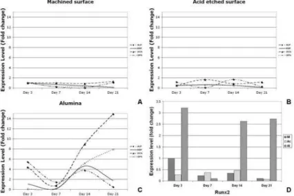
1.4.2. Comparar a expressão de genes responsáveis pela diferenciação de osteoblastos (alp, bsp, ocn, opn e runx2) em animais com implantes com revestimentos nano-estruturados, com os implantes de titânio comercialmente puro com superfície lisa e tratados com ataque ácido (Capítulos 4 e 5);
1.4.3. Avaliar o contato implante/osso em animais com implantes com revestimentos nano-estruturados, com os implantes de titânio comercialmente puro com superfície lisa e tratados com ataque ácido, por meio de análise histomorfométrica (Capítulos 3 e 4);
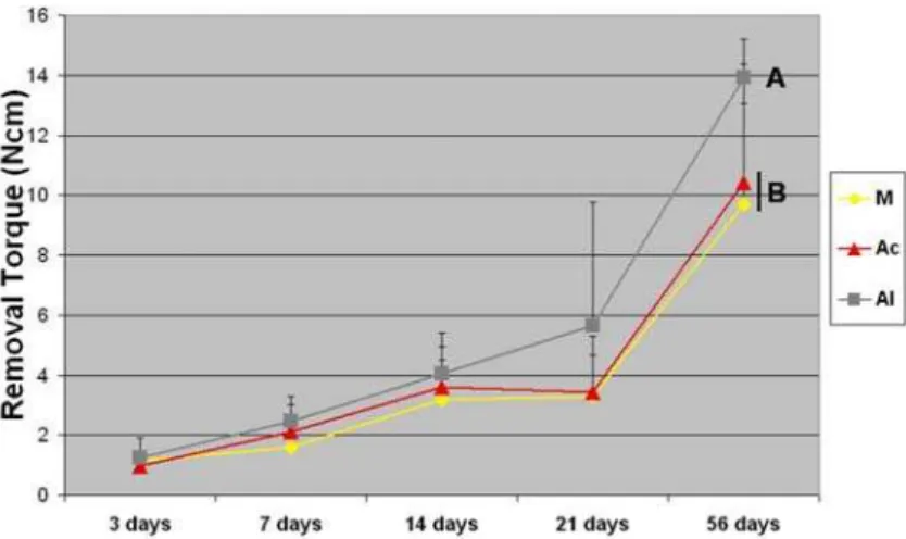
1.4.4. Avaliar o torque de remoção dos implantes com revestimentos nano-estruturados, comparados com os implantes de titânio comercialmente puro com superfície lisa e tratados com ataque ácido (Capítulos 3 e 4);
1.4.5. Avaliar a expressão de genes responsáveis pela diferenciação de osteoblastos (alp, bsp, ocn, osx, opn e runx2) em células ósseas cultivadas sobre a superfície dos implantes
com os revestimentos a base de óxido de alumínio, óxido de titânio e óxido de zircônia, e a expressão gênica em células ósseas cultivadas sobre a superfície dos implantes com os revestimentos a base de óxido de alumínio, com os implantes de titânio comercialmente puro com superfície lisa e tratados com ataque ácido, por meio de “PCR arrays” (Capítulo 5); 1.4.6. Avaliar a expressão de genes responsáveis pela diferenciação de osteoblastos (alp, bsp, ocn, osx, opn e runx2) em células ósseas cultivadas sobre a superfície dos implantes
Capítulo 2 – Tecnologia Avançada na Superfície de Implantes Dentários – da micro
para a nanotopografia
As tendências atuais na terapia com implantes odontológicos têm incluído o uso de implantes com superfícies modificadas utilizando nanotecnologia. O objetivo deste trabalho foi revisar e avaliar o papel das modificações em escala nanométrica de superfícies de implantes osseointegrados para melhorar o processo de osseointegração. Nanotecnologia oferece a engenheiros e profissionais da área de biologia e saúde novos meios para entender e melhorar funções específicas das células. As várias técnicas utilizadas para adicionar características nanométricas às superfícies de implantes osseointegrados são descritas neste trabalho. Vários trabalhos tem apresentado os efeitos da nanotecnologia na modulação de etapas fundamentais do processo de osseointegração. As vantagens e desvantagens da utilização da nanotecnologia na superfície de implantes também são discutidas nesse trabalho.
Advancing Dental Implant Surface Technology - From micron- to nano-topography
Abstract:
Current trends in clinical dental implant therapy include use of endosseous dental implant surfaces embellished with nanoscale topographies. The goal of this review is to consider the role of nanoscale topographic modification of titanium substrates for the purpose of improving osseointegration. Nanotechnology offers engineers and biologists new ways of interacting with relevant biological processes. Moreover, nanotechnology has provided means of understanding and achieving cell specific functions. The various techniques that can impart nanoscale topographic features to titanium endosseous implants are described. Existing data supporting the role of nanotopography suggests that critical steps in osseointegration can be modulated by nanoscale modification of the implant surface. Important distinctions between nanoscale and micron-scale modification of the implant surface are presently considered. The advantages and disadvantages of nanoscale modification of the dental implant surface are discussed. Finally, available data concerning the current dental implant surfaces that utilize nanotopography in clinical dentistry are described. Nanoscale modification of titanium endosseous implant surfaces can alter cellular and tissue responses that may benefit osseointegration and dental implant therapy.
Key words:
Nanotopography; Dental Implant; Surface treatment; Sol-gel techniques; Bone regeneration; Cell signaling.
Introduction:
Over two decades later, osseointegration is widely accepted in clinical dentistry as the basis for dental implant success. The low rate of implant failure in dense bone of the parasymphyseal mandible (Adell et al., 1981; Albrektsson et al., 1988; Goodacre et al., 1999; Zarb; Schmitt, 1990) has not been fully recapitulated by subsequent data from studies involving more challenging clinical situations (Morton et al., 2004; Tolstunov, 2006). Anecdotal reports of difficulty in achieving high rates of implant success in select patient populations (e.g. smokers, diabetics) were supported by initial reports (Jaffin; Berman, 1991; Bain, 1996; Fiorellini et al., 2000). The cause of these failures, while not precisely determined, was largely attributed to a failure in bone formation in support of osseointegration. Challenging osseointegration with new protocols such as immediate placement and immediate loading may require further control of bone formation and osseointegration (Morton et al., 2004).
Failure to achieve osseointegration at a high rate can be attributed to one or more implant, local anatomic, local biologic, systemic or functional factors (Adell et al., 1981; Zarb; Schmitt, 1990). Clinical control of all of these factors is represented by multidisciplinary treatment planning procedures. While it is presently acknowledged that these, as well as clinician-related factors, are important determinants of endosseous implants success, a major interest in implant design factors is evident and clinical efforts to improve implant success have been focused on increasing the amount of bone that forms at the endosseous implant surface.
Implant surface character is one implant design factor affecting the rate and extent of osseointegration (Cooper, 1998; Nanci et al., 1998; Boyan et al., 1993; Schwartz et al., 1992; Stanford et al., 2006). The process of osseointegration is now well described both histologically and at the cellular level. The adhesion of a fibrin blood clot and the population of the implant surface by blood-derived cells and mesenchymal stem cells is orchestrated in a manner that results in osteoid formation and its subsequent mineralization (Masuda et al., 1997; Meyer et al., 2004; Berglundh et al., 2003). A seamless progression of changing cell populations and elaboration and modification of the tissue / implant interface eventually results in bone forming in direct contact with the implant surface. Precisely how much of the implant surface directly contacts bone, how rapidly this bone accrual occurs, and the mechanical nature of the bone / implant connection is influenced by the nature of the implant surface itself (Le Guéhennec et al., 2007).
biocompatible nature of the cpTitanium implant (Kasemo, 1983), and revealed some pragmatic advantages for cpTitanium over other suitable materials (Johansson Albrektsson, 1991). Molecular investigations have contributed to defining cellular responses to titanium as “compatible” and advantageous. For example, Suska and colleagues (2005) showed relatively low inflammatory signaling within cells in tissues adjacent to cpTitanium implants and suggested that this is a part of the osseointegration process. During the first 10 – 20 years of applied endosseous implant experience, the concept that cpTitanium implant biocompatibility supported clinical osseointegration success dominated clinical thinking. Subsequently, experiments with surface topography encouraged new considerations of improvements in bone formation at the implant surface.
Micron-scale surface topography
The significance of micro-scale topography, was highlighted in an important report by Buser and colleagues (1991) that compared various surface preparations of cpTitanium to an electropolished surface negative control and a hydroxyapatite coated positive control group. The observation that a micron-scale rough surface prepared by grit blasting and subsequent acid etching was capable of rapid and increased bone accrual reiterated an earlier report that a TiO2-grit blasted surface also supported more rapid and increased bone accrual at
cpTitanium implants (Gotfredsen et al., 1990). These early observations indicated that the cpTitanium surface could be modified to enhance bone accrual and suggested that cpTitanium was not only “bioinert” or “biocompatible”, but could influence cellular activity or tissue responses leading to greater osteogenesis.
At least three different lines of thinking have evolved to better interpret or explain how surface topography at the micron-scale can increase bone to implant contact. One is the biomechanical theory of Hannson and Norton (Hansson; Norton, 1999), the second is the concept of contact osteogenesis (Davies, 2003), and the third is a surface signaling hypothesis supported by many cell culture investigations (Cooper, 1998; Puleo; Nanci, 1999; Schwartz et al., 1999).
performance of endosseous implants. One possible explanation is given by the adaptation of bone to mechanical loading played by the osteocytes acting as mechanosensors (Burger; Klein-Nulend, 1999; Hansson, 2006). Evidence of the important relevance of increased bone to implant contact has been provided by measurement of the physical interaction of micron level rough implants with bone using push out or torque removal assays (Wong et al., 1995; Wennerberg et al., 1997). What has not been fully elucidated is how mechanical signaling in the unmineralized tissue of forming bone and adjacent connective tissue is affected by the implant surface. The bonding of bone to the implant surface is not implicated as a mechanism of enhancing the early physical associations of the implant with bone.
A principal role for fibrin clot stabilization by the implant surface exemplifies one role that microscale surface roughness may play in improved osseointegration (Park et al., 2001). Described is a physical interlocking of fibrin fibers with the surface features which promotes the directed ongrowth of bone forming cells directly at the implant/bone interface. Topographic enhancement may aid in stabilization of fragile extracellular matrix scaffolds for conduction of cells toward and onto the implant surface (contact guidance) (Ricci et al., 2008).
Several investigators have further described surface topography-specific effects on titanium-adherent osteoblastic cell behavior (Schneider et al., 2003; Isa et al., 2006; Ogawa; Nishimura, 2003; Ogawa; Nishimura, 2006). The overriding theme of these investigations is that surface adhesion-mediated control of cell function underscores the positive influences on bone formation. Many investigations have contributed to the understanding that there is a range of micron-level surface topography that enhances the adherent osteoblasts’ differentiation and extracellular matrix formation/mineralization (Abron et al., 2001). Together these investigations have shown that increased surface topography effectively enhances extracellular matrix synthesis of adherent cells and provides a faster and more reliable osseointegration response (Ogawa; Nishimura, 2006; Buser et al., 2004; Ellingsen et al., 2004; Gutwein et al., 2004; Oh et al., 2005; Price et al., 2003a; 2003b; Webster et al., 1999; Webster et al., 2000a; Webster et al., 2001a; Webster; Ejiofor, 2004; Webster et al., 2005; Schwartz et al., 2005; Zhao et al., 2005).
human clinical histology (Trisi et al., 2003). Limited evidence that integrins are involved in cellular responses to implant surfaces has been obtained using MG63 cell culture studies (Wang et al., 2006).
Micron-scale topographic modification of the cpTitanium surface is accepted in the endosseous dental implant marketplace (Albrektsson; Wennerberg, 2004a; 2004b). The belief that micron level surface topography results in greater accrual of bone at the implant surface is supported by some clinical evidence (Cochran, 1999; Shalabi et al., 2006). Yet, these surfaces have been generally interpreted to be biocompatible devices with limited ability to directly affect the initial fate of surrounding tissues (e.g. impose bone formation or prevent bone resorption).
Today, a growing aspect of endosseous implant surface research is focused on further enhancing the activity of bone forming cells at the tissue implant interface. This desire for “bioactivity” has been addressed using a variety of different approaches. Clearly, cpTitanium surfaces can be modified to direct specific cellular responses such as osteogenesis. More specifically, cpTitanium implant surfaces can be made to direct the osteoinduction of adherent progenitor cells. While one approach is the immobilization of bioactive peptides or growth factors and notably the BMPs (Schliephake et al., 2005; Becker et al., 2006;), other approaches have embraced the use of nanoscale surface engineering to induce intrinsic osteoinductive signaling of the surface adherent cells. The purpose of this review is to explore how nanotechnology applications to the cpTitanium implant surface may provide new opportunities to create endosseous implant surfaces with greater specific control of adherent cell and adjacent tissue fate.
Nanotechnology and Surface Science
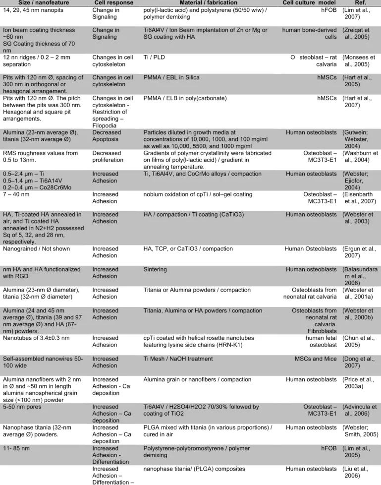
Figure 1 – Nanoscale in perspective. The scanning electron micrograph at 5000x (A) fails to represent true nanoscale features of a titanium implant surface. 100,000x image (B) shows the complex nanoscale surface; here produced by Titania sol-gel deposition.
Application of nanotechnology to the dental implant surface involves a two dimensional association of surface features (across and away from the mean surface plane) (Figure 2). These nanofeatures can be arranged in an organized manner (isotropic) or unorganized manner (anisotropic), often depending on the method of manufacture. Of the surface topographies that have been applied to a dental implant surface, the topography is often characteristically anisotropic. Isotropic features such as nanogrooves or nanopits that are created largely by optical methods are not readily applied to complex screw shaped objects. When these concepts are applied to the endosseous implant surface, implied is the embellishment of the surface with nanometer scale features that lead to novel physicochemical behavior (e.g. bone bonding) or biochemical events (e.g. altered protein adsorption, cell adhesion with changes in cell behavior).
Nanoscale modification of the titanium endosseous implant surface may affect both the topography as well as the chemistry of the surface. Specific chemical modification of cpTitanium could be the targeted goal of nanoscale modification. In fact, a complicating feature of nanoscale manipulation of any material is that there are inherent chemical changes of the bulk material surface. Albrektsson and Wennenberg (2004a), divided implant surface quality into three categories: (1) mechanical properties, (2) topographic properties, and (3) physicochemical properties. They indicated that these characteristics are related and by changing any of theses groups the others will also be affected. This important observation is likely to be even more relevant to discussions of nanotopographic modifications of the endosseous cpTitanium surface. One frequently encountered limitation to studies comparing nano- and micron-level surface topography is that it can be extremely difficult to isolate chemistry or charge effects induced by the nanotopography. When atomic level control of material assembly is approached, the surface properties are influenced by quantum phenomena that do not govern traditional bulk material behavior (Liu et al., 2006). It is very difficult but important to distinguish distinct topography-specific effects from allied changes in surface energy or chemical reactivity.
Nanotechnology requires novel ways of manipulating matter in the atomic scale. Several approaches are currently prevalent in the experimental application to endosseous implants (Table 1). One approach involves the physical method of compaction of nanoparticles of TiO2 versus micron level particles to yield surfaces with nanoscale grain
Table 1 - Methods for Creating Nanofeatures on cpTitanium Implants
Methods Characteristics
Self assembly of Monolayers
The exposed functional end group could be a molecule with different functions (an osteoinductive or cell adhesive molecule).
Compaction of nanoparticles Conserves the chemistry of the surface among different topographies. Not readily applied over implant surfaces
Physical approaches
Ion Beam Depostion Can impart nanofeatures to the surface based on the material used. Acid etching Combined with other methods (sandblasting and/or Pedoxidation) can
impart nanofeatures to the surface and remove contaminants. Peroxidation Produces a titania gel layer.
Both chemical and topography changes are imparted.
Alkali treatment (NaOH) Produces a sodium titanate gel layer allowing hydroxyapatite deposition. Both chemical and topography changes are imparted.
Chemical methods
Anodization Can impart nanofeatures to the surface creating a new oxide layer (based on the material used).
Sol-Gel (colloidal particle adsorption)
Creates a thin-film of controlled chemical characteristics. Atomic-scale interactions display strong physical interactions.
Nanoparticle
Deposition Discrete crystalline deposition
Superimposes a nanoscale surface topographical complexity on the surface.
Lithography and contact
printing technique
Many different shapes and materials can be applied over the surface. Approaches are labor intensive and require considerable development prior to clinical translation and application on implant surface.
Second is the process of molecular self-assembly. Self-assembled monolayers (SAMs) are formed by the spontaneous chemisorption and vertical close-packed positioning of molecules onto some specific substrata, exposing only the end-chain group(s) at the interface (Scotchford et al., 2002). The exposed functional end group could be an osteoinductive or cell adhesive molecule. An example of this is the use of cell adhesive peptide domains (RGD domains) appended to SAMs composed of polyethylene Glycol (PEG) and applied to the Titanium implant surfaces (Germanier et al., 2006).
A third method is the chemical treatment of different surfaces to expose reactive groups on the material surface and create nanoscale topography. This is popular among current dental implant investigators. NaOH-treatment catalyzes the production of titanium nanostructures outward from the titanium surface (Zhou et al., 2007). Treatment with a NaOH solution produces a sodium titanate gel layer on the Ti surface while H2O2 produces a
Chemical treatments (Peroxidation (H2O2) or acid oxidation, such as Hydrofluoric
acid) have also been used to create nanotopography (Nanci et al., 1998; Wang et al., 2001; Uchida et al., 2002). The use of H2O2 with acid etching has been shown to create novel
nanostructures of amorphous titanium oxide on the implant surface (Wang et al., 2002). It was found that the treatment of the implant surface with H2O2/HCl increased the adsorption
of RGD peptides onto the surface followed by passivated surfaces (30% HNO3) and
heat-treated surfaces (Mante et al., 2004). These surface treatments also increased the mineralization in the same order. Treatment with hydrofluoric acid also creates discrete nanostructures on TiO2 grit blasted surfaces (Ellingsen et al., 2006). Several cell culture
studies (Isa et al., 2006; Cooper et al., 2006; Guo et al., 2007), preclinical investigations (Ellingsen et al., 2004; Berglundh et al., 2007), and clinical studies (Stanford et al., 2006) support the observation that hydrofluoric acid treatment of TiO2 grit blasted titanium implants
is associated with rapid bone accrual at the implant surface. Complex chemical changes induced by these methods may require careful inspection.
The deposition of nanoparticles onto the titanium surface represents a fourth approach to imparting nanofeatures to a titanium dental implant (Ben-Nissan; Choi, 2006). Sol-gel transformation techniques achieve deposition of nanometer scale calcium phosphate accretions to the implant surface (Liu et al., 2001; Kim et al., 2004). Alumina, titania, zirconia and other materials can also be applied (Lee et al., 2006). Owing to their resultant atomic-scale interactions, the accretions display strong physical interactions (Ben-Nissan; Choi, 2006; Piveteau et al., 2000; Arias et al., 2003; Choi; Ben-Nissan, 2007). In a modified approach, Nishimura and colleagues (2007) recently demonstrated a directed approach to assembly of CaPO4 nanofeatures on dual acid etched cpTitanium implant surfaces. The
deposition of discrete 20-40nm nanoparticles on an acid etched titanium surface led to increased mechanical interlocking with bone and the early healing of bone at the endosseous implant surface in a rat model.
(CaP) / Discrete crystalline deposition (DCD) sol-gel coating of Ti alloy implant surfaces (Mendes et al., 2007).
A fifth approach to creating nanoscale topography on Titanium is the use of optical methods (typically lithography) reliant on wavelength specific dimensions to achieve the appropriate nanoscale modification (Zhou et al., 2007). These approaches are labor-intensive methods that require considerable development prior to clinical translation. The present use of lasers to promote micron level groove on an implant surface can produce micron level, not nanoscale, modification of the implant surface (Ricci et al., 2000). Another method of depositing nanoscale material on to the implant surface involves ion beam deposition (e.g. Hydroxyapatite) (Coelho; Suzuki, 2005). All are relevant to the endosseous dental implant surface and experimental examples of each can be identified.
Nanotopography has been shown to influence cell adhesion, proliferation, differentiation, and cell specific adhesion. Related changes in chemistry and nanostructure impart important chemical changes and permit biomimetic relationships between alloplastic surfaces and tissues. It is speculated that alloplastic nanosurfaces possess topographic elements scaled to naturally occurring substrates.
Biomimetics and Nanotechology:
Nanotopography Alters Cellular Responses
Surface nanotopography appears to affect cell interactions at surfaces and alter cell behavior when compared to conventional sized topography (Figure 3) (Klabunde et al., 1996; Wu et al., 1996; Baraton et al., 1997). Different physical relationships exist between cells and nano- versus cell and micron-scale surface features. Nanotopography specific effects on cellular behavior have been demonstrated using a wide range of different cell types including epithelial cells, fibroblasts, myocytes and osteoblasts. Nanostructured surfaces possess unique properties that alter cell adhesion by direct (cell – surface interactions) and indirect (affecting protein – surface interactions) mechanisms. Evidence has been gathered using several models and surface systems (Tables 2 and 3).
Table 2 - Reported Osteoblast responses to Nanosurfaces – In vitro
Size / nanofeature Cell response Material / fabrication Cell culture model Ref. 14, 29, 45 nm nanopits Change in
Signaling
poly(l-lactic acid) and polystyrene (50/50 w/w) / polymer demixing
hFOB (Lim et al., 2007)
Ion beam coating thickness ~60 nm
SG Coating thickness of 70 nm
Change in Signaling
Ti6Al4V / Ion Beam implantation of Zn or Mg or SG coating with HA
human bone-derived cells
(Zreiqat et al., 2005)
12 nn ridges / 0.2 – 2 mm separation
Changes in cell cytoskeleton
Ti / PLD O steoblast – rat
calvaria
(Monsees et al., 2005)
Pits with 120 nm Ø, spacing of 300 nm in orthogonal or hexagonal arrangement.
Changes in cell cytoskeleton
PMMA / EBL in Silica hMSCs (Hart et al.,
2005)
Pits with 120 nm Ø. The pitch between the pits was 300 nm. Hexagonal and square pit arrangements.
Changes in cell cytoskeleton - Restriction of spreading – Filopodia
PMMA / ELB in poly(carbonate) hMSCs (Hart et al., 2007)
Alumina (23-nm average Ø), titania (32-nm average Ø)
Decreased Apoptosis
Particles diluted in growth media at
concentrations of 10,000, 1000, and 100 mg/ml as well as 10,000, 5500, and 1000 mg/ml
Human osteoblasts (Gutwein; Webster, 2004) RMS roughness values from
0.5 to 13nm.
Decreased proliferation
Gradients of polymer crystallinity were fabricated on films of poly(l-lactic acid) / gradient in annealing temperature.
Osteoblast – MC3T3-E1
(Washburn et al., 2004)
0.5–2.4 µm – Ti 0.5–1.4 µm – Ti6A14V 0.2–0.4 µm – Co28Cr6Mo
Increased Adhesion
Ti, Ti6Al4V, and CoCrMo alloys / compaction Human osteoblasts (Webster; Ejiofor, 2004)
7 – 40 nm Increased
Adhesion
nobium oxidation of cpTi / sol–gel coating Osteoblast – MC3T3-E1
(Eisenbarth et al., 2007)
HA, Ti-coated HA annealed in air, and Ti coated HA
annealed in N2+H2 possessed Sq of 5, 32, and 28 nm, respectively.
Increased Adhesion
HA / compaction / Ti coating (CaTiO3) Human osteoblasts (Webster et al., 2003)
Nanograined / Not shown Increased Adhesion
HA, TCP, or CaTiO3 / compaction Human Osteoblasts (Ergun et al., 2007)
nm HA and HA functionalized with RGD
Increased Adhesion
Sintering Human osteoblasts (Balasundara
m et al., 2006) Alumina (23-nm Ø diameter),
titania (32-nm Ø diameter)
Increased Adhesion
Titania or Alumina powders / compaction Osteoblasts from neonatal rat calvaria
(Webster et al., 2001a)
Alumina (24 and 45 nm average Ø), titania (39 and 97 nm average Ø) and HA (67-nm) powders.
Increased Adhesion
Titania, Alumina or HA powders / compaction Osteoblasts from neonatal rat calvaria. Fibroblasts
(Webster et al., 2000b)
Nanotubes of 3.4±0.3 nm Increased Adhesion
cpTi coated with helical rosette nanotubes featuring lysine side chains (HRN-K1)
human fetal osteoblast
(Chun et al., 2005)
Self-assembled nanowires 50-100 wide
Increased Adhesion
Ti Mesh / NaOH treatment MSCs and Mice (Dong et al., 2007)
Alumina nanofibers with 2 nm in Ø and ~50 nm in length alumina nanospherical grain size (<100 nm) powder
Increased Adhesion - Ca deposition
Alumina grain or nanofibers / compaction Human osteoblasts (Price et al., 2003a)
5-50 nm pores Increased
Adhesion – Ca deposition
Ti6Al4V / H2SO4/H2O2 70/30% followed by coating of TiO2
Osteoblast – MC3T3-E1
(Advincula et al., 2006)
Nanophase titania (32-nm average Ø) powders.
Increased Adhesion – Ca deposition
PLGA mixed with titania (in various proportions) / cured in air
Human osteoblasts (Webster; Smith, 2005)
11- 85 nm Increased
Adhesion - Differentiation
Polystyrene-polybromostyrene / polymer demixing
hFOB (Lim et al., 2005)
Increased Adhesion – Differentiation – Ca deposition
∼ 100 nm / nanotubes Increased Adhesion – Proliferation - Differentiation
titania / anodization primary rat bone
marrow MSCs
(Popat et al., 2007a)
∼ 100 nm / nanopores Increased Adhesion – Proliferation - Differentiation – Ca deposition
alumina sheets / anodization primary murine bone marrow MSCs
(Popat et al., 2007b)
~100nm features on Ti Increased Differentiation
cpTi / TiO2 Blasting / HF treatment Osteoblast – MC3T3-E1 and Ratus novergicus
(Guo et al., 2007)
10 - ___ nm Increased
Differentiation
PMMA / Colloidal lithography and polymer demixing
primary human osteoprogenitors
(Dalby et al., 2006)
20 – 50 nm surface features Increased Differentiation
cpTi and Ti6Al4V / oxidation with H2SO4/H2O2 primary rat calvaria derived osteoblasts
(Oliveira; Nanci, 2004)
Elongated HA nanocrystals, with a mean length of about 100 nm.
Increased Differentiation
Ti13Nb13Zr / mechanomaking process or Ti6Al4V followed by HF/HNO3 acid etch CaP coating
hMSCs (Bigi et al., 2007)
Parallel ridges/channels (microstructured) /
nanostructured HA (100 nm).
Increased Differentiation
Photolithography / nanostructured HAP (biomimetic) on silicon microstructures
Saos-2 and MG-63 cell lines
(Tan et al., 2004)
Alumina nanofibers with 2 nm in Ø and ~50 nm in length
Increased Differentiation - Ca deposition
Alumina nanofibers / compaction / Sintered at 400oC, 600oC, 800oC, 1000oC, or 1200oC
Human osteoblast (Webster et al., 2005)
20 – 50 nm surface features Increased Differentiation – Ca deposition
cpTi / oxidation with H2SO4/H2O2 primary rat calvaria derived osteoblasts
(Oliveira et al., 2007)
Alumina (24-nm average Ø), titania (39-nm average Ø) and HA (67-nm) powders.
Increased Differentiation – Ca deposition
Titania, Alumina or HA powders / compaction Osteoblasts from neonatal rat calvaria
(Webster et al., 2000a)
island height of about 90 nm Increased Filopodia
Polystyrene and polybromostyrene/ Polymer demixing
Human Bone marrow cells
(Berry et al., 2006)
Nanofibers (60-100 nm) Increased Osteoblast Specificity
Carbon nanofibers / compaction Human osteoblasts (Price et al., 2003b; Price et al., 2004) Alumina (23-nm average Ø),
titania (49-nm average Ø) and HA (67-nm) powders.
Increased Osteoblast Specificity
PLA or PMMA powder mixed with titania, alumina or HA (in various proportions) / compaction
Neonatal rat calvaria osteoblasts. Rat skin fibroblasts
(McManus et al., 2005)
Nanophase titania (32-nm average Ø) powders.
Increased Osteoblast Specificity
PLGA mixed with titania (in various proportions) / cured in air
Human osteoblasts (Kay et al., 2002)
∼ 160 nm pores Increased Proliferation
Alumina / EBE Human osteoblasts (Briggs et al.,
2004)
AAT texture showed
micropores and an overlapped nanometric net of filaments
Increased Proliferation
cpTi / alkali etching process with CaP solution (biomimetic)
Osteoblast-like MG63
(Chiesa et al., 2007)
Table 3 - Reported Osteoblast responses to Nanosurfaces – In vivo
size / nanofeature Tissue response material / fabrication
Animal / cell culture model
ref.
3µm CaP coating Elimination of tissue fibrous encapstulation and foreing body giant cell response
PLGA / CaP coated with CaP Ratus novergicus (Lickorish et al., 2007)
8nm diameter and 100nm length
Enhanced bone formation PLGA mixed with Ti nanotubes Ratus novergicus (Kubota et al., 2004)
AAT texture showed micropores and an overlapped nanometric net of filaments
Increased Bone-to-implant contact cpTi / alkali etching process with CaP solution (biomimetic)
Sheep (Chiesa et al., 2007)
Not shown Increased Bone-to-implant contact cpTi / HA - Ion Beam Assisted Deposition (IBAD)
Rabbit (Jung et al., 2001)
~100nm features on Ti Increased Bone-to-implant contact cpTi / TiO2 Blasting / HF treatment Dog (Berglundh et al., 2007)
~100nm features on Ti Increased Differentiation cpTi / TiO2 Blasting / HF treatment Ratus novergicus (Guo et al., 2007)
Not shown Increased osseoactivity cpTi / HA - Ion Beam Assisted Deposition (IBAD)
Dog (Coelho; Suzuki, 2005) discrete deposition of
HA nanoparticles (20–40 nm) on Ti substrate
Increased Push-out test resistance cpTi / dual acid etch / coated with CaP by DCD
Ratus novergicus (Nishimura et al., 2007)
Not shown Increased removal torque – Bone-to-implant contact – Bone volume
cpTi / Sandblast / HA - Ion Beam Assisted Deposition (IBAD)
Rabbit (Park et al., 2005)
20–100 nm range of the features (HA)
Increased tensile test resistance cpTi and Ti6Al4V / acid etch / coated with CaP by DCD
Ratus novergicu (Mendes et al., 2007)
Protein/surface interactions - Surface Wettability
The changes in initial protein – surface interaction are believed to control osteoblast adhesion (Balasundaram et al., 2006). This is a critical aspect of the osseointegration process. When implants come into contact with a biological environment, protein adsorption (e.g. plasma fibronectin) that occurs immediately will mediate subsequent cell attachment and proliferation. Cell binding to protein domains of adhesive extracellular matrix proteins involves receptors termed integrin receptors that transmit signals through a collection of proteins on the cytoplasmic face of the contact, termed focal contacts (Fath et al., 1989). Surface effects are often mediated through integrins that bind the RGD motif in cell attachment proteins (Tosatti et al., 2004). The RGD motif of cell adhesive proteins such as fibronectin or vitronectin are important in mediating cell adhesion of osteoblasts and other cells to synthetic material surfaces (Sinha; Tuan, 1996). Nanofeatures could alter the conformation of theses RGD containing proteins, a phenomenon known to affect cell adhesion and behavior (Cavalcanti-Adam et al., 2007).
that hydrophobic groups are more likely to adsorb albumin and that albumin is not replaced by ECM proteins, blocking cell adhesion. Hydrophobic surfaces adsorbed fibrinogen (Rodrigues et al., 2006), while hydrophilic surfaces allowed an interchange of adsorbed albumin by ECM proteins (Arima et al., 2007).
Nanoscale topography is a powerful way of altering protein interactions with a surface. Webster and colleagues (Webster et al., 2000b; Webster et al., 2001a) observed an increased vitronectin adsorption on nanostructured surfaces when compared to conventional surfaces. They also found an increased osteoblast adhesion when compared to other cell types, such as fibroblasts, on the nanosurfaces (Webster et al., 2000a). Another study suggested higher adsorption of fibronectin on hydrophilic SAMs surfaces with greater focal adhesion formation (integrin binding) evident in the osteoblast cells adhered to the hydrophilic SAM treated surfaces (Scotchford et al., 2002). Lim and colleagues (Lim et al., 2005) more directly related protein adsorption, cell adhesion and the active process of attachment by measurement of increased focal adhesion kinase (FAK) activity. In a study using SAMs biofunctionalized with RGD, Cavalcanti-Adam and colleagues (2007) also found that the spacing among the nanofeatures modulate focal adhesion (FA) formation; cells cultured on a 58nm nanopattern formed normal FA, whereas those plated on a 108nm nanopattern failed to develop FA. Surface roughness at the nanoscale is an important determinant of protein interactions that ultimately direct cell activity in control of tissue formation at implant surfaces (Park; Webster, 2005).
Cell adhesion, spreading and motility
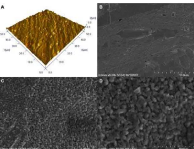
Figure 4 – Nanoscale cell interactions. There is apparent affinity of cells for nanoscale features. Here, 20-40 nm features produced by H2O2/H2SO4 treatment are interactive points for lamellipodia of spreading cells. The cause and effect relationship is a current point of investigation. A = 10,000x image of adherent cell. B and C represent 100,000x images of the same adherent cell. D = 200,000x magnification of the cell with nanofeatures.
Nanofeatures of an alloplastic surface may have unique attributes affecting cell interactions. Both the dimension and the density of the nanofeatures affect cell behavior (Cavalcanti-Adam et al., 2007). In a well-controlled investigation of Titanium nanostructure, Andersson and colleagues (2003) compared cell morphology and cytokine production on titanium substrates with 15mm wide and 185nm deep grooves versus Ti substrates with 100nm high, 168nm diameter hemispherical nanopillars. The cells appeared partially aligned to the grooves and had a cytokine release similar to that found from cells on flat surfaces. Cells on hemispherical pillars had a smaller area and had more membrane projections compared to cells on grooves. Morphological changes correlated with diminished protein secretion. It has been suggested that 70-100nm features of an implant surface are scaled to function directly with the focal adhesion of cells.