INSTITUTO DE INVESTIGAÇÃO E FORMAÇÃO AVANÇADA ÉVORA, MARCH 2017 ANO SUPERVISORS: Professor Ana Teresa Caldeira Professor António Candeias Professor Dora Martins Teixeira
r
Thesis presented to obtain PhD degree in
Biochemistry by the University of Évora
Mara Teresa Caldeira da Silva
Novel Biocides for Cultural Heritage
“The very essence of instinct is that it's followed
independently of reason.”
Aos meus pais e Pedro
To my parents and Pedro
“A ciência descreve as coisas como são; a arte, como
são sentidas, como se sente que são.”
V
Acknowledgments
Having completed this PhD project, I can only express my sincere thanks to the several people, organisations and institutions that have contributed in various ways to make this project become a reality, because a PhD is not an individual work.
First of all, I would like to express my gratitude to my supervisors Professor Ana Teresa Caldeira, Dora Teixeira and António Candeias that have always provided their knowledge, experience and friendships, for their unconditional support, tireless help and confidence throughout this journey.
I would like to thank to Professor António Pereira for his full availability, the help given and the knowledge shared during a key part of this work, including for the suggestions and comments on the respective scientific paper already published.
I am thankful to Doctor Maria de Fátima Candeias willingness to support me and this work, as well as to share her knowledge during the toxicological assays with biological models performed in the final part of this thesis.
My thanks go also to Professor Maria do Rosário Félix and Doctor Carla Varanda, who, during the first year of this PhD, have collaborated by sharing the laboratory space and equipment needed. Thank you for the tremendous support, knowledge sharing, caring and friendship that welcomed me from the first minute.
I also want to express my sincere thanks to all teachers and colleagues from HERCULES Laboratory, which always showed the greatest support, generosity, availability and friendship during these three years. It is much easier to work when you are part of a cohesive and dynamic team that give you all the support you need.
To all the laboratory technicians of Luís António Verney College - Fase III of the Universidade de Évora that were an important part of this project; my thanks go to D.
Lena, D. Esperança, D. Jesuína, Custódia and Anabela who spent all their good will, kindness and friendship, during laboratory work.
VI
The Biotechnology students Sofia Silva and Marina Ramalho who cooperated with me at different stages of this work. I would like to thank them for allowing me to grow as a researcher, individual and friend throughout their senior year in which participated on this project.
I cannot forget my colleagues and friends, daily companions in the anguishes and joys, successes and failures that have always supported me. To all of them, Margarida Pires, Patrícia Nunes, Ana Carolina Fialho, Sílvia Arantes, Ana Filipa Branco, Ricardo Vieira, Cátia Salvador, Tânia Rosado e Marina González-Peréz, thank you for giving me all the comfort and friendship in the darkest hours. I have no words to express my sincere gratitude and friendship which I return. But mostly, to Marina and Tânia that were my pillar, friendly shoulder and my greatest encouragement, to them I reciprocate my admiration.
To my friends (they know who they are) that geographically near or far have always been present, giving all the support, friendship, understanding and patience, because I know that, especially in recent times, I have not been as present and available as they deserve.
I would like to thank to my colleagues of the Instituto Clínico de Évora, D. Inácia, Paulo, Roberto, Mafalda e Sara, that in several ways, helped me in the senior year of this PhD, but mainly to Joana Garcia who always helped, facilitated, understood and give me strength in the more lonely days and difficult situations, that I face behind a clinic reception.
To the Biotechnology Laboratory of Chemistry Centre, Universidade de Évora, and HERCULES Laboratory, that have seen me growing as a person and professional, since I took the decision of joining into the world of research and science; but mainly for providing me all the equipment, reagents and materials needed for the lab work, for all the facilities and resources for the work dissemination. To the Instituto de Investigação e Formação Avançada (IIFA) da Universidade de Évora for the bureaucratic support.
VII
To my family, especially to the people to whom I owe my life, without them, I would never have come this far. I must be grateful for all they have given me, as well as for the efforts and hard work always done it has taken. They gave me love and affection, education, moral values and principles. Without what they have taught me, I would not be who I am today, because I am proud of how I grew up and of the person I have become. To my parents!
Of course I could not forget, my best friend, companion, colleague and boyfriend, the person who gives me courage when I am down, that calms me when I think that everything all goes wrong, that gives me strength when I am exhausted. To you, Pedro, thank you for EVERYTHING. Everything that is not necessary to enumerate or explain, because it is known, because it is seen, because it is felt.
IX
Agradecimentos
A terminar esta tese de doutoramento resta-me apenas registar e expressar os meus sinceros agradecimentos às várias pessoas, entidades e instituições que de várias formas contribuíram para que este projeto se tornasse numa realidade, pois um Doutoramento não se faz sozinho.
Em primeiro lugar aos meus orientadores Professora Doutora Ana Teresa Caldeira, Professora Doutora Dora Teixeira e Professor Doutor António Candeias que sempre me disponibilizaram todo o seu conhecimento, experiência e amizade; expresso o meu enorme agradecimento pelo apoio incondicional, incansável ajuda e confiança demonstrada durante todo este percurso.
Ao Professor Doutor António Pereira pela ajuda, conhecimento partilhado e total disponibilidade dada durante uma parte fundamental deste trabalho, inclusive sugestões e correções do respetivo artigo científico já publicado.
À Doutora Maria de Fátima Candeias pelo apoio, colaboração, disponibilidade e transmissão de conhecimento facultado durante os ensaios de toxicidade com modelos animais efetuada na parte final deste trabalho.
À Professora Doutora Maria do Rosário Félix e Doutora Carla Varanda, que durante a realização da primeira unidade curricular deste Doutoramento, além da disponibilização da utilização do laboratório e equipamentos necessários, agradeço o enorme apoio, partilha de conhecimento, carinho e amizade com que me receberam desde o primeiro minuto.
Quero também expressar o mais sincero agradecimento a todos os docentes e colegas do Laboratório HERCULES, que sempre demonstraram o maior apoio, generosidade, disponibilidade e amizade durante estes três anos de trabalho. É muito mais fácil trabalhar quando temos uma equipa coesa e dinâmica a dar-nos todo o suporte que necessitamos.
X
A todas as técnicas de laboratório da Fase III do Colégio Luís António Verney da Universidade de Évora que foram uma parte fundamental deste trabalho; agradeço à D. Lena, D. Esperança, D. Jesuína, Custódia e Anabela que de uma forma ou de outra disponibilizaram sempre toda a sua boa vontade, simpatia e amizade durante todo o trabalho realizado em laboratório.
Às estagiárias de licenciatura em Biotecnologia Sofia Silva e Marina Ramalho que cooperaram comigo em diferentes fases deste trabalho, e me permitiram que eu crescesse como investigadora, colaboradora e amiga ao longo do ano letivo em que participaram neste projeto.
Não posso esquecer sem dúvida os meus colegas e amigos, companheiros de todos os dias, agonias e alegrias, sucessos e insucessos que sempre me apoiaram e deram todo o conforto e amizade nas horas mais difíceis. A todos eles, Margarida Pires, Patrícia Nunes, Ana Carolina Fialho, Sílvia Arantes, Ana Filipa Branco, Ricardo Vieira, Cátia Salvador, Tânia Rosado e Marina González-Peréz, nem encontro palavras sequer para expressar a minha sincera gratidão e amizade que retribuo de igual intensidade. Mas principalmente à Marina e Tânia que foram o meu pilar, o ombro amigo e o meu maior incentivo, a elas retribuo a minha admiração.
Aos meus amigos (eles sabem quem são) que geograficamente mais longe ou perto, estiveram sempre presentes, dando todo o apoio, amizade, compreensão e paciência, pois sei que principalmente nos últimos tempos não tenho sido a amiga mais presente e disponível que eles merecem.
Aos meus colegas do Instituto Clínico de Évora, D. Inácia, Paulo, Roberto, Mafalda e Sara, que da forma que lhes foi possível, me ajudaram e compreenderam nesta última fase do doutoramento, mas principalmente à Joana Garcia que sempre ajudou, facilitou, compreendeu, deu força e me animou nos dias e situações mais solitárias e difíceis, com que me confrontei atrás de uma receção de uma clínica.
XI
Ao Laboratório de Biotecnologia do Centro de Química, Universidade de Évora e Laboratório Hercules, que me viram crescer como pessoa e profissional, nestes últimos anos e desde que tomei a decisão de enveredar pelo mundo da investigação e da ciência; mas principalmente por terem permitido a realização deste trabalho, facultando todos os equipamentos, reagentes e material necessário para a sua execução, e todas as facilidades e meios atribuídos para a divulgação do trabalho produzido. Ao Instituto de Investigação e Formação Avançada (IIFA) da Universidade de Évora pelo suporte e apoio burocrático.
À minha família e principalmente, às pessoas a quem eu devo a minha vida que sem elas não teria chegado até aqui nem a lado nenhum, que me deram tudo o que uma criança, adolescente e jovem adulta pode precisar, sempre com muito sacrifício, que me deram amor e carinho, educação, valores e princípios morais, pois sem o que eles me ensinaram eu não seria metade do que sou hoje, pois tenho orgulho da forma como cresci e da pessoa que me tornei. Aos meus pais!
Claro que não me podia esquecer, do meu melhor amigo, companheiro, cúmplice e namorado, à pessoa que me dá incentivo quando estou mais em baixo, que me acalma quando acho que tudo corre mal, que me dá força quando elas estão prestes a esgotar. A ti, Pedro, obrigada por TUDO. Tudo o que não é preciso enumerar nem explicar, porque se sabe, porque se vê, porque se sente.
XIII
Abstract
Many microorganisms, influenced by environmental conditions, are the main responsible for biological contamination in built heritage. Biocides based on chemical toxic compounds have been the most often used to mitigate this problem. Thus, it is of vital importance to develop proper remediation actions based on environmentally innocuous alternatives. Bacteria of the genera Bacillus are emerging as an optimistic alternative due to their capacity to produce secondary metabolites with antagonistic activities against many fungal pathogens.
This work aimed to develop ground-breaking research in the area of cultural and built heritage rehabilitation, by the development of natural and green safe biocides for biodegradation/biodeterioration treatment of Cultural Heritage.
A complementary methodology, including antifungal tests and molecular approaches was used, in combination with microscopic and analytical techniques to detect, characterise and study the efficiency of the biological active compounds produced by Bacillus sp. strains.
Flow cytometry allowed a comprehensive study of the physiological mechanism behind the bioactive compounds production in order to understand and improve the strategic approaches for process optimisation and scale up production. Moreover, according to the results of the toxicological tests, these compounds have proven to be a real environmental safe and innocuous alternative to the chemical biocides commonly used during the conservative interventions. Thus, they have shown a great potential for their future application in cultural and built heritage rehabilitation.
Keywords
Biodegradation/Biodeterioration, Bacillus sp., Bioactive compounds, Lipopeptides, Iturin, Antifungal activity, Sporulation, Green Biocides, Cultural Heritage rehabilitation
XV
Novos Biocidas para o Património Cultural
Resumo
Vários microrganismos influenciados pelas condições ambientais são os principais responsáveis pela contaminação biológica do património cultural edificado. Na tentativa de mitigação destes agentes, compostos geralmente tóxicos têm sido os mais utilizados. Assim, é de enorme importância desenvolver ações de remediação dirigidas aos agentes efetivamente biodeteriogénicos, baseados em alternativas inócuas para o meio ambiente. As bactérias do género Bacillus surgem, como uma viável alternativa devido à capacidade de produzir metabolitos secundários com atividade antagonista, contra diversos fungos.
Este trabalho teve como objetivo desenvolver uma investigação inovadora que possa vir a ser útil na área de reabilitação do património cultural edificado, através da produção de novos biocidas naturais e mais ecológicos.
Utilizou-se uma abordagem metodológica, que incluiu testes antifúngicos e abordagens moleculares, combinadas com técnicas microscópicas e analíticas, de forma a detetar, caracterizar e estudar a eficiência de compostos biologicamente ativos produzidos por estripes de Bacillus sp.. Foram ainda utilizados os mecanismos fisiológicos por detrás da produção destes compostos, de forma a perceber e melhorar as abordagens estratégicas no processo de otimização da produção. Em testes toxicológicos, compostos produzidos por estirpes de Bacillus sp. selecionados, provaram ser uma alternativa ecológica aos biocidas químicos, comumente utilizados em intervenções de conservação. Desta forma, estes demonstram um elevado potencial para futura utilização na reabilitação do património cultural edificado.
Palavras-chave
Biodegradação/Biodeterioração, Bacillus sp., Compostos bioativos, Lipopéptidos, Iturina, Atividade antifúngica, Esporulação, Biocidas ecológicos, Reabilitação do Património Cultural
XVII
List of Publications
Silva M., Rosado T., Teixeira, D., Candeias A., Caldeira A. T. (2017). Green mitigation strategy for Cultural Heritage: Bacterial potential for biocide production. Environmental Science and Pollution Research, 24:4871–4881
Silva M., Pereira A., Candeias M. F., Teixeira D., Candeias A., Caldeira A.T. (2016). Combined use of NMR, LC–ESI-MS and antifungal tests for rapid detection of bioactive lipopeptides produced by Bacillus. Advances in Microbiology, 6 (10): 788-796.
Silva M., Salvador C., Candeias M. F., Teixeira D., Candeias A., Caldeira A.T. (2016). Toxicological Assessment of Novel Green Biocides for Cultural Heritage. International Journal of Conservation Science 7 (SI1) 7: 223-230.
Silva M., Rosado T., Teixeira D., Candeias A., Caldeira A. T. (2015). Production of green biocides for cultural heritage- Novel biotechnological solutions. International Journal of Conservation Science 6 (SI): 519-530.
Silva M., Silva S., Teixeira D., Candeias A., Caldeira A.T. (2014). Production of novel biocides for cultural heritage from Bacillus sp.. in Science, Technology and Cultural Heritage, Taylor and Francis Group, London, UK, 223-229 ISBN: 978-1-138-027-44-2.
XIX
Table of Contents
Acknowledgments ... V Agradecimentos ... IX Abstract ... XIII Resumo ... XV List of Publications ... XVII Table of Contents ... XIX List of Figures ... XXIV List of Tables ... XXIX Abbreviations ... XXXI Units ... XXXIIIAims and Methodology ... 1
Chapter I. Introduction ... 3
1.1. Cultural Heritage biodeterioration/biodegradation ... 5
1.1.1. Mitigation approaches ... 9
1.1.2. Biocides treatment ... 13
1.2. Biosurfactants compounds – a natural solution ... 17
1.2.1. Lipopeptides production ... 19
1.2.2. Bacillus sporulation ... 26
1.2.3. Lipopeptides biosynthesis... 33
XX
Chapter II. Microorganisms, producers of Lipopeptides ... 43
1. Overview ... 45
2. Introduction ... 46
3. Materials and Methods ... 48
3.1. Bacterial strains and culture media ... 48
3.2. Bioactive compounds production ... 49
3.3. Antifungal activity assessment ... 49
3.4. Liquid cultures growth conditions ... 49
3.5. Bacterial DNA extraction ... 50
3.6. 16S ribosomal DNA sequence analysis ... 51
3.7. Amplification of Bacillus lipopeptide genes ... 52
3.8. Real-time PCR analysis ... 53
3.9. LC-ESI-MS analysis ... 54
4. Results and Discussion ... 55
4.1. Selection of bacterial strains with antifungal potential ... 55
4.1.1. Screening antifungal activity ... 55
4.1.2. Amplification of Bacillus lipopeptide genes ... 57
4.2. Quantification of iturin genetic expression ... 59
4.3. Identification and characterisation of bacterial strains with antifungal potential ... 61
4.4. Molecular phylogeny analysis ... 67
4.5. Physiological characterisation ... 69
XXI
Chapter III . Bioactive compounds detection: Antifungal potential in cultural heritage context ... 73
1. Overview ... 75 2. Introduction ... 76 3. Materials and Methods ... 78 3.1. Microorganisms maintenance ... 78 3.2. Growth conditions and bioactive compounds production ... 79 3.3. Antifungal activity of bioactive compounds ... 79 3.3.1. Antifungal paper disks diffusion assay ... 79 3.3.2. Interaction between novel biocides and heritage biodeteriogenic fungi 80 3.3.3. Statistical analyses ... 80 3.2. Column chromatography ... 81 3.3. LC–ESI-MS analysis ... 81 3.4. Nuclear magnetic resonance (NMR) ... 82 3.5. Bioautographic detection ... 82 4. Results and Discussion ... 83
4.1. Antifungal activity of bioactive compounds ... 83 4.2. Characterisation of the antifungal compounds ... 88 5. Conclusions ... 97
Chapter IV. Physiology of Bacillus with antifungal proprieties: lipopeptides production ... 99
1. Overview ... 101 2. Introduction ... 102 3. Materials and Methods ... 105 3.1. Microorganism and culture media ... 105 3.2. Bacillus growth monitoring ... 105 3.3. Spores formation assessment ... 105 3.4. Physiological monitorisation ... 106
XXII
3.5. Monitorisation of LP production ... 107 3.6. Spores germination of Bacillus sp. CCLBH 1053 ... 108 4. Results and Discussion ... 110 4.1. Cell growth dynamics of Bacillus sp. CCLBH 1053 ... 110 4.2. Bacillus sporulation assessment ... 111 4.3. Antifungal compounds production ... 113 4.4. Cells population analysis by flow cytometry ... 116 4.3.1. NB culture medium analysis ... 117 4.3.2. LAPM culture medium analysis ... 122 4.5. Spore germination study ... 126 5. Conclusions ... 132
Chapter V. Toxicological assessment and antifungal efficiency of the bioactive compounds ... 135
1. Overview ... 137 2. Introduction ... 138 3. Materials and Methods ... 141 3.1. Microorganisms and Bioactive compounds production ... 141 3.2. Acute toxicity assessment ... 141 3.2.1. Toxicity in Artemia salina ... 141 3.2.2. Animals ... 142 3.2.3. Acute toxicity in Swiss mice ... 143 3.2.4. Statistical analyses ... 144 3.3. Simulation assays with mortar samples ... 145 3.3.1. In vitro assay using mortars slabs ... 145 3.3.2. Real mortars under the presence of the new biocides... 146 4. Results and Discussion ... 147 4.1. Toxicological evaluation ... 148 4.1.1. Toxicity in Artemia salina ... 149
XXIII
4.1.2. Acute toxicity in Swiss mice ... 151 4.2. New compounds effectiveness in cultural heritage context ... 152
4.2.1. Simulation in vitro assays using mortars slabs ... 152 4.2.2. Monitorisation of artificially inoculated fragments of mortars from mural painting ... 155 5. Conclusion ... 158
Chapter VI . Concluding Remarks ... 159
References ... 165
Annexes ... 189 Annexe A . Culture media composition ... 191 Annexe B. Solutions composition ... 192 Annexe C. Bacillus 16S ribosomal DNA amplification and agarose gel electrophoresis ... 193 Annexe D. Determination of DNA extracted from the bacteria cells ... 194 Annexe E.Multiple alignment of the rDNA 16s sequence of the Bacillus sp. strains and the most similar Bacillus sequence found for each one in the Gene Bank sequence database. ... 195 Annexe F. Toxicity assay ... 199 Annexe G. Statistical analysis ... 200
XXIV
List of Figures
Chapter I.
Figure I-1: Different artworks with signals of deterioration caused fungal communities. A - Renaissance Frescoes from Santo Aleixo Church, Montemor-o-Novo, Portugal, B- Mural paintings from Casa Pintadas, Évora, Portugal, C – Mural paintings from Santa Clara Church (Sabugueiro, Arraiolos, Portugal), D- Gilded woodcarving from altars of the Espírito Santo Church, Évora, Portugal. ... 7 Figure I-2: Work of preventive cultural heritage conservation. A- Application of
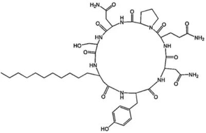
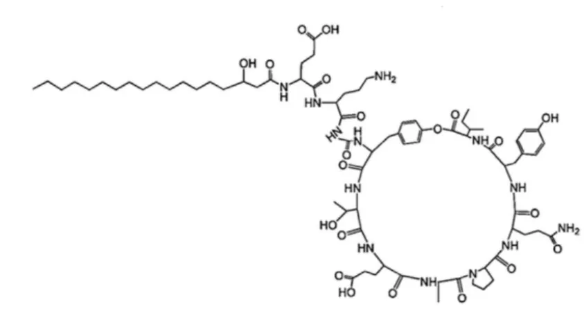
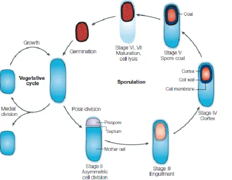
mechanical methods in a rock wall with prehistoric paintings at the Serra da Capivara, National Park, Brasil. B- Conservator-restorer cleaning mold with chemical toxic biocide. ... 10 Figure I-3: Structure of a surfactin member synthesised by Bacillus species.. ... 22 Figure I-4: Structure of an iturin member synthesised by Bacillus species. ... 24 Figure I-5: Structure of fengycin member synthesised by Bacillus species. ... 25 Figure I-6: The key stages of the sporulation cycle of Bacillus subtilis.. ... 27 Figure I-7: Cell division during Bacillus subtillis growth. ... 28 Figure I-8: Chromosome partitioning and Bacillus subtillis asymmetric division. ... 29 Figure I-9: Bacillus cell-specific activation of σF. ... 30
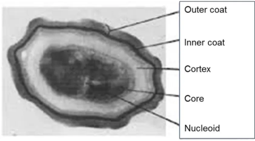
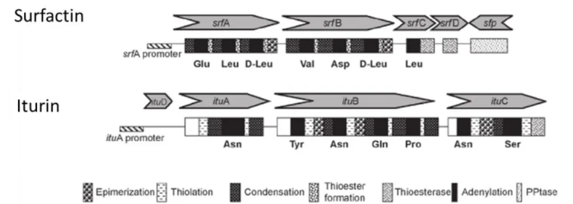
Figure I-10: Cross-section of a spore of Bacillus subtilis, showing the cortex and coat layers surrounding the core (dark central area). The spore is 1.2 microns across, about 100 times smaller than the width of a human hair. ... 32 Figure I-11: Organisational structure of peptide synthetases located outside of the ribosome. ... 33 Figure I-12: Structural organisation of the genes encoding surfactin and iturin biosurfactant synthetases family.. ... 36 Figure I-13: Structural organisation of the genes encoding fengycin synthetases family. ... 37 Figure I-14: Multianalytical approaches to Bacillus lipopeptides detection. ... 38
Chapter II.
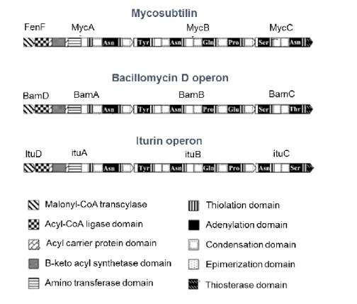
Figure II-1: Schematic diagram of mycosubtilin, bacillomycin D and iturin A operon from Moyne et al., 2004. ... 47 Figure II-2: Agarose gel electrophoresis of DNA isolated from different selected strains. A- CCLBH 1051; B- CCLBH 1052; C- CCLBH 1053; D- CCLBH 1054; Lane
TABLE OF CONTENTS
XXV
1- 100 bp Ladder (Nzytech Ladder VII); Lane 2- PCR product of bacterial amplification with ituA primer; Lane 3- PCR product of bacterial amplification with ituB primer; Lane 4- PCR product of bacterial amplification with ituC primer; Lane 5- PCR product of bacterial amplification with ituD primer. ... 58 Figure II-3: Iturinic ORF’s amplified DNA copies in the genome of the selected bacteria
strain determined with real time PCR. ( itu A; itu B; ituC; ituD). ... 59 Figure II-4: Phylogenetic relationships of Bacillus species based on nucleotide
sequences of the 16S rDNA. The tree was generated by the neighbor-joining method (MEGA 4.0 software)... 68 Figure II-5: Time course profiles of Bacillus sp.. Bacillus CCLBH sp. 1051 (–●–); Bacillus sp. CCLBH 1052 (–о–); Bacillus sp. CCLBH 1053 (– –); Bacillus CCLBH sp. 1054 (–Δ–). ... 69 Figure II-6: Total ion chromatogram of the supernatant liquid culture of Bacillus sp. CCLBH 1051 and mass spectra corresponding to the peaks (a) m/z 1031.36 and (b) m/z 1045.37. ... 71
Chapter III.
Figure III-1. Antifungal activity of biocides against Cladosporium sp. using the paper disk diffusion assay. A- Control. B- In the presence of 10 μL of Bacillus sp. CCLBH 1053 cell-free culture Broth. ... 85 Figure III-2: Percentage of inhibition growth resulted from interaction assays between
biodeteriogenic fungi and Bacillus sp. cell-free culture broth. ( Penicillium sp.1, Alternaria sp., Mucor sp., Fusarium oxysporum, Cladosporium sp., Penicillium sp. 2, Aspergillus niger). Different letters (a-m) following the values indicate significant differences (p<0.05). Values of each determination represents means ± SD (n=3)... 87 Figure III-3: Total ion chromatogram of the supernatant liquid culture of Bacillus sp. CCLBH 1051 and mass spectra obtained for the retention times (a) m/z 1031.35 and (b) m/z 1045.40. ... 90 Figure III-4: Total ion chromatogram of the supernatant liquid culture of Bacillus sp.
CCLBH 1052 and mass spectra obtained for the retention times (a) m/z 1031.27, (b) m/z 1045.31 and (c) m/z 1464.07. ... 91 Figure III-5: Total ion chromatogram of the supernatant liquid culture of Bacillus sp.
CCLBH 1053 and mass spectra obtained for the retention times (a) m/z 1031.37, (b) m/z 1045.37 and (c) m/z 1463.53. ... 92 Time (h) 0 5 10 15 20 25 30 35 L n ( N /N 0 ) 0 1 2 3 4 D2 C2 B10 x column 4 vs y column 4 x column 5 vs y column 5 x column 6 vs y column 6
XXVI
Figure III-6: 1H-NMR spectra of the bioactive compounds produced by Bacillus sp.
CCLBH 1053. ... 94 Figure III-7: Autobiographical results. Chromatographic scheme profile. A- The Bacillus sp. CCLBH 1053 extracted cell-free supernatant shows the presence of two antifungal compounds against biodeteriogenic fungi Cladosporium sp. B – Scheme of TLC plate in Petri dish with the two layers (I- Rf = 0.429 and II- Rf = 0.364 imagem); C- Bioautograms with TLC layers applied separately from Bacillus CCLBH 1053. ... 95 Figure III-8: FTIR-ATR analyses of the biologically active compounds produced by Bacillus CCLBH 1053 (I- Rf = 0.429) on the bioautographic plate. ( Bacillus sp. CCLBH 1053; Silica) ... 96
Chapter IV.
Figure IV-1: Methodological scheme of inducing germination assay ... 109 Figure IV-2: Time course profiles of Bacillus sp. CCLBH 1053. NB medium ( ); LAPM medium ( ). All data was determined in triplicate. ... 111 Figure IV-3: Concentration of particles in Bacillus sp. CCLBH 1053 in NB (A) and
LAPM (B) liquid culture. ( ) [Cell] (cell/mL); ( ) [Spo] (CFU/mL); ( ) [Spo]/[Cell] (CFU/cell). The marked area evidence the strongest variations observed... 112 Figure IV-4: Total ion chromatogram of the cell free supernatant of Bacillus sp. CCLBH 1053 and mass spectra corresponding to the peaks (a) m/z 1031.33, (b) m/z 1045.37 and (c) m/z 1463.56, after 48 h of culture. ... 113 Figure IV-5: Lipopeptide production of Bacillus sp. CCLBH 1053 culture along culture
time for NB culture medium (A) and LAPM medium (B) ( m/z 1463; m/z 1031; m/z 1045). ... 115 Figure IV-6: Variation of cell population distribution, of Bacillus sp. CCLBH 1053 growth in NB, along time assessed by staining with 7-AAD and Annexin V followed and flow cytometry analysis. A- ( ) Late apoptotic cells; ( ) Early apoptotic cells; ( ) Total apoptotic cells. B- ( ) Live cells; (●) Dead cells. The marked area evidence the strongest variations observed. ... 118 Figure IV-7: Time course of the two sub-populations of Bacillus sp. CCLBH 1053 growing in NB medium. A, B- Dot-plot cytometry and histograms at 1 and 96 hours of incubation, respectively. (a)-(e)- No stained cells; (f)-(j) - Cells stained with a mixture of Annexin V and 7-AAD dyes. (a), (c), (f), (h) - Cells sensed with Annexin V detector; (b), (d), (g), (i) - Cells sensed with 7-AAD detector. (e), (j) - Flow cytometer forward scatter (FSC) for cell size analysis.. ... 120
XXVII
Figure IV-7 (continued): Time course of the two sub-populations of Bacillus sp. CCLBH 1053 growing in NB medium. C, D- Dot-plot cytometry and histograms at 168 and 720 hours of incubation, respectively. (a)-(e)- No stained cells; (f)-(j) - Cells stained with a mixture of Annexin V and 7-AAD dyes. (a), (c), (f), (h) - Cells sensed with Annexin V detector; (b), (d), (g), (i) - Cells sensed with 7-AAD detector. (e), (j) - Flow cytometer forward scatter (FSC) for cell size analysis. ... 121 Figure IV-8: Variation of cell population distribution, of Bacillus sp. CCLBH 1053 growth in LAPM medium, along time assessed by staining with 7-AAD and Annexin V followed and flow cytometry analysis. A- ( ) Late apoptotic cells; ( ) Early apoptotic cells; ( ) Total apoptotic cells. B- ( ) Live cells; (●) Dead cells. ... 123 Figure IV-9: Time course of the two sub-populations of Bacillus sp. CCLBH 1053 growing in LAPM medium. A, B- Dot-plot cytometry and histograms at 1 and 120 hours of incubation, respectively. (a)-(e)- No stained cells; (f)-(j) - Cells stained with a mixture of Annexin V and 7-AAD dyes. (a), (c), (f), (h) - Cells sensed with Annexin V detector; (b), (d), (g), (i) - Cells sensed with 7-AAD detector. (e), (j) - Flow cytometer forward scatter (FSC) for cell size analysis.. ... 124 Figure IV-9 (continued): Time course of the two sub-populations of Bacillus sp. CCLBH 1053 growing in LAPM medium. C, D- Dot-plot cytometry and histograms at 168h and 720 hours of incubation, respectively. (a)-(e)- No stained cells; (f)-(j) - Cells stained with a mixture of Annexin V and 7-AAD dyes. (a), (c), (f), (h) - Cells sensed with Annexin V detector; (b), (d), (g), (i) - Cells sensed with 7-AAD detector. (e), (j) - Flow cytometer forward scatter (FSC) for cell size analysis.. ... 125 Figure IV-10: Time course profile of Bacillus cells (A) and spores formation (B), before
and after the peptone supplementation and heat activation assays. ( ) Normal growth in NB culture medium; ( ) NBPSHA assay; ( ) NBPS assay. ... 127 Figure IV-11: Lipopeptide production increment for NBPS assay and NBPSHA assay.
The relative percentage of increase was calculated considered as 100% the area of peak corresponding to 0 h of incubation time after treatment. ( ) NBPSHA; (
XXVIII Chapter V.
Figure V-1: Schematic representation of the acute toxicity assay on Swiss mice. 143 Figure V-2: Swiss mice toxicity assay. A- Pineal reflexes tests, B- Motor activity teste, C- Traction tests and D- Behaviour observation in toxicological evaluation assay. ... 144 Figure V-3: Methodological scheme of simulation in vitro assays using mortars slabs
and real samples under the presence of bioactive compounds. ... 147 Figure V-4: % of Artemia salina mortality for the bioactive compounds produced and for the commercial biocides at a concentration of 1000 μg/mL in each well. Different letters (a-b) following the values indicate significant differences (p<0.05). Values of each determination represents means ± SD (n=3). ... 149 Figure V-5: Slabs mortars, SEM micrographs of the MS1 microfragments and EDS analysis. A-E: Control slabs inoculated with Cladosporium sp.; F-H: MS with CB 3 (bioactive compounds produced by Bacillus sp. CCLBH 1053). ... 153 Figure V-6: Bi-dimensional projection of International Commission on Illumination a*
b* chromatic coordinates of the 12 measurements that were performed on the real mortars of the Santo Aleixo’s mural painting. Control; RM1; RM2. ... 157
Annexes.
Figure C-1: Agarose gel electrophoresis of 16S ribosomal DNA isolated from different Bacillus sp. strains. Legend: Lane 1- 100 bp Ladder (Nzytech Ladder VII); Lane 2- PCR product of 16S rDNA amplification of Bacillus sp. CCLBH 1051; Lane 3- PCR product of 16S rDNA amplification of Bacillus sp. CCLBH 1052; Lane 4- PCR product of 16S rDNA amplification of Bacillus sp. CCLBH 1053; Lane 5- PCR product of 16S rDNA amplification of Bacillus sp. CCLBH 1054. ... 193
XXIX
List of Tables
Chapter I.
Table I-1: Chemical biocides used in artworks treatment. ... 14 Table I-2: Structural variants diversity within the surfatin lipopeptide family synthesised by Bacillus species. ... 21 Table I-3: Structural variants diversity within the iturin lipopeptide family synthesised by Bacillus species. ... 23 Table I-4: Structural variants diversity within the fengycin lipopeptide family synthesised by Bacillus species. ... 25
Chapter II.
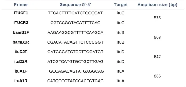
Table II-1: Oligonucleotide primers used to detect genetic markers for biological activity in bacteria with antifungal potential. ... 52 Table II-2: Antifungal activity assay with 21 bacteria strains isolated against
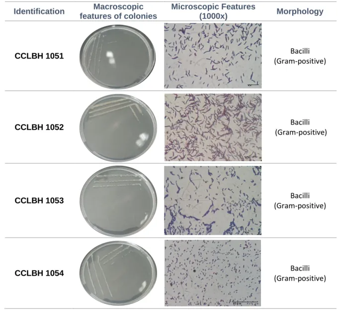
biodeteriogenic fungi. ... 56 Table II-3: Macroscopic and microscopic appearance of the selected microorganisms
in analysis. ... 62 Table II-4: Homology search from the Ribosomal database (RDP) of the CCLBH 1051
16S rDNA. ... 63 Table II-5: Homology search from the Ribosomal database (RDP) of the CCLBH 1052
16S rDNA. ... 64 Table II-6: Homology search from the Ribosomal database (RDP) of the CCLBH 1053
16S rDNA. ... 64 Table II-7: Homology search from the Ribosomal database (RDP) of the CCLBH 1054
16s rDNA. ... 64 Table II-8: Specific growth rate and generation time values for the Bacillus sp. strains
selected. ... 70
Chapter III.
Table III-1. Antifungal activity of bacterial bioactive compounds and commercial antifungal drugs against the biodeteriogenic fungi, isolated from deteriorated mural paintings. ... 84
XXX Chapter IV.
Table IV-1: Flow cytometer forward scatter (FSC), fluorescence histograms of Bacillus sp. CCLBH 1053 culture in NB and of two different sporulating assays in the same medium with Peptone Supplementation (NBPS) only, or followed by heat shock (NBPSHA) in different time of incubation. ... 129
Chapter V.
Table V-1: LC50 parameter of biocides tested. ... 150 Table V-2: Schematic representation of the lab experiments conducted on real mortars samples. RM1- Mortar impregnated with extracted compounds from Bacillus sp. CCLBH 1053; RM2- Mortar impregnated with CB 3. ... 156
Annexes.
Table A-1: Composition of the culture media used for microbiological growth. ... 191 Table D-1: DNA concentration extracted from of the bacteria strains in analysis. . 194 Table F-1: Composition of the saline medium used to Artemia salina toxicity assay.
... 199 Table G-1: Analysis of variance (ANOVA) of interaction assay against biodeteriogenic fungi. ... 200 Table G-2: Average multiple comparison of interaction liquid assay against biodeteriogenic fungi, by Tukey HSD test. ... 201 Table G-3: Analysis of variance (ANOVA) of the Artemia salina toxicity assay for bioactive compounds produced and commercial biocides. ... 202 Table G-4: Average multiple comparison of the Artemia salina toxicity assay for bioactive compounds produced and commercial biocides. ... 203 Table G-5: Analysis of variance (ANOVA) of the bi-dimensional projection of CIElab chromatic coordinates of the real mortars from mural painting. ... 204 Table G-6: Average multiple comparison of the bi-dimensional projection of CIEla*b* chromatic coordinates, by Tukey HSD test. ... 204
XXXI
Abbreviations
Specific growth rate
7-ADD 7-aminoactinomycin D
A Adenine nucleotide
A Adenylation domain
Ala Alanine
ANOVA Analysis of variance
Asn Asparagine
Asp Aspartic acid
ATP Adenosine triphosphate BDP Biocidal Product Directive
BLAST Basic Local Alignment Search Tool BSA Bovine serum albumin
C Condensation domain
C Cytosine nucleotide
CB Compounds Bioactive
CF Cycle fluorescence
cLPP Cyclic Lipopeptide CRB Cook Rose Bengal
Ct Threshold cycle
DNA Deoxyribonucleic acid
dNTPs Deoxynucleotide triphosphates DPA Dipicolinic acid
e Neper number
E Epimerization
EC European Comission
EDS Energy Dispersive X-ray Spectroscopy EDTA Ethylenediaminetetraacetic acid ELISA Enzyme-linked immunosorbent assay
FCM Flow cytometry
FELASA Federation of European Laboratory Animal Science Associations FSC Flow cytometer forward scatter forward scatter
FTIR-ATR Fourier Transform Infrared spectroscopy-Attenuated Total Reflection
g Time of generation
G Guanine nucleotide
GC Gas chromatography
Glu Glutamic acid
HPLC High performance liquid chromatography
Ileu Isoleucine
IR Infrared spectroscopy
LAPM Lipopeptide Antibiotic Production medium LC50 Lethal concentration 50%
LC–ESI-MS Liquid chromatography coupled with mass spectrometry/ Electrospray ionisation
XXXII
LD50 Lethal dose 50%
Leu Leucine
LPP Lipopeptide
MALDI-TOF Matrix-Assisted Laser Desorption Ionisation/Time-Of-Flight MEA Malt Extract Agar
MS Mass spectroscopy
MS Mortar Slabs
NA Nutrient Agar
NaOH Sodium hydroxide
NB Nutrient Broth
NBPS Nutrient Broth Peptone Supplementation assay
NBPSHA Nutrient Broth Peptone Supplementation and Heat-Activation assay NCBI National Center for Biotechnology Information
NH4 Ammonium
NMR Nuclear magnetic resonance NRPS Nonribosomal peptide synthetase
OECD Organisation for Economic Co‑operation and Development
ORF Open Reading Frame
Orn Ornithine
p p-value
PBS Phosphate-buffered saline PCR Polymerase Chain Reaction
PI Propidium iodide
PKS Polyketide synthases
Pro Proline
PS Phosphatidylserine
rDNA Ribosomal Deoxyribonucleic Acid RDP Ribosomal Database Project
Rf Retention Factor
RFU Relative fluorescence units
RM Real mortar
RNA Ribonucleic Acid
rRNA Ribossomal Ribonucleic Acid SDS Sodium dodecyl sulfate
SEM Scanning Electron Microscopy
SEM-EDS Scanning Electron Microscopy coupled with Energy Dispersive X-ray Spectroscopy T Thymine nucleotide T Thiolation domain Thr Threonine TLC Thin-layer chromatography Tyr Tyrosine UV Ultraviolet light Val Valine
XXXIII
Units
% Percentage
A Ampere
Bp Base pairs
CFU Colony-forming unit
cm Centimetre
Da Dalton
g Gram
h Hour
Hz Hertz
m/z Mass to charge ratio
min Minute
nm Nanometer
ºC Celsius degree
ppm Part per million
rpm Rotation per minute
s Second
V Volt
v Volume
1
Aims and Methodology
Biodeterioration is an undesirable process, triggered by living organisms, which can affect cultural and built heritage and economically important materials. The importance of carrying out proper remediation actions for microbiologically contaminated historic materials is of vital importance. The growth control of microflora in cultural and built heritage is usually done by treatments using chemical compounds that have high toxicity to humans. Bacillus species can be worth for these treatments because they produce a great diversity of secondary metabolites known to possess antagonistic activity against many fungal pathogens.
Thus, the main goal of this project is to obtain and study new bioactive molecules produced by different strains of Bacillus sp., to prove their remediation potential and to establish preventive approaches in heritage and construction context. For this reason, several methodological approaches, including microorganism and DNA manipulations, microscopic, spectrometric, spectroscopic and cytometric techniques, in combination with simulation assays, were developed in order to establish effective tools for the study of active compounds obtained from natural sources, against biodeteriogenic agents of artistic heritage.
The methodology defined for this work intended:
To select microorganism producers of compounds with antifungal potential; To identify and characterise the bacterial strains with higher activity against
biodeteriogenic fungi isolated from biodegraded cultural heritage artefacts; To determine kinetic features for the selected bacterial strains;
To develop a combined methodology for quick identification of bioactive compounds-producing strains;
2
To evaluate the antifungal potential against heritage biodeteriogenic fungi using different antifungal activity approaches;
To define methodological basis for bioactive compounds detection directed to heritage biodeteriogenic fungi;
To characterise the bioactive metabolites using spectroscopic analyses, including FTIR-ATR, 1H- NMR and LC–ESI-MS analysis;
To monitor the physiology of the Bacillus sp. cells by multi-parameter flow cytometry and the physiological response to nutrient starvation and re-supplementation;
To characterise, interpret and understand the compounds production and the relation with cell viability and sporulation;
To evaluate the toxicological properties of the new bioactive compounds produced using two different biological models: brine shrimp (Artemia salina) and Swiss mice (Mus Muculus);
To study the real life efficiency and influence of these new compounds in the growth of biodeteriogenic fungi, using simulation assays.
CHAPTER I
5
1.1. Cultural heritage biodeterioration/biodegradation
The preservation of historic monuments and buildings, which represent the cultural heritage of a country, constitutes a high societal priority in order to give the opportunity for the future generations to witness their ancestors achievements (Steinbauer et al., 2013).
The problems caused by lack of proper preservation of historical built heritage often only come to attention when a tragedy, such as a fire or collapse, occurs. Unfortunately silence threats lurking permanently in our cultural heritage, far from the eyes of the great majority of the people.
Nowadays, science and technology interact with art in several ways. In fact, the combination of biotechnological an analytical approaches can play an important role in protecting and preserving cultural heritage for future generations (Fernandes, 2006).
Environmental factors (humidity, temperature, light, CO2 concentration,
atmospheric pressure and pH), geological conditions of the ground, chemical composition (organic and inorganic nutrient sources), quality and ageing of the materials, internal mechanical stress and biological agents constituted the main parameters that influence artworks decay (Nugari et al., 2009; Pangallo et al., 2009; Capodicasa et al., 2010; Gaylarde et al., 2011; Tran et al., 2012; Rosado et al., 2014).
Whereas several biotic and abiotic factors can induce degradation/deterioration, the action of the biotic factors was neglected for a long time, and the abiotic factors were the only ones taken into account (Rojas et al., 2009).
Biodegradation/biodeterioration can be defined as “any undesirable change in a material brought about by the vital activities of organisms” (Sterflinger and Piñar, 2013). This phenomenon is an undesirable process, triggered by living organisms, which can affect cultural and built heritage and economically important materials (Sterflinger, 2010; López-Miras et al., 2013; Sterflinger and Piñar, 2013)
6
Microorganisms, including bacteria, fungi, algae and lichens as well as insect pests, influenced by environmental conditions, are the main biodeteriogenic agents responsible for aesthetical and structural damage of cultural heritage (Rosado et al., 2013a), causing problems in its conservation. This holds true for all types of historic artefacts and even for art made of modern materials (e.g., polymers) from public museums and from private art collections (Sterflinger and Piñar, 2013).
In the specific case of mural paintings, the development of microorganisms may cause discolouration of pigments and mortars, formation of stains and biofilms, salt efflorescence appearance, exfoliation of paint layers, formation of paint blisters, cracking and disintegration of paint layers, and degradation of binders that results in detachment of the paint layer (Guiamet et al., 2011; Borrego et al., 2012; López-Miras et al., 2013; Sterflinger and Piñar, 2013).
Despite the involvement of microorganisms in the deterioration process is well known, the specific role of the different groups and species that integrate the microbial communities is not yet well understood. A wide diversity of microorganisms are involved in artworks deterioration. Among them, fungi are particularly dangerous because their hyphae may have high level of proliferation in the materials and their spores, in a dormant state, are commonly present and available for germination but also because fungal derived carboxylic acids (e.g., oxalic, citric, succinic, formic, malic, acetic, fumaric, glyoxylic, gluconic, and tartaric acids) can induce chemical attack. Fungi of the genera Penicillium, Cladosporium, Alternaria, Curvularia, Dreschlera, Chaetomium, Fusarium,
Trichoderma, Gliomastix, Aureobasidium, are the most abundant in degraded mural
7
Figure I-1: Different artworks with signals of deterioration caused fungal communities. A - Renaissance Frescoes from Santo Aleixo Church, Montemor-o-Novo, Portugal, B- Mural paintings from Casa Pintadas, Évora, Portugal, C – Mural paintings from Santa Clara Church (Sabugueiro, Arraiolos, Portugal), D- Gilded woodcarving from altars of the Espírito Santo Church, Évora, Portugal. Adapted from (Rosado et al., 2013b; Rosado et al., 2014; Rosado et al., 2015a; Rosado et al., 2015b).
Stone and other building materials, such as concrete, mortar, slurries, paint coatings, glass and metals used in architecture are object of overall deterioration phenomena in which different sorts of microorganisms are involved (Scheerer et al., 2009). On building stone exposed to open air, fungi may also be the most important biodeteriorative organisms because of their extremely erosive potential (Scheerer et al., 2009; Sterflinger and Piñar, 2013).
Depending on the physical properties of the material, fungi may penetrate or not inside the stone. There are two major morphological and ecological groups of stone-inhabiting and stone-dwelling fungi. These have adapted to different environmental conditions. In moderate or wet weather, the fungal communities on rock are dominated by hyphomycetes that form mycelia (hyphal networks) in the porous space of the stones (Sterflinger, 2010). Since the first step for fungal colonisation is the settlement of spores
8
from the air, the species diversity of stone fungi is rather similar to the diversity of common airborne spores. Alternaria, Cladosporium, Epicoccum, Aureobasidium and Phoma are the most important fungal strains (Sterflinger, 2000). However, due to their
thick walls, fungi can resist to chemical attack and, therefore, resist biocides and other anti-microbial treatments.
Cyanobacteria, algae and lichens contribute to the weathering of stone in humid as well as in semi-arid and arid environments, producing a characteristic phenomenon consisting of large green-black stains (Cutler et al., 2013).
Additionally, the role of chemoheterotrophic bacteria in the weathering of rock probably depends largely on the environmental conditions. While bacteria might growth in humid environments and form biofilms within the porous space of building stone, the occurrence of chemoheterotrophic bacteria might be more limited (Lamprinou et al., 2013).
The excretion of inorganic acids (e.g. nitric and sulfuric acids) on rock surfaces, by chemolithotrophic bacteria, that use carbon dioxide as carbon source, produce energy through the oxidation of inorganic compounds (electron donors) such as ammonium (NH4), nitrogen dioxide and hydrogen sulphide (Sáiz-Jiménez and Laiz, 2000). Another
way consist in the excretion of organic acids in surfaces of the monuments by fungi and bacteria chemoorganotrophic - those obtain energy by oxidising organic molecules or other living beings. Microorganisms use these organic substrates to increase their population and activity - and consequently, the biodeterioration process (Gaylarde et al., 2011; Cutler et al., 2013).
Moreover, a well-known phenomenon often observed on buildings and wall paintings is the formation of salt efflorescence on surfaces (Sáiz-Jiménez and Laiz, 2000). Salt may be available in the wall itself, from biological processes (ammonium salts) or simply due to co-migration with infiltrating water. Due to changes in physical parameters (i.e., temperature or humidity) salts can precipitate on the exposed surfaces. The crystallisation on walls and wall paintings results in a destructive effect, leading to
9
material loss and destruction due to cracking and detachment of the walls (Saiz‑Jimenez et al., 2012; López-Miras et al., 2013)
1.1.1. Mitigation approaches
The recent field, Science for Conservation, has developed following two main streams: i) the characterisation of the technique used by the artists, the analytical characterisation of the materials constituting the works of art and the chemical reactions involved in their degradation; ii) the search for new scientific methods for the restoration/conservation, allowing the safeguard of our Cultural Heritage for its transmission to future generations (Giorgi et al., 2010).
In general, the restoration of a work of art consists in:
1. Cleaning, which is a transient treatment, meant to remove the materials not originally belonging to the work of art;
2. Consolidation, which is a durable intervention that should remediate, prevent, or slow down further degradation due to aging or external agents (Giorgi et al., 2010).
According to these purposes, during several years the conservation and restoration process was done without taken into account the presence of microbiological contamination in cultural heritage artefacts. In fact, many of the restoration procedures were short term processes, providing to microorganism all the nutrients needed for their development and proliferation. Thus, several efforts have been made to solve this problem.
In order to control the biodeterioration process the most suitable methods and products must be used. With regard to the principles and the nature of the means employed, biodeterioration control methods can be classified as: mechanical, physical, biological or biochemical. However, the chemical methods based on active principles in
10
solution are the most frequently applied (either as wide-spectrum active principles, or more specific, narrow-spectrum biocides such fungicides, algaecides, herbicides, insecticides and repellents for birds) (Allsopp et al., 2004).
a) Mechanical Methods
The application of irradiation treatment for microbial elimination and cultural heritage artefacts protection has been used in several studies.
All the techniques described as "mechanical" have in common the method of displacing the biodeteriogens: physical removal. It can be carried out by hand or with tools such as scalpels, spatulas, scrapers, air abrasive or vacuum cleaners (Figure I-2A) Although they were frequently used in the past, these methods do not produce lasting results and the eradication of contaminants could do not completely stop its vegetative activity. Moreover, the use of mechanical methods can damage the substrate, even if they have the advantage to avoid the addition of any substance that might cause further deterioration (Savulescu and Ionita, 1971).
Figure I-2: Work of preventive cultural heritage conservation. A- Application of mechanical methods in a rock wall with prehistoric paintings at the Serra da Capivara, National Park, Brasil. B- Conservator-restorer cleaning mold with chemical toxic biocide.
(Adapted from http://www.wikiwand.com/en/Conservation-restoration_of_cultural_heritage and https://www.epa.gov/mold/brief-guide-mold-moisture-and-your-home)
11 b) Physical Methods
The physical methods used in some mitigation approaches are: ultraviolet radiation (UV), gamma-radiation, low frequency electrical systems, heat, deep-freeze temperatures, ultrasonic, titanium dioxide and laser techniques (Tiano, 2002; Scheerer et al., 2009). Ultraviolet radiation has been used especially against bacteria, algae and
fungi colonising of renders and plasters, however its handling can be dangerous and could cause injuries to the conservator-restorer. Gamma-radiation has been used for sterilising microflora and killing insects, especially on organic materials such as paper, parchment and wood. However, gamma irradiation does not have a long-lasting effect and can promote possible deterioration of the object to preserve (Abdel-Haliem et al., 2013).
High-frequency can be used to kill wood-destroying insects (Anobiidae), but only in the absence of metals (Van Der Molen et al., 1980; Cutler et al., 2013). Low-frequency electrical current systems have recently been used to keep birds from roosting on monuments (Caneva et al., 2008). This method is completely harmless for animals and humans. Heat (dry or wet) is used in the disinfestations and disinfections of organic materials (Gaylarde et al., 2011).
Titanium dioxide is a photo-catalytic nanoparticle with antibacterial and antifungal abilities due to the production of reactive redox species (hydroxyl radicals, superoxide anions and hydrogen peroxide) which induce damages in the cell membrane and can inactivate a wide range of organisms like bacteria, viruses, fungi and algae. Titanium dioxide was proposed for preventing biodeterioration of mortars in cultural heritage buildings such as Palacio da Pena (Sintra, Portugal). However, despite these good indications it is necessary to take into account risks to humans as well as for paint materials because these particles are not as well studied neither their effect (De Filpo et al., 2013).
12
Also, the use of laser techniques has been emergent in the last few years, due to their promising results not only for diagnostic but also for restoration procedures. Laser ablation takes place when a pulse of laser radiation is absorbed at the surface of a material, determining a sudden transition of its solid phase to another phase (gas, vapor, plasma). The sudden phase transition is mainly due to photothermal effects rising the temperature of the material up to a hot vapor that expands quickly in the surroundings, producing a material removal. However, this technique still present some inconvenient because do not completely remove the microorganism and could contribute for the deterioration of artworks (Salimbeni, 2006).
c) Biological and Biochemical Methods
The application of biological means is based on the existence of parasitic or antagonistic organisms of the biodeteriogens. Bacteria, insects and bacteriophage might be used as predators species and introduced on the environment in order to eliminate the coloniser microorganism (Caneva et al., 2008).
The group of biochemical methods encompass biodeterioration control systems by the use of chemical compounds of biological origin. Antibiotics, enzymes and pheromones are the substances most commonly used (Ranalli et al., 2005; Webster and May, 2006; Roig et al., 2013)
d) Chemical Methods
Many organic and inorganic compounds have been used as biocide agents, to eliminate the biodeteriogens from cultural objects (Figure I-2B). The chemical biocides are classified in different ways depending on their chemical nature (e.g., organic or inorganic) or on the target species.
13
Pesticides, for example, are commonly used due to their biocide action with specific toxicity for the species to be eliminated. However, a problem associated with the use of pesticides is the persistence of the product in the soil or water. This issue is especially prevalent with herbicides that are applied or dispersed in external environments, with high risk for soil and water contamination (Blazquez et al., 2000; Moreau et al., 2008; Young et al., 2008; Camara et al., 2011).
Disinfectants constituted another example of biocides commonly used in conservation treatment. These can destroy vegetative forms but are not always effective against survival resistant or quiescent phase structures such as bacterial spores, fungal conidia and insect eggs. In addition to these biocides, other constituents, such as additives to improve the effectiveness of the product or to facilitate its application, are present in the chemical formulation. These are called co-formulants and could have negative effects on the objects to be treated (Nugari and Salvadori, 2003; Fonseca et al., 2010; De los Ríos et al., 2012).
1.1.2. Biocides treatment
According to the European Commission’s Biocidal Product Directive 98/8/EC,1 (BPD) (http://ec.europa.eu/environment/biocides/index.htm) biocides can be divided into four main groups – disinfectant, preservative, pest control and other biocidal products – and further classified into 23 product types, including approximately 955 substances and 372 notified substances (Ashraf et al., 2014). However, the choice of an appropriate biocide in Europe is limited by the single Directive previously cited. There are fundamental requirements for industrial biocides suitable for protection of materials like: effective antimicrobial activity, economical feasibility, very low human toxicity and compatibility with the environment (Singer et al., 2010).
Although the number of chemical classes with biocide activity includes a wide variety of compounds, such as alcohols, aldehydes, phenols, acids, acid esters, amides,
14
carbamates, dibenzamidines, pyridines, azoles, heterocyclics, activated halogen compounds, surface active agents, organometallics and oxidising agents (Table I-1), the number of products suitable for cultural heritage is comparatively limited. This is due to the fact that only a small number of biocides have been tested with respect to their compatibility with historic materials, and only a very few studies exist about their long term effects (possible promotion of colour alterations or degradation products appearance) (Nittérus, 2000; Paulus, 2005; Sterflinger and Piñar, 2013). Despite the well-established short-term biocides efficiency, the removal of the microbial community may give rise to a new succession of microorganisms, which may be more damaging than the old microbial surface populations (Singer et al., 2010).
Table I-1: Chemical biocides used in artworks treatment.
Biocide Class/
Active principle Action form Reference
Preventol R50 Ammonium quaternary compounds/ Na and Ca hypochlorite Active transport and membrane integrity
disruption (Paulus, 2005; Nugari et al., 2009; Maxim et al., 2012; Silva et al., 2016) New-Des Diuron Urea derivatives/ aromatic halide Photosynthetic process blocking (Blazquez et al., 2000; Rosado et al., 2014) Karmex
Wikamol Murosol Organometallics/
Tributyltin oxide Metabolism inhibitor (Rosado et al., 2014)
Igran 500FW Triazines/ Terbutryn Photosynthesis inhibition; electron transport alterations (Fernandes, 2006; Maxim et al., 2012)
Panacide Chlorinated Phenol/
Dichlorophene
Clearing of intestinal contents increased by
veterinary fungicide
(Silva et al., 2016)
Against microbial recolonisation, some studies have suggested that the combined application of hydrophobic compounds and biocides is more effective than only of the biocide. The application can be done in a single step when the water-repellent and the biocide are mixed together, or in two steps when the biocide is applied before or after the water-repellent (Urzì and De Leo, 2007; Moreau et al., 2008).
15
Therefore, the approach to control biodeterioration must be polyphasic and interdisciplinary, and must consider the history and condition of the artefact as well as the physical and chemical damaging factors (Scheerer et al., 2009).
However, the mode of action of antimicrobial biocides and the associated resistance mechanisms are generally poorly understood (Mavri and Možina, 2013). The historical view that antimicrobial biocides possess broad-spectrum activity has led to their false association with low-target specificity (Ashraf et al., 2014).
Moreover, a substantial part of all biocidal products currently available on the market act through toxic mechanisms exhibiting numerous pharmacological activities toward a number of specific cellular targets, including damaging or inhibiting the synthesis of cell walls and affecting DNA or RNA, proteins or metabolic pathways (Singer et al., 2010; Ashraf et al., 2014).
For these reasons, the chemical biocides with high toxicity can jeopardizes the health of any human beings, especially those that can easily handle them. In fact, conservators-restorers are the most direct persons exposed to these hazards, but few data are available regarding their health (Varnai et al., 2011). This enhance the problem of the inadequate consideration of health and safety risks of biocides use. Despite the duty of each biocide manufacturer to have a Safety Data Sheet with all the health risk implicit, the importance of Cultural Heritage for Europe are still stressed to be central (Caminiti et al., 2016) due to the real problems in term of safety and health that chemical biocides can bring.
Therefore, green alternatives based on natural biocides or innocuous solutions with very low toxicity, environmental acceptability, easy and safe to handling and storage, are currently essential in order to irradiate the potential risk associated with chemical biocides used (Silva et al., 2016). This goal can be achieved by proposing adequate alternative materials, products and methods of intervention instead of the traditional ones.
16
Therefore, developing proper remediation actions for microbiologically contaminated historic materials based on environmentally safe solution is of vital importance.
Recently, biological cleaning is recognised as being a viable alternative to traditional chemical treatments such as organic solvents or mechanical treatments. In fact, microorganisms can act as new bioagents for the recovery and conservation of artwork and historical architectural monuments. These innovative biological methods (biocleaning, bioconsolidation) is encouraged by the fact that only a few known microorganisms play a destructive role (causing deterioration) in the natural processes, while the majority of them are responsible for positive processes (Azevedo et al., 2009; Roig et al., 2013). Careful selection of the appropriate (not pathogenic) microorganisms with the requisite characteristics for the removal of undesirable substances (nitrates, sulfates, organic matter, etc.) is one of the first steps to be taken in formulating the best biorestoration strategy. Biotechnological approach have been able to resolve a range of problems on various artistic materials (including monumental stone, wall paintings, marble statues, etc.) and to combat diverse pathologies that affect artworks (such as the bioremoval of organic substances, black crusts, and mineral salts) by using different cultures of viable bacteria; such as sulphate-reducing bacteria like Desulfovibrio desulfuricans and Desulfovibrio vulgaris; nitrate-reducing bacteria like Pseudomonas
stutzeri, and others (Cappitelli et al., 2005; Cappitelli et al., 2007; Bosch-Roig et al., 2012;
Bosch Roig et al., 2013).
However, the use of P. stutzeri has only been reported in the removing of organic matter in wall paintings (Sorlini and Cappitelli, 2008; Ranalli et al., 2009; Polo et al., 2010). Studies have described the different ways to expose the artwork to the bacteria beginning from its immersion in a solution, followed by it direct application using delivery systems like cotton, Carbogel and multilayer systems (Alfano et al., 2011). The reports further emphasise that the most efficient method to apply the bacteria is by using a
17
delivery system where it provides enough water for their survival but not too much for not to be able to produce damage in the artwork (Cappitelli et al., 2007; Roig et al., 2013). Hence, the use of viable microorganisms in biocleaning treatment has given rise to a number of questions about the risks of these methods. Restorers, public and private sector restoration committees are asking the scientific community for more information about the safety of this new technology.
In this way, importance must be given to process monitorisation in order to confirm and validate the biocleaning process, developing at the same time suitable strategies for inspection of any new microbial interactions on biocleaned artworks. When possible, these should include adequate on-site technologies based on non- invasive tools to understand the potential risks on biocleaned tangible heritage and include physical-chemical, biological, and aesthetic analyses (Roig et al., 2013).
Similarly, further research and clear demonstrations of the complete safety of biocleaning is of fundamental importance because this technology has a very significant role to play in the introduction and diffusion of a new approach to the application of human-friendly, environmentally- sustainable techniques and technologies for the conservation and restoration of heritage properties (Bosch-Roig and Ranalli, 2014).
1.2. Biosurfactants compounds – a natural solution
Due to the limitations related with the use of chemical biocides, such as those mentioned above, it is urgent to find new products and antimicrobial compounds based on natural sources. This will allow to replace the risks associated with the use of chemicals products, avoiding the hazards associated to chemical products application.
Some solutions such as water-based micelles or microemulsions, (Giorgi et al., 2010) and essential oils (Rakotonirainy and Lavédrine, 2005; Stupar et al., 2014), are used in the replacement of chemical methods for cultural heritage safeguard.