INTRODUCTION
Article/Artigo
1. Laboratório de Esquistossomose, Centro de Pesquisas René Rachou, Fundação Oswaldo Cruz, Belo Horizonte, MG. 2. Departamento de Ciências Básicas, Laboratório de Bioquímica, Universidade Federal dos Vales do Jequitinhonha e Mucuri, Diamantina, MG 3. Laboratório de Pesquisas Clínicas, Centro de Pesquisas René Rachou, Belo Horizonte, MG.
Address to: Dr. Paulo Marcos Zech Coelho. Lab. Esquistossomose/CPqRR/FIOCRUZ. Av. Augusto de Lima 1715/sala 205, 30190-002 Belo Horizonte, MG, Brasil.
Phone: 55 31 3349-7740; Fax: 55 31 3295-3115 e-mail: [email protected]
Received in 31/10/2011 Accepted in 13/02/2012
Antigens of worms and eggs showed a diferentiated detection of speciic
IgG according to the time of
Schistosoma mansoni
infection in mice
Antígenos de vermes e ovos demonstraram detecção diferenciada de IgG baseado no tempo
de infecção pelo
Schistosoma mansoni
em camundongos
Rafaella Fortini Queiroz Grenfell
1, Watson Hermann Martins
1, Vanessa Silva-Moraes
1, Suedali Villas-Boas Barata
2,
Elizandra Giani Ribeiro
1, Edward Oliveira
3and Paulo Marcos Zech Coelho
1ABSTACT
Introduction: he correlation between the immunological assay and the antibody titer can ofer a tool for the experimental analysis of diferent phases of the disease. Methods: Two simple immunological assays for Schistosoma mansoni in mice sera samples based on speciic IgG detection for worms soluble antigens and eggs soluble antigens were standardized and evaluated in our laboratory. Fity mice were used in negative and positive groups and the results obtained by enzyme-linked immunosorbent assays (ELISA) assays were compared with the number of worms counted and the IgG titers at diferent times of infection. Results: Data showed that ELISA using adult worm antigens (ELISA-SWAP) presented a satisfactory correlation between the absorbance value of IgG titers and the individual number of worms counted ater perfusion technique (R2=0.62). In addition, ELISA-SWAP diferentially detected
positive samples with 30 and 60 days post infection (p=0.011 and 0.003, respectively), whereas ELISA using egg antigens (ELISA-SEA) detected samples ater 140 days (p=0.03).
Conclusions: hese data show that the use of diferent antigens in immunological methods can be used as potential tools for the analysis of the chronological evolution of S. mansoni infection in murine schistosomiasis. Correlations with human schistosomiasis are discussed.
Keywords: Schistosomiasis mansoni. IgG. Worm/egg antigens. Acute/chronic phases.
RESUMO
Introdução: A correlação entre o ensaio imunológico e o título de anticorpos serve como ferramenta para a determinação das diferentes fases da doença. Métodos: Dois ensaios imunológicos simples para detecção de IgG especíico para antígenos de vermes adultos e ovos do Schistosoma mansoni com amostras de soro murino foram padronizados e avaliados em nosso laboratório. Cinquenta camundongos negativos e positivos foram avaliados e os resultados obtidos por enzyme-linked immunosorbent assays (ELISA) foram comparados com o número de vermes adultos contados em tempos diferentes de infecção. Resultados: Os dados mostraram que a ELISA com antígenos de vermes adultos (ELISA-SWAP) apresentou uma correlação satisfatória entre a absorbância obtida para os títulos de IgG e o número individual de vermes contados por perfusão do sistema porta hepático (R2=0,62). Adicionalmente, a ELISA-SWAP foi capaz de detectar diferencialmente amostras positivas com 30 e 60 dias de infecção (p=0,011 e 0,003, respectivamente), enquanto a ELISA com antígenos de ovos (ELISA-SEA) detectou amostras positivas com 140 dias de infecção (p=0,03). Conclusões:
Estes dados mostram que o uso de antígenos diferentes em métodos imunológicos pode ser usado como ferramentas potenciais para a análise da evolução cronológica da infecção por S. mansoni na esquistossomose murina. Correlações com a esquistossomose humana devem ser discutidas.
Palavras-chaves: Esquistossomose mansônica. IgG. Antígenos de vermes/ovos. Fases aguda/ crônica.
Schistosomiasis is the most important of the human helminthiases in terms of morbidity and mortality associated with subtle but persistent morbidities1-4. During schistosomiasis progression, schistosomes mature to adults in the hepatic circulation and then in pairs migrate to inhabit in the mesenteric veins, where they mate and lay a large number of eggs in the vessels of the intestinal wall5 and other tissues, mainly in the liver. he major pathological consequences of chronic schistosomiasis are associated with soluble egg antigens secreted from schistosome egg that produces a vigorous inlammatory response resulting in granuloma formation6. Understanding the dynamic responses of the hosts with schistosomiasis related to the time of infection is important to provide insights into the mechanisms underlying disease progression and thus could be potentially useful for the diferential diagnosis in schistosomiasis evolution. Patent schistosome infections are highly immunogenic and there is not difficulty to demonstrate the presence of anti-schistosome antibodies7. Together, diferent levels of immunoglobulin G (IgG) isotypes against Schistosoma mansoni antigens have been demonstrated before and ater chemotherapy in experimental schistosomiasis8-10, but there have been no studies that evaluated the IgG levels against diferent antigens at diferent times ater infection.
METHODS
RESULTS
Mice sera
hirty swiss female mice (4-6 weeks), purchased at the Animal Facility of René Rachou Research Center, Oswaldo Cruz Foundation (FIOCRUZ), were exposed to an average of 40 cercariae of S. mansoni (LE strain) per animal, by subcutaneous route12. Serum samples were collected by retro orbital sinus puncture at 30, 60 and 140 days ater infection, when mice were sacriiced by cervical dislocation and submited to perfusion of the hepatic portal system using saline solution (0.85% NaCl) plus 50U/L heparin13. Adult worms were counted in a stereoscopic microscope (Zeiss Stemi DV4, New Jersey, USA). Twenty swiss female mice were used as negative control of infection. he use of animals was approved by the Ethical Commission in the Use of Animals, FIOCRUZ, Brazil (CEUA No. L-0023/08).
Preparation of Schistosoma mansoni adult
worm soluble antigen
Swiss female mice (4-6 weeks) were subcutaneously infected by syringe injection, with 100 cercariae12. Aiming to recover the adult worms, ater 45 days the animals were sacriiced by cervical dislocation, and underwent perfusion of the hepatic portal system using saline solution (0.85% NaCl) plus 50U/L heparin13. Adult worms were washed three times with 0.15M phosphate bufer saline pH7.2, submited to mechanical grinding (Virtiz Precisa, Dietikon, Switzerland), and centrifuged at 9,500g for 1h at 4ºC (Eppendorf AG, Hamburg, Germany). he supernatant obtained was dialyzed in cellulose membrane (Sigma-Aldrich D9777, St Louis, USA) against saline solution (0.9% NaCl) for 48h at 4ºC. he antigen was centrifuged at 1,250g for 15min at 4ºC and the supernatant was aliquoted and stored in freezer at -20ºC. An aliquot was submited to protein assessment following Bradford method14 and the obtained concentration was used as parameter in the standardization of the immunoassay for detection of murine antibody, called here as enzyme-linked immunosorbent assay-soluble adult worm antigens (ELISA-SWAP).
Preparation of Schistosoma mansoni eggs soluble antigen
After performing the perfusion of hepatic portal system of infected mice, the livers of these animals were removed to provide eggs recovery. The antigen used in this study was prepared by the method described by Colley et al.15. Eggs of S. mansoni were homogenized and ground in Virtiz (Virtiz Precisa, Dietikon, Switzerland) with 0.85% saline solution for 40 minutes. The homogenate was centrifuged at 9,500g for an hour at 4ºC. Ater 48h of dialysis in cellulose membrane (Sigma-Aldrich D9777, St Louis, USA) against 0.9% saline solution, the supernatant was submited to protein assessment14. he inal concentration was used as parameter in the standardization of the immunoassay for detection of human and murine antibody, named here as enzyme-linked immunosorbent assay-soluble egg antigens (ELISA-SEA).
Enzyme-linked immunosorbent assays
ELISA-SWAP: microtiter plates MaxiSorpTM Surface (NUNC Brand Products, Roskilde, Denmark) were sensitized with 100µl/ well of 1µg/mL of SWAP diluted in buffer 0.05M carbonate-bicarbonate pH 9.6 for 16h at 4ºC. he plates were washed three times with 0.15M phosphate bufer saline pH 7.2 with 0.05% of
Tween 20 (PBS-T) (LGC Biotecnologia São Paulo, SP, BR) and, the non speciic sites were blocked with 10% fetal bovine serum in PBS-T 0.05% at 37ºC for an hour. Ater new washing steps, 100µl of serum samples diluted 1:100 in PBS were added in triplicate into each well and the plates were incubated at room temperature for one hour. hen, the plates were submited to washing steps and incubated at room temperature for one hour with conjugated anti-IgG mouse-peroxidase (Southern Biotech, Birmingham, USA) diluted 1:5,000 in PBS-T 0.05%. he plates were again washed and 100µl of substrate 3,3',5,5-tetramethylbenzidine solution (TMB (Invitrogen, Carlsbad, USA) were added to each well. he reaction was stopped ater 20 minutes of incubation in the dark by addition of 50µl/well of 2N sulfuric acid. he results were obtained as absorbance values at 450nm in microplate reader (Model 3550, Bio-Rad Laboratories, Tokyo, JA). he cut of value was determinate as 0.250 of absorbance, using Roc curve (area=0.997). Positive and negative sera were added to the plates, and wells without antigen and antibodies were kept as controls of the assay. he standard dilution used in each technique was determined by a dilution curve, performed with the same reagents and equipment16.
ELISA-SEA: Microtiter plates MaxiSorpTM Surface (NUNC Brand Products, Roskilde, Denmark) were sensitized with 100µL/ well of 3µg/mL of SEA antigen diluted in bufer 0.05M carbonate-bicarbonate bufer pH 9.6 for 16h at 4ºC. he plates were washed three times with 0.15M phosphate bufer saline pH 7.2 with PBS-T 0.05% of Tween 20 and the non speciic sites were blocked with 10% fetal bovine serum in PBS-T 0.05% at 37ºC for 1h. Ater new washing steps, 100µL of serum samples diluted 1:100 in PBS were added in triplicate into each well and the plates were incubated at room temperature for one hour. Following, the plates were submited to washing steps and incubated at room temperature for one hour with conjugated anti-IgG mouse-peroxidase (Southern Biotech, Birmingham, USA) diluted 1:15,000 in PBS-T 0.05%. he plates were washed and the results were obtained as described for ELISA-SWAP. he cut of value was determined as 0.544, using Roc curve (Area=0.828). Positive and negative sera were added to the plates, and wells without antigen and antibodies were used as controls of the assay. The standard dilution used in each technique was determined by a dilution curve, performed with the same reagents and equipment16.
Statistical analysis
Data deriving from absorbance values were analyzed with Minitab sotware by Kolmogorov-Smirnov normality test. Normal distributed data were analyzed by Student’s t test and non-normal distributed data were analyzed by Mann-Whitney test, p<0.05 as signiicance level. Signiicance levels for percentages were determined by Chi-square test (x2). he cut of values and likelihood ratio were determined with Prism 4.0 sotware.
Ethical considerations
All the experiments on animals reported were performed ater the Oswaldo Cruz Foundation ethics commitee approval (L-0023/08).
performed using cut of values determined by the ROC curve, and compared with worms recovered by perfusion technique. he cut of values of 0.250 and 0.544 were determined for SWAP and ELISA-SEA, respectively. he geometric mean of the number of worms per mouse previously infected with 40 cercariae, estimated by the perfusion technique, was 8.
Analysis of results showed that 7 samples of infected mice were negative for both ELISA-SWAP and ELISA-SEA assays. Four of these samples were from infected mice with low parasite burden (3-5 worms recovered) (p=0.001) while 1 sample was from an infected mice with absence of worms. Two other samples were from mice with 10 and 14 worms recovered ater perfusion technique.
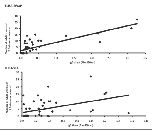
The relationship between individual values of the number of worms after perfusion and speciic IgG levels in mice sera ater both ELISA assays was evaluated.
As showed in Figure 1, ELISA-SWAP
showed a more conident association with a R2 of 0.62 in comparison with ELISA-SEA with a R2 of 0.21.
To analyze the capability of both assays in detecting IgG titers in diferent times of infection by S. mansoni, replicates of the same samples were assessed on three diferent days post infection (30, 60 and 140 dpi). As shown in Figure 2, ELISA-SWAP was capable of detecting speciic IgG antibodies in 50% of serum samples from mice with 30 days of infection (p=0.01) and in 58% of samples with 60 days (p=0.003). On the other hand, only 33% of the positive serum from mice ater 140 days of infection was detected.
Whereas, ELISA-SEA was sensible for detecting speciic IgG titers in only 17% of serum samples from mice with 30 or 60 days of infection, but showed a much beter result for 140 dpi samples with 50% of positive detection (p=0.03). All negative samples were also negative for both ELISA assays. 0 5 10 15 20 25 30
0.0 0.5 1.0 1.5 2.0 2.5 3.0 3.5
N u m b e r o f a d u lt w o rm s o f Sc h is to s o m a m a n s o n i
IgG ters (Abs 450nm) ELISA-SWAP 0 5 10 15 20 25 30
0.0 0.2 0.4 0.6 0.8 1.0 1.2 1.4 1.6 1.8
N u m b e r o f a d u lt w o rm s o f S c h is to s o m a m a n s o n i
IgG ters (Abs 450nm)
ELISA-SEA
FIGURE 1 - Relationship between individual values of the number of worms ater perfusion of hepatic portal system and IgG levels in mice serum detected by ELISA assays at 140 days ater infection. ELISA-SWAP with a cut of value of 0.250 showed a linear regression equation of y = 6.683x + 1.923 (R2=0.62). ELISA-SEA with a cut of value of 0.544 showed a linear regression equation of y = 7.755x + 2.266 (R2=0.21).
ELISA-SWAP: enzyme-linked immunosorbent assay-soluble adult worm antigens; ELISA-SEA: enzyme-linked immunosorbent assay-soluble egg antigens.
0 10 20 30 40 50 60 70 80 90 100
Infected 140 dpi Infected 60 dpi Infected 30 dpi Non infected ELISA-SWAP
*
*
0 10 20 30 40 50 60 70 80 90 100
Infected 140 dpi Infected 60 dpi Infected 30 dpi Non infected ELISA-SEA
*
FIGURE 2 - Percentage of positive mice sera for the detection of IgG levels by SWAP and ELISA-SEA. Mice were exposed to 40 cercariae of S. mansoni and serum samples were collected at 30, 60 and 140 days ater infection. Each column represents the percentage of mice sera that were positive for the presence of speciic IgG anti-SWAP (*p=0.011, 0.003 respectively for 30 and 60 days ater infection) and anti-SEA (*p=0.029 for 140 dpi) for Chi-square test. he Cut of values were 0.250 and 0.544, respectively.
ELISA-SWAP: enzyme-linked immunosorbent assay-soluble adult worm antigens; ELISA-SEA: enzyme-linked immunosorbent assay-soluble egg antigens.
DISCUSSION
he authors declare that there is no conlict of interest.
CONFLICT OF INTEREST
FINANCIAL SUPPORT
REFERENCES
injected antigens, complement ixation, indirect immunoluorescence, indirect haemagglutination, radioimmunoassay, and various locculation and precipitation tests17-20. But thus far, all these methods revealed low sensitivity, demonstrating the remaining ineiciency correlation between results from direct and indirect methods.
In this study, we evaluated the eiciency of two diferent ELISA assays based on the detection of IgG antibodies (against worm soluble antigens and egg soluble antigens) as a tool to conirm the results obtained by the portal system perfusion technique considering the number of worms from mice infected with 40 cercariae. Worm antigens are most abundant and easily obtained source of antigenic material. Crude extracts of worms work well in ELISA11,21, and worm antigens generally give higher sensitivity and speciicity than those from larvae16. Collectively, antigens from schistosome eggs are highly immunogenic. heir exit from the host ater all depends on it21 and, in consequence, anti-Schistosoma antibody titers rise ater the onset of infection patency, as deined by the detection of eggs in clinical specimens.
For that propose, infected mice were submited to perfusion technique ater 140 days ater infection and the number of worms were counted. We notice that ELISA-SWAP showed a better correlation between the number of adult worms and the titer of IgG for each individual mouse with R2=0.62. All the non infected mice samples analyzed were diagnosed as real negative reaching a 100% of speciicity. Two samples showed incoherent results with 10 and 14 worms and low absorbance for IgG speciic for SWAP antigens.
he same analysis was performed for ELISA-SEA that showed a very low correlation with the individual number of adult worms (R2=0.21). Seven serum samples from mice with 10-20 worms recovered from perfusion technique presented disjointed correlation with IgG titers for soluble egg antigens.
Pathologic lesions of schistosomiasis are mainly caused by the eggs deposited in various tissues22-24. Host immune response to antigens excreted from the embryonated mature eggs results in the formation of granulomas that in chronic infections lead to ibrotic changes. he female adult worms start laying eggs 35 days postinfection, and about 5 weeks postinfection, the eggs may be seen in host liver tissues, and 6 weeks postinfection, egg granulomas appear in infected liver. If S. mansoni infections could be detected in the early stage the chemotherapy could abort the pathology8,25.
In order to determine the ability of both assays in detecting IgG titers in diferent times of the infection, we assessed the titers of IgG on three diferent days post-infection (30, 60 and 140dpi). Data showed that IgG against adult worms antigens were easily detected in most of the positive samples 30 and 60 days post-infection. In this case, 50% of serum samples from mice infected with 40 cercariae ater 30 days of infection (p=0.01) and 58% of samples with 60 days (p = 0.003) were properly detected. In contrast, ELISA-SWAP assay was able to detect positive serum samples collected ater 140 days of infection in only 33% of the cases. When we checked the IgG levels against eggs antigens by ELISA-SEA, the assay showed low detection capability for serum ater 30 and 60 days of infection since only 17% of serum samples were detected as positive samples. On the other hand, the ELISA-SEA showed a much beter result for 140 dpi samples with 50% of positive detection (p=0.03). Accordingly, all negative samples were also negative for both ELISA assays.
Results from individual laboratories and, from multicentre trials, suggest that egg antigens provide greater diagnostic sensitivity and
he present work was inancially supported by he National Council for Scientiic and Technological Development (CNPq-DECIT 576026/2008-5), Research Support Foundation of the State of Minas Gerais (FAPEMIG), Oswaldo Cruz Foundation (FIOCRUZ), Rene Rachou Research Center (CPqRR), Research Program for SUS: shared management in health (PPSUS). CNPq also supported Coelho PMZ as a Senior Fellow and Grenfell RFQ as a Doctoral Fellow.
1. King CH, Sturrock RF, Kariuki HC, Hamburger J. Transmission control for schistosomiasis - why it maters now. Trends Parasitol 2006; 22:575-582. 2. Utzinger J, N’Goran EK, N’Dri A, Lengeler C, Xiao S, Tanner M. Oral artemether
for prevention of Schistosoma mansoni infection: randomized controlled trial. Lancet2000; 355:1320-1325.
3. Prata A, Ruiz-Guevara R, Antunes CM, Marinho CC, Queiroz LC, Voieta I, et al. Comparison between clinical and ultrasonographic indings in cases of periportal ibrosis in an endemic area for schistosomiasis mansoni in Brazil. Rev Soc Bras Med Trop 2010; 43:129-134.
4. Lambertucci JR, Silva LC, Amaral RS. Guidelines for the diagnosis and treatment of schistosomal myeloradiculopathy. Rev Soc Bras Med Trop 2007; 40:574-581. 5. Wu J, Xu W, Ming Z, Dong H, Tang H, Wang Y. Metabolic changes reveal the
development of schistosomiasis in mice. PLoS Negl Trop Dis 2010; 4:e807. 6. Kasinathan RS, Greenberg RM. Schistosoma mansoni soluble egg antigens trigger
erythrocyte cell Death. Cell Physiol Biochem 2010; 26:767-774.
speciicity than worm antigens for the detection of an infection11,16,26. In the present study, we show that diagnostic tests based on egg antigens should be postponed until egg laying is started. To obtain a positive result, the parasite cycle must be completed within the deinitive host with the development of males and female adult worms, which reproduce and produce the oviposition27. Also, although extracts prepared by homogenizing Schistosoma eggs contain a large number of molecules, only a minority of the constituents of SEA might be released by viable eggs in vivo, as demonstrated in vitro28 which can explain the low detection capability of ELISA-SEA for the detection of IgG from positive mice sera. On the contrary, others have shown that during the acute phase of the disease there is an increase in anti-worm antibody titers and this fact may be due to the production (at least initially) of antibodies speciic for glycanic epitopes which schistosome larvae and worms, and probably also other parasites29, have in common.
Briely, the use of ELISA-SWAP or SEA as a possible tool for the diagnosis of S. mansoni infection has been reairmed. Importantly, although both methods showed limitations, a higher ratio of
7. Beltran S, Gourbal B, Boissier J, Duval D, Kiefer-Jaquinod S, Pierce RJ, et al. Vertebrate host protective immunity drives genetic diversity and antigenic polymorphism in Schistosoma mansoni. J Evol Biol 2011; 24:554-572. 8. Coelho PM, Tavares CA. ELISA detection of speciic circulating antibodies
against Schistosoma mansoni in mice after treatment with oxamniquine. Braz J Med Biol Res 1991; 24:485-493.
9. Ribeiro F, Mello RT, Tavares CA, Kusel JR, Coelho PM. Synergistic action of praziquantel and host speciic immune response against Schistosoma mansoni at diferent phases of infection. Rev Inst Med Trop Sao Paulo 2004; 46:231-233. 10. Ruppel A, Xing Y, Dell R, Numrich P, Shi YE. Schistosoma mansoni and
S. japonicum: decline of antibodies against diagnostic adult worm antigens (Sm31/32) following praziquantel treatment of mice. Trop Med Parasitol 1991; 42:325-331.
11. Mot KE, Dixon H. Collaborative study on antigens for immunodiagnosis of schistosomiasis. Bull World Health Organ 1982; 60:729-753.
12. Peters PA, Warren K. A rapid method of infecting mice and other laboratory animals with Schistosoma mansoni: subcutaneous injection. J Parasitol 1969; 55:558. 13. Pellegrino J, Siqueira A. Técnica de perfusão para colheita de Schistosoma
mansoni em cobaias experimentalmente infestadas. Rev Bras Malar Doenças Trop 1956; 8:589-597.
14. Bradford MM. A rapid and sensitive method for the quantitation of microgram quantities of protein utilizing the principle of protein-dye binding. Anal Biochem 1976; 7:248-254.
15. Colley DG, Lewis FA, Goodgame RW. Immune Responses During Human Schistosomiasis. IV. Induction Of Suppressor Cell Activity By Schistosome Antigen Preparations And Concanavalin. A J Immunol 1978; 120:1225-1232. 16. McLaren M, Draper CC, Roberts JM, Minter-Goedbloed E, Ligthart GS, Teesdale CH,
Amin et al. Studies on the enzyme linked immunosorbent assay (ELISA) test for
Schistosoma mansoni infections. Ann Trop Med Parasitol 1978; 72:243-253. 17. Bergquist NR. Present aspects of immunodiagnosis of schistosomiasis. Mem Inst
Oswaldo Cruz 1992; 87 (supl IV):29-38.
18. Hamilton JV, Klinkert M, Doenhof MJ. Diagnosis of schistosomiasis: antibody detection, with notes on parasitological and antigen detection methods. Parasitology 1998; 117:S41-S57.
19. Kagan IG, Cahill KM. Parasitic serologic studies in Somaliland. Am J Trop Med Hyg 1968; 17:392-396.
20. Maddison SE. Serodiagnosis of parasitic infections. Clin Microbiol Rev 1991; 4:457-469.
21. Maddison SE, Slemenda SB, Tsang VC, Pollard A. Serodiagnosis of Schistosoma mansoni with adult worm antigen in an enzyme-linked immunosorbent assay using a standard curve developed with a reference serum pool. Am J Trop Med Hyg 1985; 34:484-494.
22. Doenhof MJ. Granulomatous inlammation and the transmission of infection: schistosomiasis - and TB too? Immunol Today 1998; 19:462-467.
23. Asahi H, Stadecker MJ. Analysis of egg antigens inducing hepatic lesions in schistosome infection. Parasitol Int 2003; 52:361-367.
24. Ross AG, Bartley PB, Sleigh AC, Olds GR , Li Y, Williams GM, et al. Schistosomiasis. N Engl J Med 2002; 346:1212-1220.
25. Helmy MM. Touchdown PCR, ELISA and stool examination for early diagnosing of Schistosoma mansoni in mice. J Egypt Soc Parasitol 2007; 37:903-913. 26. Mot KE, Dixon H, Carter CE, Garcia E, Ishii A, Matsuda H, et al. Collaborative
study on antigens for immunodiagnosis of Schistosoma japonicum infection. Bull World Health Organ 1987; 65:233-244.
27. Chaiworaporn R, Maneerat Y, Rojekitikhun W, Ramasoota P, Janecharut T, Matsuda H, et al. Therapeutic effect of subcurative dose praziquantel on
Schistosoma mansoni infected mice and resistance to challenge infection ater treatment. Southeast Asian J Trop Med Public Health 2005; 36:846-852. 28. Ashton PD, Harrop R, Shah B, Wilson A. he schistosome egg: development
and secretions. Parasitology 2001; 122:329-338.