ORIENTADOR
François-Guillaume Saulnier-Troff
CO-ORIENTADOR
Doutor Luis Miguel Alves Carreira UNIVERSIDADE DE LISBOA
Faculdade de Medicina Veterinária
SURGICAL TREATMENT OF DEGENERATIVE LUMBOSACRAL STENOSIS IN THE DOG: A CRITICAL APPRAISAL
SARA FILIPA GONÇALVES REIS SILVA
CONSTITUIÇÃO DO JÚRI:
Doutor António José de Almeida Ferreira Doutor Luis Miguel Alves Carreira
Doutora Sandra de Oliveira Tavares de Sousa Jesus
2016 LISBOA
ORIENTADOR
François-Guillaume Saulnier-Troff
CO-ORIENTADOR
Doutor Luis Miguel Alves Carreira UNIVERSIDADE DE LISBOA
Faculdade de Medicina Veterinária
SURGICAL TREATMENT OF DEGENERATIVE LUMBOSACRAL STENOSIS IN THE DOG: A CRITICAL APPRAISAL
SARA FILIPA GONÇALVES REIS SILVA
DISSERTAÇÃO DE MESTRADO INTEGRADO EM MEDICINA VETERINÁRIA
CONSTITUIÇÃO DO JÚRI:
Doutor António José de Almeida Ferreira Doutor Luis Miguel Alves Carreira
Doutora Sandra de Oliveira Tavares de Sousa Jesus
2016 LISBOA
i
Acknowledgements
Words will never suffice when it comes to say “thank you”. Regardless, and risking a potential and probable failure, I will hereby attempt to demonstrate my gratitude to everyone involved in this dissertation and important in my life.
I would firstly like to thank François for accepting a complete stranger under his wing, and for kindly supervising my training, while patiently teaching me through what was a wonderful opportunity and learning experience.
Secondly, I would like to thank Professor Miguel Carreira, who, despite his busy schedule, kindly and willingly accepted to co-supervise and help me through the challenging process that was writing this dissertation, as if I was his only student.
To Dr. Telmo Nunes, who always took the time (and so much of it) to settle my anxieties and fears and guide me into the right path, I am endlessly grateful. Also, for always making me see things in a more objective, pragmatic, and achievable way. More importantly, for seeing promise in my ideas and being essential in the process of making them feasible.
To my parents, thank you for supporting and allowing my 24 years of parasitism. I can only hope I am worthy of your efforts in raising me. To my big brother, without whom I cannot imagine achieving what I have, and for being present at the happiest and saddest times. To Carla, for always having a gentle word of advice and making me see things in a different perspective. To Tia Bela and Avó Fernanda, for being incredible rocks and making me feel proud.
To everyone at Chestergates, for welcoming and helping me through my training. Particularly Virginie, Miguel, Alex, Marlene, Luca, Ben, John, Sarah, and Becca, for kindly and patiently teaching me and answering my never-ending questions.
To my friends from FMV: Lili, João, Clari, Maria, Catarina, Mariana, Pon, and so many others; you were responsible for the best 6 years of my life. I have lived and experienced with you what one can only dream of. I can only hope of continuing to be as happy as I were with you, and to be able to look back 40 years from now and still have this family I chose near me. To my childhood friends, Fábio and Nuno, for accompanying me since the football fields with ripped knee trousers to where I am now.
To my friends from FNK: Bazi, Cátia, Gerson, JB, Sof, Sofia, Rita, Myléne, Tânia, Tiago; whom I only regret not knowing sooner, and with whom I have had unforgettably irrational moments of laughter and the best road trips.
To everyone in VETuna, especially Madeira, Daniela, Toja, Gonçalo, Sandra, Pecho, Joana, Menino, Lídia, Zé, and so many others; for being part of a marvellous group which helped me grow in unmeasurable ways, and for the joy that accompanied everything we lived together.
ii
Resumo
A estenose lombossagrada degenerativa é a compressão da cauda equina causada pela protusão de tecidos de suporte no canal vertebral. Apesar de também poder afetar gatos, cães de raça grande, machos e jovens adultos parecem ter predisposição para esta doença. Quando o tratamento médico não promove o alívio dos sinais clínicos associados à estenose lombossagrada degenerativa, o tratamento cirúrgico é uma escolha frequente. Apesar de já terem sido descritas diversas técnicas cirúrgicas para o tratamento de estenose lombossagrada degenerativa, não há critérios definitivos para a escolha de uma ou outra técnica.
A análise crítica é o exame sistemático da evidência científica para verificar a sua fiabilidade, o seu valor e relevância em determinado contexto. É, portanto, essencial para a tomada de decisões fundadas em prática clínica. Os principais objetivos deste estudo foram os de apreciar criticamente a literatura que relata os resultados do tratamento cirúrgico de estenose lombossagrada degenerativa, de identificar lacunas no conhecimento e justificar a necessidade de mais investigação acerca do tema e ainda propôr elementos que poderão valorizar a execução e o relato de informação destes estudos. Foi construída uma ferramenta de análise crítica que examina a execução e o relato de informação de cada estudo e, após uma pesquisa sistemática e seleção da literatura, 17 artigos foram analisados criticamente. Os resultados mostraram que 94% dos estudos incluídos não relataram claramente critérios de inclusão e exclusão e em 71% não foi claro se a inclusão dos participantes foi feita de forma consecutiva. 94% dos estudos relatou a idade, a raça e o sexo dos participantes e 65% não relatou a duração e prevalência dos sinais clínicos. Em 76% dos estudos, a doença não foi medida de forma padronizada e repetível. O diagnóstico da doença foi auxiliado por radiografia em 88% dos estudos, tomografia computorizada em 29%, e ressonância magnética em 53%. Foi considerado que 47% dos estudos incluídos não descreveram claramente a intervenção cirúrgica. Em 35% dos estudos foram usadas medidas objetivas para avaliação dos resultados e todos os estudos incluídos forneceram orientação clínica prática ao leitor. Apesar de se situarem numa posição relativamente baixa na hierarquia da evidência e possuírem diversas limitações, os resultados demonstram que há lugar para melhorar a execução e o relato de informação em séries de caso, de forma a que se possam gerar e analisar dados rigorosos, para informar a investigação científica, guiar a prática clínica e, acima de tudo, melhorar a prestação de cuidados de saúde em medicina veterinária.
Palavras-chave: cão, estenose lombossagrada degenerativa, síndrome de cauda equina, doença lombossagrada, análise crítica, tratamento cirúrgico.
iv
Abstract
Degenerative lumbosacral stenosis (DLSS) is the compression of the cauda equina caused by protrusion of supportive tissues into the vertebral canal. Young adult, male, and large-breed dogs seem to be predisposed to this condition, although it can also affect cats. When medical treatment fails to provide adequate relief of clinical signs associated with DLSS, surgical treatment is often performed. Although several surgical modalities to treat DLSS have been reported, definitive criteria for surgical technique preference are currently lacking.
Critical appraisal is the process of systematically examining research evidence to judge its trustworthiness, its value and relevance in a particular context. Therefore it is essential to make informed decisions in clinical practice. The main objective of this study was to critically appraise the literature reporting the results of surgical treatment of DLSS; to identify gaps in current knowledge and ensure there is justification for future research on the subject; and to propose study characteristics that would enrich the conduct and reporting of these studies. A critical appraisal tool that examined the conduct and reporting of each study was designed. After a systematic search and screening of the literature, 17 papers were critically appraised. Results showed that 94% of included studies did not clearly report inclusion and exclusion criteria and in 71% it was unclear whether consecutive inclusion of participants was applied. 94% of studies reported age, breed, and sex of the participants, and 65% did not report duration and prevalence of clinical signs. In 76% of studies, the condition was not measured in a standard and reliable way. Identification of the condition was performed using radiography in 88% of studies, CT in 29%, and MRI in 53%. It was considered that 47% of included studies did not clearly describe the intervention. Objective outcome measures were used in 35% of studies and clinical practical guidance was provided by all included studies. Although case series rank relatively low in the evidence hierarchy and have several limitations, results demonstrate that there is room for improvement of the conduct and reporting quality of case series so that rigorous data can be generated and analysed, to inform research design, guide clinical practice, and improve veterinary healthcare delivery.
Keywords: dog, degenerative lumbosacral stenosis, cauda equina syndrome, lumbosacral disease, critical appraisal, surgical treatment.
vi
Table of Contents
Acknowledgements ... i
Resumo ... ii
Abstract ... iv
List of Figures ...viii
List of Tables ... ix
List of Annexes ... ix
List of Abbreviations and Symbols ... ix
Training report ... x
I. Introduction ... 1
1. Embryology of the vertebral column ... 1
2. Anatomy of the vertebral column ... 7
3. Embryology of the spinal cord ... 13
4. Anatomy of the spinal cord ... 18
5. Degenerative lumbosacral stenosis ... 24
5.1. Pathophysiology ... 25
5.2. Clinical signs and physical examination ... 27
5.3. Differential diagnoses ... 30
5.4. Diagnosis ... 30
5.5. Medical Treatment ... 39
5.6. Surgical Treatment ... 40
5.7. Postoperative care ... 46
5.8. Outcome and prognosis ... 46
6. Case reports, case series and critical appraisals ... 48
II. Aims ... 49
III. Methods ... 49
1. Literature search... 49
vii
3. Screening process... 50
4. Critical appraisal ... 50
III. Results ... 51
1. Search and screening process... 51
2. Critical appraisal ... 51
IV. Discussion ... 57
1. Critical appraisal ... 57
2. Limitations ... 63
V. Conclusions... 64
1. Proposed study characteristics ... 64
2. Future prospects ... 66
References ... 67
viii
List of Figures
Figure 1 - Dorsal view of the germ disc ... 1
Figure 2 - Transverse sections of the germ disc showing formation of the notochord ... 2
Figure 3 - Somite differentiation ... 3
Figure 4 - Vertebrae formation ... 4
Figure 5 - Cranial view of the first lumbar vertebra ... 8
Figure 6 - Dorsal view of the sacrum ... 10
Figure 7 - Long and short ligaments of the vertebral column ... 12
Figure 8 - Dorsal view of the developing embryo ... 14
Figure 9 - Dorsal view of the embryo undergoing neurulation ... 15
Figure 10 - Development of the spinal cord ... 17
Figure 11 - Transverse section of the spinal cord at the level of C2. ... 20
Figure 12 - Dorsal view of the relationship between spinal cord segments and vertebrae after laminectomy and reflection of the dura mater. ... 22
Figure 13 - Pathological changes associated with DLSS that may contribute to cauda equina compression ... 28
Figure 14 - Clinical signs and physical examination ... 29
Figure 15 - Lateral radiograph of the lumbosacral region in a dog ... 32
Figure 16 - Transverse CT image of the L7-S1 intervertebral disc in a dog ... 36
Figure 17 - T2-weighted midsagittal MR image of the lumbosacral region of a dog ... 38
Figure 18 - Fenestration after dorsal laminectomy ... 42
Figure 19 - Dorsal view of the lumbosacral region after dorsal laminectomy and pedicle screw and rod fixation ... 44
Figure 20 - Dorsal view of the lumbosacral region after iliac osteotomy and L7-S1 foraminotomy ... 45
ix
List of Tables
Table 1 – Key criteria to answer each question of the critical appraisal tool. ... 52 Table 2 – Summary of appraisal of the 17 studies which met the inclusion criteria (close-ended questions) ... 54 Table 3 - Summary of appraisal of the 17 studies which met the inclusion criteria (open-ended questions) ... 55
List of Annexes
Annex 1 – Search terms ... 79 Annex 2 - Critical appraisal tool for case series assessing the results of surgical treatment for degenerative lumbosacral stenosis in the dog... 80
List of abbreviations and symbols
% - Percent
C1-7 – cervical vertebrae 1 through 7.
T1-13 –thoracic vertebrae 1 through 13.
L1-7 – lumbar vertebrae 1 through 7.
S1-3 – Sacral vertebrae 1 through 3.
Ca1-20(23) - Caudal vertebrae 1 through 20(23).
CARE – Case report
CT – Computed tomography Cm - Centimetre
DLSS – Degenerative lumbosacral stenosis IL-6 - Interleukin 6
IL-β - Interleukin beta
MRI – Magnetic resonance imaging TNF-α - Tumor necrosis factor alpha
x
Training report
I underwent my curricular training for the Integrated Masters in Veterinary Medicine between the period of 28th September 2015 and 18th December 2015 at Chestergates Veterinary Specialists, a small animal referral practice in Chester, UK, under the supervision of Dr. François Saulnier-Troff and co-supervision of Prof. Luís Miguel Carreira, summing up to a total of approximately 600 hours. During that period, I rotated through the internal medicine, diagnostic imaging, neurology, and surgery departments, as well as the intensive care unit of the practice.
The emphasis of my training resided in the surgery department. There, I had the opportunity to participate in the preoperative preparation of the patient, surgical procedures, and postoperative patient care. Among the various orthopaedic procedures I collaborated in are: tibial tuberosity advancement, closing wedge ostectomy, lateral suture, tibial plateau leveling osteotomy, stifle and elbow arthroscopy, total hip replacement, tibial crest transposition, and diverse fracture repairs; neeurosurgery procedures included: mini-hemilaminectomy, hemilaminectomy, ventral slot, and lateral foraminotomy by partial iliac osteotomy; and also aided in the following soft tissue surgery procedures: gastropexy, tumor excisions, porto-systemic shunt ligation, unilateral arytenoid lateralisation, soft palate and nare resection, perineal urethrostomy, ovariohysterectomy, mastectomy, cholecystectomy, laparotomy, cystopexy, total ear canal ablation, and anal sacculectomy; amongst others. Patient induction, intubation, and anesthetic monitoring were also part of my routine activities, under the supervision of an experienced anesthetist. In the diagnostic imaging department, I was able to assist and interpret exams mainly from conventional and contrast-enhanced radiography, ultrasound, and magnetic resonance imaging, as well as some computed tomography images, under the supervision of experienced veterinary diagnostic imagers. I participated in patient consultations in the neurology and internal medicine departments, in diagnostic imaging procedures such as endoscopy and colonoscopy, and collection of cerebrospinal fluid.
I also had the opportunity to attend two lectures held by Chestergates Veterinary Specialists, as part of their continuous professional development program. One was a thoracic and abdominal interactive x-ray film reading session, given by Virginie De Busscher (DVM, DipECVDI, MRCVS); the other by François Saulnier-Troff (DVM, DipECVS, MRCVS), entitled “Pathophysiology of lumbosacral stenosis, and advances in surgical management”, which inspired me to write this dissertation.
1
I. Introduction
1. Embryology of the vertebral column
Development of the body’s form begins during gastrulation (Kaplan, Spivak & Bendo, 2005). Gastrulation, or germ layer formation, is a stage of embryological development during which the single layered blastula is converted into a trilaminar structure consisting of an outer ectodermal, a middle mesodermal, and an inner endodermal layer (McGeady, Quinn, Fitzpatrick, Ryan & Cahalan, 2006a). Cells in these three layers will originate all of the tissues and organs in the embryo. The primitive streak, well defined germ layers and the notochord also develop during gastrulation. The notochord and somites are the most significant structures responsible for the development of the future vertebral column. Epiblastic cells migrate from the deep surface of the primitive streak and form the embryonic endoderm. Furthermore, cells continue to migrate from the primitive streak, originating the embryonic mesoderm. The embryonic ectoderm consists of the cells that remain on the epiblastic side of the embryonic disc. The migration of these cells from the primitive streak is thought to be induced by multiple embryonic growth factors. A group of specialised cells that migrate through the primitive node, which is located at the cranial end of the primitive streak, gives rise to the prechordal plate and notochordal process (figure 1). Cells of the notochordal process align themselves to create a notochordal plate, which will subsequently fold to form the notochord (figure 2). The notochord will be an early representation of the future vertebrae and bony skeleton. On both sides of the notochord, the mesoderm differentiates into three main areas: paraxial, intermediate and lateral mesoderm (Kaplan et al., 2005; Sadler, 2012d).
Figure 1 - A. Dorsal view of the germ disc. Illustration of the migration of surface epiblast cells from
the primitive node and streak (black lines) and between the hypoblast and epiblast (broken lines). B. Transverse view of the germ disc, showing inward migration of epiblast cells to create the endoderm. The remaining epiblast cells form the mesoderm (adapted from Sadler, 2012).
2
Cells with a mesenchymal morphology of different origins give rise to all skeletal tissues. The sclerotomal portion of the mesodermal somites originates the segmented axial skeleton in the trunk. The appendicular skeleton arises from mesenchyme of the lateral plate mesoderm (Carlson, 2009b).
Figure 2 - Transverse sections of the germ disc showing formation of the notochord A. The
notochordal plate folds and detaches from the endoderm to form the definitive notochord. B. The definitive notochord (in black), endoderm (in yellow), mesoderm (in red), and ectoderm (in blue) of the embryonic disc (adapted from Sadler, 2012).
Somites are bilateral segmental transient structures, which derive from paraxial mesoderm and are located laterally to the developing neural tube and notochord. These structures are formed in a cranio-caudal sequence, and are essential for the segmental arrangement of the spinal column and the associated spinal nerves. The outlines of somites first become visible during the third week of gestation in domestic animals (McGeady et al., 2006b). The important components of somite formation are periodicity, epithelialisation, specification, and differentiation. The first somites appear in the anterior portion of the trunk, and new somites “bud off” from the rostral end of the paraxial mesoderm at regular intervals. Although all the somites look identical, they will form different structures at different positions along the anterior-posterior axis (Gilbert, 2000). The number of somite pairs, which is constant for a given species, is usually one pair per vertebra. In domestic animals, differentiation of somites starts around the fourth week of gestation. When somite formation is complete, by the fifth week of gestation, somites formed at an earlier stage have already undergone further differentiation (McGeady et al., 2006b). When the somite is first separated from the
3
presomitic mesoderm, its cells can become any of the somite-derived structures. However, as maturation occurs, its various regions become committed to forming only certain cell types (Gilbert, 2000). Initially, cells located in the centre of a somite are arranged in an undefined pattern and those at the periphery have the appearance of epithelial cells. The epithelial-like cells of the medial and ventral walls of each somite lose their epithelial appearance and differentiate into mesenchymal cells. Each somite develops into two parts: a sclerotome and a dermomyotome (Kaplan et al., 2005; McGeady et al., 2006b). Cells of the ventral and medial walls of somites undergo differentiation and give rise to connective tissue, such as cartilage and bone, and are called sclerotomes. The epithelial-like cells of the dorsal and lateral walls of each somite give rise to the dermomyotomes. The myotomes originate from the dorso-medial and dorso-lateral borders of the dermomyotomes, and will contribute to the development of the skeletal muscles of the trunk, neck and limbs; the central region of the dermomyotome gives rise to the dermatome, which will form the dermis of the skin (figure 3). The bodies of vertebrae develop from mesenchymal cells derived from the sclerotomal division of somites, but the actual manner of formation is not fully understood. Differentiation of the somites is influenced by factors produced by adjacent structures, including the notochord, neural tube, lateral plate mesoderm and surface ectoderm. Sclerotomal cells lose their intercellular adhesion molecules and undergo transformation into mesenchymal cells (McGeady et al., 2006b). These cells migrate around the spinal cord and notochord to unite with cells from the opposing somite on the other side of the neural tube (Sadler, 2012b).
Figure 3 - Somite differentiation. A. Somites are located laterally to the developing neural tube and
notochord. B. Myotomes, dermatomes and sclerotomes develop from the differentiating somite (adapted from Sadler, 2012).
4
Sadler (2012b) suggested that as development continues, the caudal half of each sclerotome grows into and fuses with the cephalic half of each subjacent sclerotome in a process called resegmentation. Each vertebra is formed from the combination of the caudal half of one somite and the cranial half of its adjacent somite. Mesenchymal cells between cephalic and caudal parts of the original sclerotome segment fill the space between two precartilaginous vertebral bodies, thus contributing to the arrangement of the intervertebral disc. Resegmentation results in the bridging of the intervertebral discs by the myotomes, allowing them to move the spine. For the same reason, intersegmental arteries, which originally lie between the sclerotomes, now pass midway over the vertebral bodies. Spinal nerves come to lie near the intervertebral discs and leave the vertebral column through the intervertebral foramina (figure 4) (Sadler, 2012b).
Figure 4 - Vertebrae formation. A. Resegmentation, in which adjacent sclerotome segments fuse
(arrows). B. Arrangement of the intervertebral disc and relocation of intersegmental arteries and nerves. C. Complete formation of successive vertebrae, interposed with intervertebral discs (adapted from Sadler, 2012).
Currently, it is proposed that no resegmentation occurs and that vertebral bodies arise from chondrogenic centres originating in unsegmented sclerotomally-derived mesoderm which surrounds the notochord throughout its entire length. Cells, which migrate medially and ventrally from the sclerotomes on either side of the neural tube, form a continuous tube of mesenchymal cells, the perichordal tube, which completely surrounds the notochord. Initially, these cells are uniformly distributed but later they undergo differential proliferation and organise, within the sclerotomes, in alternating series of dense caudal accumulations of cells and less dense rostral accumulations (McGeady et al., 2006b). The intervertebral disc will form between these two layers of cells. The cells of the dense accumulations migrate to form
5
the annuli fibrosi of the intervertebral discs. The developing intervertebral discs divide each sclerotome level and force the remaining cells from a given densely packed layer to fuse with the loosely packed cells of the adjacent caudal level. Thus, one complete vertebra requires two somites to interact properly with each other in order to develop normally (Kaplan et al., 2005). The bodies of the vertebrae and the intervertebral ligaments develop from the less dense cellular accumulations of the perichordal tube. Cells from the dense regions of the sclerotomes on either side of the perichordal tube, which migrate and surround the neural tube, meet dorsally forming each primordial vertebral arch. Each arch, in turn, fuses with its corresponding vertebral body. The primordia of the vertebral processes and, in the thoracic region, ribs, also originate from cells in the dense regions of the sclerotomes. The lower cell density of rostral regions facilitates neural crest cell migration and also permeation by spinal nerves and intersegmental blood vessels (McGeady et al., 2006b). Each vertebra has a complex and unique morphology specified by controls operating at several levels and during several developmental periods.
Mesenchyme of the primordial vertebrae is replaced by cartilaginous templates which, in turn, suffer endochondral ossification at specific periods during embryogenesis (McGeady et al., 2006b). By day 25 of gestation the vertebral column consists of individual chondrified elements similar to definitive vertebrae (Evans & De Lahunta, 2013b). Each vertebra (except the atlas and the axis) is ossified from three primary centres, one for each neural arch or half of the vertebral arch and one for the vertebral body or centrum (Evans & De Lahunta, 2013b; McGeady et al., 2006b). In early stages, formation of centra and neural arches differs. However, later growth and ossification implies perichondral and endochondral ossification. Endochondral ossification of the Beagle vertebral column is possible to observe at 38 days in the thoracic and lumbar regions. Ossification of intervening centra occurs rapidly in both directions from C2 through L6. By day 40, lumbar and the first sacral centra are present and by
day 43 all three sacral centra are present. Lateral to S1 and S2 there are ancestral forms of
sacral “ribs” which suffer supplementary ossifications. Growth and fusion, subsequently, result in a combined sacrum dominated by S1, with its large auricular surface for articulation
with the ilium. Perichondral neural arch ossifications occur in pairs. They first appear at 38 days in the cervical region and increase in a craniocaudal sequence. Right and left ossification centres rarely differ in their time of formation. By day 42 the sequence of neural arch ossifications becomes discontinuous because some caudal neural arches show premature ossification before sacral neural arches (Evans & De Lahunta, 2013b). Secondary centres of ossification develop cranial and caudally within the body of each vertebra. Prior to complete
6
osseous fusion between the body and arch of each vertebra, which occurs after birth, proliferation of the cartilage between the centres of ossification facilitates growth of the vertebrae. Each vertebral process has a separate centre of ossification. Remnants of the notochord become incorporated into the body of each vertebra (McGeady et al., 2006b). The portions of the notochord which persist in each intervertebral region expand, contributing to the nuclei pulposi of the intervertebral discs (Sadler, 2012b; McGeady et al., 2006b). Mesenchymal cells arranged around each nucleus pulposus form an annulus fibrosus (McGeady et al., 2006b).
Muscle embryology may be studied by determination and differentiation of individual muscle cells, through muscle tissue histogenesis or by the morphogenesis of entire muscles (Carlson, 2009b). Myotomes form in close association with the development of their corresponding vertebrae. By overlapping the intervertebral joint, vertebral musculature contributes to the stabilisation of the vertebral column. Myoblasts are progenitor muscle cells that originate from the myotomes. The myoblasts derived from the dorso-medial region of the myotomes form a structure referred to as the epimere, while a group of myoblasts from the dorso-lateral region of the myotomes form the hypomere. The epimere is located dorsal to the transverse process of the developing vertebra, while the hypomere is located ventral to the process. Spinal nerves develop in association with each developing somite, and each nerve gives off a dorsal branch to an epimere and a ventral branch to a hypomere. Each muscle is innervated by more than one spinal nerve because most skeletal muscles of the body derive from more than one myotome. Because they derive from somites, epimeres and hypomeres give rise to muscle groups initially arranged along the cranio-caudal axis. Subsequently, the segmentally arranged epimeric muscles fuse, forming the extensor muscles of the vertebral column, which are referred to as the epaxial muscles of the body. The hypomeric muscle bundles proliferate and extend ventrally into the somatopleure of the body wall, forming its primordial musculature, which initially remains segmented. Subsequently, with the exception of those in the thoracic region, the hypomeres fuse. Myoblasts from the hypomeres in the lumbosacral region give rise to the sub-lumbar muscles, the psoas major and minor muscles and the quadratus lumborum muscles. In the sacro-caudal region, myoblasts give rise to the muscles of the pelvic diaphragm, the coccygeus and the levator ani muscles. Muscles which derive from hypomeres are referred to as hypaxial muscles (McGeady et al., 2006b). Several studies have reported differences in cellular properties between the cellular precursors of limb muscles and axial muscles (Carlson, 2009b).
7
2. Anatomy of the vertebral column
The vertebral column consists of approximately 50 vertebrae. Vertebrae are irregular bones arranged in five different groups: cervical, thoracic, lumbar, sacral and caudal. The first letter of the word designating each group by means of abbreviation, followed by a digit designating the number of vertebrae in the specific group, constitutes the vertebral formula (Evans & De Lahunta, 2013c). The canine vertebral formula is C7T13L7S3Ca20-23 (Sisson, 2002). Although it
varies within the species, the number of caudal vertebrae for the Beagle is frequently constant at 20. All vertebrae except the sacral vertebrae remain separate and articulate with adjacent vertebrae, thus forming movable joints. The vertebrae aid in the support of the head, provide attachment for the muscles governing body movements and protect the spinal cord and roots of the spinal nerves. The vertebral column as a whole possesses considerable flexibility (Badoux, 2005). With the exception of the sacral region, where there is some delay, longitudinal growth of the vertebral column continues until approximately 12 months of age, when the epiphyses fuse with the bodies of the vertebrae (Dyce, Sack & Wensing, 2010a). A typical vertebra consists of a body and an arch that completes the enclosure of a vertebral foramen (Dyce et al., 2010a). The vertebral arch consists of right and left pedicles and laminae. Each vertebra also possesses various processes for muscular or articular connections, which may be transverse, spinous, articular, accessory and mammillary (Evans & De Lahunta, 2013c). The body of a typical vertebra is mostly cylindrical, fairly flattened on its dorsal surface, which faces into the vertebral canal and it may present, ventrally, a median crest. It has a slightly convex cranial articular surface and a centrally depressed caudal articular surface (Dyce et al., 2010a). The intervertebral disc consists of fibrocartilage located between adjacent vertebrae. In its centre there are laminae of organised fibrous tissue that constitute the annulus fibrosus, which surrounds the nucleus pulposus, a gelatinous material. The vertebral arch consists of two pedicles and two laminae. The vertebral foramen is the tube that results from the union of the vertebral arch with the vertebral body. The many vertebral foramina coalesce to form the vertebral canal. On each side of the vertebra, the pedicle extends dorsally, from the dorsolateral surface of the body, presenting the cranial and caudal vertebral notches. When the vertebral column articulates, the notches of each side of adjacent vertebrae form the intervertebral foramina, through which pass spinal nerves, arteries and veins (Evans & De Lahunta, 2013c). From lateral to medial, these structures course through exit, middle and entrance zones of each intervertebral foramen (Lanz & Rossmeisl, 2012). Sometimes, an additional lateral vertebral foramen is present in the pedicle near the intervertebral foramen (Dyce et al., 2010a). The dorsal portion of the vertebral arch is
8
composed of right and left laminae, which unite in the middorsal line to form a spinous process. Each typical vertebra has, on either side, an irregularly shaped transverse process, which projects laterally (Evans & De Lahunta, 2013c). These processes divide the muscles of the trunk into dorsal and ventral divisions (Dyce et al., 2010a). At the cranial and caudal surfaces of the vertebra, at the junction of the pedicle and the lamina, there are paired articular processes. The cranial articular process faces craniodorsally or medially and the caudal process faces caudoventrally or laterally. The interarcuate space is the interval between adjacent arches. The yellow ligament is located dorsally in this space (Evans & De Lahunta, 2013c).
The lumbar vertebrae have longer and more uniformly shaped bodies than the thoracic vertebrae (Dyce, Sack & Wensing, 2010b). Their bodies are dorsoventrally flattened and their width increases from L1 to L7, and length from L1 to L6 (Sisson, 2002). The body of the
seventh lumbar vertebra is approximately the same length as the first. The pedicles and laminae of the lumbar vertebrae resemble those of typical vertebrae of the other regions, however, they are longer and more massive. The spinous processes are highest and most massive in the midlumbar region (Evans & De Lahunta, 2013c). Their height decreases caudally, starting at L4 (Sisson, 2002). They are approximately half as long, and the dorsal
borders are about twice as wide as those of the vertebrae of the cranial thoracic region (Evans & De Lahunta, 2013c). With the exception of the last lumbar vertebra, the lumbar spinous processes are discretely inclined cranially. The transverse processes of the lumbar vertebrae are directed cranially and slightly ventrally, their length increases until L5 and L6 and they do
not articulate with the sacrum or the adjacent transverse processes. With the exception of L7,
the extremities of the lumbar transverse processes are elongated (Sisson, 2002). The accessory processes are well developed on the first three or four lumbar vertebrae, and absent on the fifth or sixth. They overlie the caudal vertebral notches and extend caudally lateral to the articular processes of the succeeding vertebrae. The articular processes lie predominantly in sagittal planes (Evans & De Lahunta, 2013c). The cranial articular processes are big, compressed laterally and bear mammillary processes (Sisson, 2002). The caudal processes restrict lateral flexion and lie between the cranial processes of succeeding vertebrae (Evans & De Lahunta, 2013c). Other regional features of the lumbar vertebrae are prominent mammillary, and sometimes also accessory, processes (figure 5) (Dyce et al., 2010a).
The sacrum results from the early fusion of the three sacral vertebrae (Sisson, 2002). It is short, wide and wedge-shaped, lies between the ilia and articulates with them. The body of the first sacral segment is larger than the bodies of the other two segments combined.
9
Figure 5 - Cranial view of the first lumbar vertebra (adapted from Evans & De Lahunta, 2013).
The arched mass with a concave ventral surface that results from the fusion of the three sacral vertebrae is an important obstetric feature (Evans & De Lahunta, 2013c). The median sacral crest develops from the fusion of the three spinous processes of the sacral vertebrae (Sisson, 2002). The crest has two indentations which indicate the areas of fusion. There are two pairs of dorsal sacral foramina in the dorsal surface, through which the dorsal divisions of the sacral spinal nerves and spinal vessels pass. Medial to these foramina are low projections which represent the remnants of the fused mamilloarticular processes of adjacent segments. The collection of the processes and the connective ridges between them forms the intermediate sacral crest. The sacral caudal articular processes are small and articulate with the first caudal vertebra. The cranial articular processes are large, face dorsomedially, and form joints with L7. The pelvic surface bears two pairs of foramina larger than the corresponding dorsal
foramina, situated lateral to the fused sacral bodies. They transmit not only blood vessels, but also the ventral branches of the first two sacral nerves. Lateral to the pelvic sacral foramina there are fused transverse processes. They are greatly enlarged and modified in the first and part of the second segments of the sacrum, in order to articulate with the ilium. The thin lateral sacral crest is formed by the transverse processes of the third and part of the second segments of the sacrum. It terminates caudally in the caudolateral angle, which is flattened and pointed (Evans & De Lahunta, 2013c). The transverse processes of the third sacral segment project caudally and they can fuse or articulate with the first caudal vertebra (Sisson, 2002). The wing of the sacrum is the enlarged, prismatic-shaped lateral portion, which has a rough auricular surface that articulates with the ilium. The base of the sacrum faces cranially, articulating with the last lumbar vertebra, and above its articular surface is the beginning of the sacral canal, which is formed by the coalescence of the three vertebral foramina (Evans & De Lahunta, 2013c). The sacral canal is compressed dorsoventrally (Sisson, 2002). The cranioventral part of the base of the sacrum has a transverse ridge, the promontory. During
10
birth, the fetuses pass through the pelvic inlet. The dorsal boundary of the smallest part of this bony ring is formed by the promontory and the ilia. The caudal extremity of the sacrum is known as the apex and articulates with the first caudal vertebra. Occasionally, the first caudal vertebra is fused to the sacrum (figure 6) (Evans & De Lahunta, 2013c).
Two adjacent vertebrae with the interposed cartilaginous disc, the articulations between them and the connecting ligaments form a functional unit. This functional unit is complemented by the nerves and blood vessels, leaving the vertebral canal through the intervertebral foramina, and the muscles, covering the cervical, thoracic, lumbar and sacral regions (Liebich & König, 2004a).
Figure 6 - Dorsal view of the sacrum. Note the presence of the dorsal sacral foramina lateral to the
intermediate sacral crest (arrows) (adapted from Evans & De Lahunta, 2013).
There are two types of joints between vertebrae: one cartilaginous (involving the direct connection of the vertebral bodies) and a synovial one (between articular processes carried on vertebral arches). The space at the lumbosacral junction is about 1 cm in diameter, in a medium-sized dog, and is located, in a transverse plane, about 2 cm deeper than the highest palpable points on the wings of the ilia (Dyce et al., 2010a). With the exception of the intervertebral space between C1 and C2, there are thick pads called intervertebral discs located
in every intervertebral space, uniting the bodies of adjacent vertebrae. The thickness of the discs is greatest in the cervical and lumbar regions (Evans & De Lahunta, 2013a). The intervertebral discs represent about 16% of the length of the vertebral column of the dog (Dyce et al., 2010a). Each intervertebral disc has an outer fibrous ring, the annulus fibrosus, and a gelatinous, amorphous centre, the nucleus pulposus. The annulus fibrosus is made of bands of parallel fibers that run obliquely from one vertebral body to the next. Stresses and strains required by lateral and dorsoventral movements of the vertebral column are transmitted by this fibrous ring. Near the nucleus pulposus, the annulus fibrosus becomes
11
more cartilaginous and less fibrous. The annulus fibrosus is one and a half to three times thicker ventrally than dorsally. The nucleus pulposus is a semifluid remnant of the notochord which lies on each end of the vertebral body, surrounded by a line displaced dorsally off-center. Bulging occurs because it is under pressure due to vertebral body movement, when the annulus fibrosus ruptures or degenerates (Evans & De Lahunta, 2013a). There are regional and species variations of the mobility of the synovial joints between the vertebral articular processes. In the caudal thoracic and lumbar regions movement is more or less restricted to flexion and extension due to the radial alignment of the surfaces. The interarcuate ligaments fill the dorsal spaces between the arches of successive vertebrae and may be considered accessory to these joints (Dyce et al., 2010a).
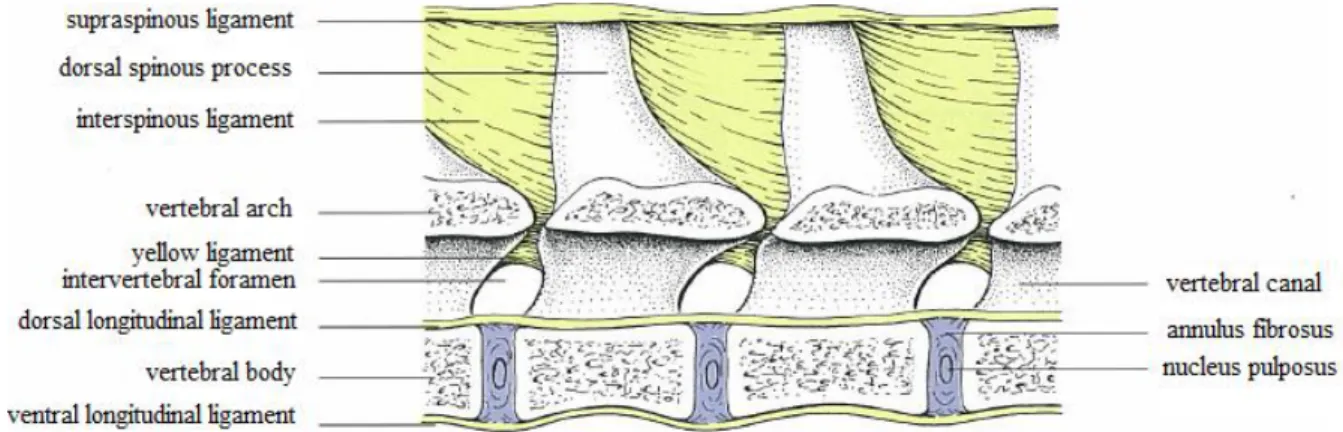
The ligaments of the vertebral column can be grouped into short ligaments, connecting successive vertebrae and long ligaments, spanning several vertebrae, thus forming functional units (Liebich & König, 2004a). Three long ligaments extend along considerable portions of the vertebral column: the supraspinous, the ventral longitudinal and the dorsal longitudinal ligaments (Dyce et al., 2010a). The supraspinous ligament is a thick band, especially in the thoracic region. It attaches to the apices of the spines as it passes through them (Evans & De Lahunta, 2013a), merges with the tendons of the epaxial muscles (Dyce et al., 2010b) and extends caudally from the spinous process of the first thoracic vertebra to the third caudal vertebra. During flexion of the vertebral column, the supraspinous ligament prevents abnormal separation of the spines, along with the thin interspinous ligaments, which send some strands to its ventral surface (Evans & De Lahunta, 2013a). The ventral longitudinal ligament is located ventral to the surfaces of the vertebral bodies from the axis to the sacrum and attaches to each of the intervertebral discs (Liebich & König, 2004a). The dorsal longitudinal ligament is thicker than the ventral longitudinal ligament and lies on the dorsal surfaces of the bodies of the vertebrae, forming part of the floor of the vertebral canal (Evans & De Lahunta, 2013a). It is narrow over the middle of each vertebral body, widens where it crosses each intervertebral disc and extends from the dens of the axis to the sacrum (Dyce et al., 2010b). The intertransverse ligaments are only distinct in the lumbar region, unite the transverse processes of the lumbar vertebrae, and are tensed during lateral flexion and rotation (Liebich & König, 2004a). The yellow ligaments lie between the arches of adjacent vertebrae. They blend with the articular capsules surrounding the articular processes. Ventral to this ligament is the epidural space, which separates the ligaments and arches of the vertebrae from the dura covering the spinal cord (figure 7) (Evans & De Lahunta, 2013a).
12
Figure 7 - Long and short ligaments of the vertebral column (adapted from Liebich & König, 2004).
The epaxial muscles, or dorsal musculature, associated with the vertebral column and ribs, provide extension and allow lateral movement of the trunk when acting only on one side of the vertebral column. The organisation of these epaxial muscles is complex and there are considerable variations in the literature. The erector spinae muscles are the dorsal muscles that include the epaxial muscles located on the dorsal surface of the vertebral column and ribs. They encompass three longitudinal systems: iliocostalis, longissimus and spinalis. The
iliocostalis muscles are located laterally to the other epaxial muscles, forming a narrow
longitudinal mass that runs cranioventrally over many segments (Hermanson, 2013). These muscles are relatively thin and have only lumbar and thoracic parts (Liebich, Maierl & König, 2004b; Dyce et al., 2010b). The caudal fascicle of these muscles constitutes the lumbar portion, represented by the iliocostalis lumborum muscle, which arises from the ilium and contributes to the fixation of the vertebral column and lateral movement when only one side contracts. The longissimus muscle is the major portion of the epaxial muscle mass and lies medial to the iliocostalis muscle, extending from the ilium to the head. It has lumbar, thoracic, cervical, atlantal and capital regional divisions (Hermanson, 2013). Its lumbar portion arises from the wings of the ilium and the lumbar spinous processes (Dyce et al., 2010b) and corresponds to the longissimus lumborum muscle, which allows extension and stabilisation of the vertebral column (Liebich et al., 2004b). The transversospinalis muscle is a muscle mass composed of many different fascicles that join one or more vertebrae, primarily located medial to the iliocostalis and longissimus muscles and lateral to the spinalis, interspinalis, and intertransversarius groups. It is composed by the semispinalis, the multifidus and the rotatores
muscles. The lumbar part of the multifidus muscle contributes to the fixation of the vertebral
column, especially in bilateral action. It consists of a series of muscle bundles that arise from the mammillary process of the first caudal vertebra, the rudimentary articular processes of the sacrum, and the mammillary processes of the lumbar vertebrae and last two thoracic vertebrae
13
(Hermanson, 2013). Each bundle passes over two segments, thus the insertions are to the spinous processes of the sixth lumbar to the tenth thoracic vertebrae (Dyce et al., 2010b). The
interspinalis muscles support ventroflexion of the vertebral column (Liebich et al., 2004b) and
are separable into lumbar, thoracic and cervical portions. The lumbar division is covered by the multifidus muscle. The intertransversarii muscles are segments split off from the longissimus system. They have caudal, lumbar, thoracic and cervical parts and pass over one, two or three vertebrae, between transverse processes, between articular and transverse processes or between mammillary and transverse processes. These muscles overlap in the lumbar (intertransversarii lumborum muscles) and thoracic (intertransversarii thoracis
muscles) regions (Hermanson, 2013). The lumbar hypaxial muscles lie on the ventral surfaces
of the lumbar vertebrae and ilium, insert on the os coxae and femur, and include the psoas
minor, psoas major, and quadratus lumborum. The psoas minor muscle runs ventromedially
towards the pelvis, lies between the iliac fascia and the peritoneum ventrally and the psoas
major and quadratus lumborum muscles dorsally. It arises from the bodies of the last thoracic
and first four to five lumbar vertebrae (Dyce et al., 2010b). The psoas major muscle is located dorsal to the psoas minor and ventral to the quadratus lumborum. It arises from the transverse processes of L2 and L3, lying medial to the quadratus lumborum muscle. It attaches ventrally
to L3 and L4, and to the ventral and lateral surfaces of L4 to L7. As it passes the cranioventral
border of the ilium, the caudal portion of the psoas major muscle receives the iliacus muscle from the ventral surface of the ilium. These two muscles compose the iliopsoas muscle. When the femur is fixed in position, these three muscles allow flexion and fixation of the vertebral column (Hermanson, 2013). The quadratus lumborum muscle is the most dorsal of the lumbar hypaxial muscles and lies directly ventral to the bodies of the last three thoracic vertebrae and the bodies and transverse processes of all the lumbar vertebrae and inserts on the medial surface of the wing of the ilium (Dyce et al., 2010b). It is covered ventrally by the psoas
minor caudal to L1 and caudal to L4 by the psoas major. The quadratus lumborum is active in
flexion and fixation of the lumbar vertebral column (Hermanson, 2013).
3. Embryology of the spinal cord
Throughout much of its development, the spinal cord preserves its fundamental organisation, making it a useful tool to study the structural and functional features of the central nervous system (Carlson, 2009c). After gastrulation, the ectodermal germ layer has the shape of a disc, broader in the cephalic than in the caudal region (Sadler, 2012d). In domestic animals, at the end of the third week of embryological development, the notochord induces thickening of the overlying columnar ectodermal cells to become pseudostratified neuroepithelial cells, thus
14
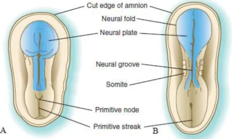
forming the neural plate (McGeady et al., 2006c). Cells of the plate arrange to make up the neuroectoderm, and this phenomenon induces neurulation. The process through which the neural plate originates the neural tube is called neurulation (Sadler, 2012c). Progessive changes in the columnar neuroepithelium originate folding of the neural plate (McGeady et al., 2006c). Notochord-induced changes at the median angle seem to largely influence plate bending. Many explanations have been proposed for lateral folding of the neural plate and it is now apparent that it is the result of numerous region-specific mechanisms intrinsic and extrinsic to the neural plate (Carlson, 2009a). In this phenomenon, the edges of the neural plate elevate to form the neural folds, originating the neural groove at the depressed midregion that forms during that process (Sadler, 2012c). Cellular proliferation at the medial aspects of the neural folds causes their median approach and results in fusion, which begins in the cervical region, at the level of the fourth somite, and then progresses cranially and caudally (figure 8) (McGeady et al., 2006c).
Figure 8 - Dorsal view of the developing embryo. A. At the beginning of the third week of
development, the central nervous system is a plate of thickened ectoderm. B. By the end of the third week of development, neurulation commences (adapted from Carlson, 2009).
As the neural folds suffer elevation, cells at the lateral border or crest of the neuroectoderm, begin to dissociate from their neighbors. This cell population, the neural crest, suffers an epithelial-to-mesenchymal transition and leaves the neuroectoderm by migration and displacement (Carlson, 2009c; Sadler, 2012c). Migration of neural crest cells is influenced by intrinsic properties of these cells and also features of the external environment encountered during this movement (Carlson, 2009c). After migration from the neural tube, neural crest cells form cellular aggregations in a dorsal position, extending along the length of the neural tube on either side. A single pluripotent neural crest cell can differentiate into many cell types
15
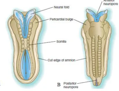
depending on its location within the early embryo (McGeady et al., 2006c). Until complete fusion is accomplished, the cephalic and caudal ends of the neural tube communicate with the amniotic cavity by an anterior or cranial and posterior or caudal neuropores, respectively (Sadler, 2012c). At this stage, the developing central nervous system has limited vascular supply, so it has been suggested that these structures receive their supply of nutrients from the amniotic fluid through the neuropores (figure 9). Closure of the anterior neuropore occurs at approximately midway through the embryonic period and the posterior neuropore closes shortly afterwards, thus completing neurulation (McGeady et al., 2006c). Prior to this phenomenon, the future spinal cord and brain are already recognisable (Carlson, 2009c). Neural tube formation in the sacral and caudal regions occurs through secondary neurulation.
Figure 9 - Dorsal view of the embryo undergoing neurulation. A. The neural folds suffer elevation and
start fusing in the cervical region. B. Formation of the extremities of the neural tube, the anterior and posterior neuropores (adapted from Carlson, 2009).
A column of mesenchymal cells, derived from the primitive streak in the caudal region of the embryo, fuses with the closed caudal end of the neural tube, forming a cavitary central canal in this cord of cells. This structure becomes continuous with the neural canal that resulted from the primary neurulation (Dellman & McClure, 1986; McGeady et al., 2006c; Carlson, 2009a). Besides giving rise to subcutaneous, mammary and pituitary glands and enamel of the teeth, the ectodermal germ layer is also responsible for the development of the central and peripheral nervous systems, the sensory epithelium of the ear, nose and eye; and the epidermis, including hair and nails (Sadler, 2012d). By the end of neurulation, the central nervous system is represented by a tubular structure with a narrow caudal portion, the spinal cord, and a much broader cephalic portion with a number of dilations, the brain vesicles
16
(Sadler, 2012c). These vesicles are continuous with the lumen of the spinal cord, the central canal.
Neural tube neuroepithelial cells give rise to two cell types: neuronal and glial progenitor cells (McGeady et al., 2006c). Gliablasts originate supporting cells, while neuroblasts suffer differentiation and further development into adult nerve cells, the neurons of the central nervous system (McGeady et al., 2006c; Sadler, 2012a). Upon differentiation of its cells, the neural tube is composed of three layers: an inner ependymal or ventricular layer, a middle mantle or intermediate layer and an outer marginal layer. Neuroblasts, through outward migration from the ependymal layer, give rise to the mantle layer. This area is later responsible for the formation of the gray matter of the spinal cord (McGeady et al., 2006c; Carlson, 2009c). Nerve fibers emerging from neuroblasts in the mantle layer give rise to the marginal layer. After myelination of its nerve fibers, this layer acquires a white appearance, making it the white matter of the spinal cord. Proliferation of neuroblasts in the mantle layer causes ventral and dorsal thickening of each side of the neural tube (McGeady et al., 2006c; Carlson, 2009c; Sadler, 2012a). The dorsal thickenings, referred to as alar plates, form sensory areas. Axons of the alar plates penetrate into the marginal layer of the cord and ascend to higher or lower levels to form association neurons. The ventral thickenings or basal plates form the motor areas of the spinal cord and contain ventral motor horn cells. Axons of neurons in these plates break through the marginal zone and become visible on the ventral aspect of the cord. They conduct impulses from the spinal cord to the muscles and are known as the ventral motor root of the spinal nerves. The boundary between the two is a longitudinal groove called the Sulcus limitans (Sadler, 2012a). Cell division causes expansion of the alar and basal plates, which results in fusion of these four plates, forming the butterfly-shaped gray area characteristic of the cross-section of the spinal cord (McGeady et al., 2006c). The dorsal and ventral midline areas of the neural tube, known as the roof and floor plates, respectively, serve as pathways for nerve fibers crossing from one side of the spinal cord to the other and do not have neuroblasts (Sadler, 2012a). The notochord exerts a very important effect on the organisation of the dorsal and ventral roots that enter and leave the spinal cord, due to its action on the floor plate. If it is absent, recognisable dorsal and ventral roots will also be absent (Carlson, 2009c). A group of neurons accumulates between the dorsal and ventral horns, becoming the intermediate horn. This horn contains neurons of the sympathetic portion of the autonomic nervous system and is present only at thoracic (T1- T12) and upper
17
After production of neuroblasts ceases, neuroepithelial cells form the gliablasts, which migrate from the neuroepithelial layer to the mantle and marginal layers. In the mantle layer, they differentiate into protoplasmic astrocytes and fibrillar astrocytes, located between blood vessels and neurons, providing support and having metabolic functions (Sadler, 2012a). Oligodendroglial cells, originally found in the marginal layer, form myelin sheaths around the ascending and descending marginal layer axons, and are also thought to originate from gliablasts (McGeady et al., 2006c; Carlson, 2009c; Sadler, 2012a). Microglial cells are highly phagocytic cells derived from vascular mesenchyme when blood vessels grow into the nervous system. They appear in the second half of development. After ceasing to produce neuroblasts and gliablasts, neuroepithelial cells differentiate into ependymal cells lining the central canal of the spinal cord and the brain ventricles (McGeady et al., 2006c).
Figure 10 - Development of the spinal cord. A. Neural tube layers after differentiation: marginal layer,
mantle layer and ependymal layer (neuroepithelial layer). B. Formation of the white and gray areas of the spinal cord. The alar plates form sensory areas and the basal plates form motor areas (adapted from Sadler, 2012).
Sensory ganglia or dorsal root ganglia of the spinal nerves, the sensory components of the peripheral nervous system, arise from lateral migration of neural crest cells (McGeady et al., 2006c; Sadler, 2012a). Neuroblasts of the sensory ganglia form centrally and peripherally growing processes. The first penetrate the dorsal portion of the neural tube to, in the spinal cord, either end in the dorsal horn or ascend through the marginal layer to one of the higher brain centres. These processes are called dorsal sensory root of the spinal nerve. The peripherally growing processes participate in formation of the trunk of the spinal nerve by joining fibers of the ventral motor roots. These processes terminate in the sensory receptor organs. Dorsal root neurons therefore originate from neuroblasts of the sensory ganglia derived from neural crest cells. Neural crest cells also differentiate into sympathetic
18
neuroblasts, Schwann cells, pigment cells, odontoblasts, meninges and mesenchyme of the pharyngeal arches. Peripheral nerves are myelinated by Schwann cells originated from the neural crest (McGeady et al., 2006c; Sadler, 2012a). Motor nerve fibers arise from the basal plate nerve cells, collecting in bundles to form ventral nerve roots (Sadler, 2012a). During this developmental period, few neurons differentiate in the caudal end of the cord, which results in the tapering of the spinal cord and gives rise to the conus medullaris. Caudal to this structure, the spinal cord is composed of a strand of glial and ependymal cells, which attaches the conus medullaris to the caudal vertebrae. As a result of the difference in growth rates of the spinal cord (derived from the ectoderm) and the vertebral canal (derived from the mesoderm) during the foetal period, the newborn animal has a longer spinal column than the spinal cord. Therefore, the roots of the spinal nerves arising from the lumbar, sacral and caudal regions of the cord, referred to as cauda equina, must pass caudally within the vertebral canal before emerging through the intervertebral foramina (Dellman & McClure, 1986; McGeady et al., 2006c; Budras et al., 2007; Carlson, 2009c; Dyce, Sack & Wensing, 2010c; Meij & Bergknut, 2010).
4. Anatomy of the spinal cord
The central nervous system is composed of the spinal cord and the brain. The vertebral canal encloses the dorsal and ventral spinal roots that belong to the peripheral nervous system and the spinal cord. The spinal cord is elongated, approximately cylindrical (Fletcher, 2013) and extends cranially into the medulla oblongata at the level of the foramen magnum (Dellman & McClure, 1986). It exhibits some degree of dorsoventral flattening and presents anatomical variations throughout its length (Fletcher, 2013). Two thickenings, or intumescences, that give rise to the nerve supply of the limbs, and the final caudal tapering, or conus medullaris, are present in the spinal cord (Dyce et al., 2010b). The neck, trunk and tail, the limbs, and the caudal and dorsal surfaces of the head are innervated by the spinal cord. It has three purposes: to act as a reflex centre, producing subconscious responses of muscles and glands to stimuli; to process afferent information from muscles, tendons, joints, ligaments, blood vessels, skin and viscera, and it discharges efferent commands that control muscles and regulate glands; to conduct information to and from the brain, through a system of axonal tracts, by which the brain receives status information about the neck, trunk and limbs while sending out information that controls posture, movement, and the visceral aspects of behavior (Fletcher, 2013). Throughout the entire length of the spinal cord, the ventral spinal artery runs along the ventral median fissure, providing important arterial supply (Budras et al., 2007). The spinal
19
cord may be divided into two parts: gray matter, located deeply in transverse section; and superficially, white matter (Dellman & McClure, 1986).
Three protective layers, the meninges, surround the spinal cord and spinal roots within the vertebral canal. The outer layer, the dura mater is a thick and fibrous envelope which, along with the other meningeal layers, through lateral extensions, ensheathes spinal roots (Fletcher, 2013). The dura mater is separated from the periosteum of the skull bones at the level of the foramen magnum to form a tube separated from the margin of the vertebral canal by the epidural space. This space is occupied by fat and the internal vertebral venous plexus, which cushion the spinal cord passively deformed by the curvatures of the vertebral column (Budras et al., 2007; Dyce et al., 2010b). The venous sinuses, one on each side of the floor of the vertebral canal, collect blood from vertebrae, meninges, and nerve roots (Sjöström, 2003). The arachnoid membrane is the thin intermediate meninx, which attaches to the inner surface of the dura mater. It is joined to the pia mater by arachnoid trabeculae that cross the subarachnoid space, which is filled by cerebrospinal fluid. The pia mater is the deepest meninx. It has bilateral thickenings along the spinal cord forming denticulate ligaments. The arachnoid membrane and the pia mater are designated leptomeninges because they are delicate relative to the dura mater. The central canal is filled with cerebrospinal fluid and lined with ependymal cells. Cerebrospinal fluid is usually a clear, colorless, and slightly alkaline liquid derived from blood, mostly of the choroid plexus vessels, and therefore must be returned to the circulating blood. Arachnoid villi and lymphatics associated with nerves are the two major drainage routes for that purpose (Fletcher, 2013).
Gray matter, relatively rich in capillary supply and composed of cell bodies and processes of neurons and glial cells surrounds the central canal (Fletcher, 2013). In transverse sections it resembles the shape of a butterfly or an H (Dellman & McClure, 1986; Budras et al., 2007; Dyce et al., 2010c), having bilateral wings connected across the midline by central intermediate substance. The lateral intermediate substance is the lateral extension of intermediate substance into each gray matter wing. It projects into the surrounding white matter as a lateral horn in the thoracolumbar segments of the spinal cord (Dellman & McClure, 1986; Dyce et al., 2010c; Fletcher, 2013). The dorsal horn is the dorsal extension of the lateral intermediate substance of the gray matter and the ventral horn is the corresponding ventral extension. Sensory or afferent neurons are situated in the dorsal root and have their origin in the dorsal horn. Motor or efferent neurons are located in the ventral root and arise from the ventral horn (Budras et al., 2007). The neurons contained in each horn are grouped according to their functional and topical associations. However, this is not fully perceptible
20
(Dyce et al., 2010c). The white matter surrounds the gray matter externally in the spinal cord. It includes myelinated, as well as nonmyelinated axons, oligodendrocytes, astrocytes, and blood vessels. The white matter of each half of the spinal cord is divided into three funiculi: dorsal, ventral and lateral (Dyce et al., 2010c; Fletcher, 2013). There is a dorsolateral sulcus where dorsal roots enter the spinal cord and a corresponding ventrolateral sulcus where ventral roots leave the spinal cord. The latter, however, is frequently imperceptible (Dellman & McClure, 1986; Fletcher, 2013). The dorsal funiculus is located between the dorsolateral sulcus, where dorsal rootlets enter the spinal cord, and the dorsal median sulcus. The ventral funiculus is found medial to the ventral root attachments. The lateral funiculus is located between the dorsal and ventral root attachments (Budras et al., 2007; Dyce et al., 2010c; Fletcher, 2013). Ascending and descending nerve fibres, frequently accumulated in bundles of common origin, destination and function, compose the three funiculi (Dyce et al., 2010b). The white commisure connects right and left ventral funiculi. Septae, sulci, and fissures are also featured in the spinal cord (Fletcher, 2013). The spinal cord is divided bilateral and symmetrically by a ventral median fissure and a dorsal median sulcus. The dorsal median septum extends ventrally from the dorsal median sulcus to the central intermediate substance where the central canal is located (Figure 11).
Figure 11 - Transverse section of the spinal cord at the level of C2. The dorsal funiculus is located
between the dorsolateral sulcus and the dorsal median sulcus. The ventral funiculus is located between the ventrolateral sulcus and the ventral median fissure. The lateral funiculus is located between the dorsal and ventral root attachments (adapted from Evans & De Lahunta, 2013).
The spinal cord may be divided into segments. A segment corresponds to an area of the spinal cord where a pair of spinal roots from a particular spinal nerve exits or enters the spinal cord (Dellman & McClure, 1986). Spinal cord segments, spinal roots, and spinal nerves are identified numerically, similarly to vertebrae, according to their region. The lumbar intumescence innervates the pelvic cavity and pelvic limbs and involves segments L5, L6 and