CLINICOPATHOLOGICAL
CHARACTERISTICS OF
BREAST CANCERS TREATED
IN A HOSPITAL:
IS SCREENING HAVING AN IMPACT?
ANABELA FARIA DE SOUSA
DISSERTAÇÃO DE MESTRADO APRESENTADA
AO INSTITUTO DE CIÊNCIAS BIOMÉDICAS ABEL SALAZAR DA UNIVERSIDADE DO PORTO EM
ONCOLOGIA
ANABELA FARIA DE SOUSA
CLINICOPATHOLOGICAL
CHARACTERISTICS
OF
BREAST
CANCERS TREATED IN A HOSPITAL:
IS SCREENING HAVING AN IMPACT?
Dissertação de Candidatura ao grau de Mestre
em Oncologia - Especialização em Oncologia
Molecular - submetida ao Instituto de Ciências
Biomédicas de Abel Salazar da Universidade
do Porto.
Orientador – Doutor Guilherme Gonçalves
Categoria
–
Professor
Associado
com
Agregação
Afiliação
– Instituto de Ciências Biomédicas
Abel Salazar da Universidade do Porto.
Coorientador – Doutora Maria José Bento
Categoria – Professora Associada
Afiliação
– Instituto de Ciências Biomédicas
Contents
Figure Index ... iv Table Index ... v Resumo ... vii Abstract ... ix Abbreviations ... xi Introduction ... 1Epidemiology of Breast Cancer ...1
Histopathology ...2
Risk factors for Breast Cancer ...3
Breast Cancer Treatments ...4
Breast Cancer Screening Programme ...4
Aims ... 9
Materials and Methods ... 11
Results ... 15
Discussion ... 23
Conclusion and Future Perspectives ... 27
References ... 29
Attachments ... 34
Figure Index
iv
Figure Index
Figure 1 – Estimated breast cancer incidence worldwide in 2012 ...1
Figure 2 - Estimated age-standardised incidence and mortality rates among women (A) and both sexes (B) in 2012 in Portugal ...2
Figure 3 - AJCC breast cancer staging – Primary tumour (T) ...3
Figure 4 - AJCC breast cancer staging - Regional lymph nodes (N) ...3
Table Index
Table 1 - Personal characteristics of invasive breast cancers from LPCC screening, Opportunistic screening or Symptomatic tumours diagnosed in 2011 and treated exclusively at IPO. ...16 Table 2 - Clinicopathological characteristics of invasive breast cancers from LPCC screening, Opportunistic screening or Symptomatic tumours diagnosed in 2011 and treated exclusively at IPO. ...19 Table 3 - Treatments applied to women with invasive breast cancers diagnosed in 2011 and treated exclusively at IPO. ...20 Table 4 - Multivariate logistic regression for the association between clinicopathological characteristics of breast cancers and diagnostic modality. Three models comparing pairwise the three studied groups. ...21
Resumo
O cancro da mama constitui a neoplasia com mais elevada taxa de incidência entre as mulheres portuguesas, sendo também a principal causa de morte por cancro no sexo feminino. O rastreio organizado, em implementação em Portugal para mulheres com idades compreendidas entre os 45 e os 69 anos, é uma das formas usadas para reduzir a mortalidade associada a este cancro. Contudo, existem ainda mulheres que não estão abrangidas por este meio de prevenção secundária, mulheres estas que recorrem ao rastreio oportunista ou apenas procuram ajuda médica quando apresentam já sintomas da doença.
O objetivo principal deste estudo consistiu em comparar as características clínico -patológicas de tumores mamários de mulheres provenientes do rastreio organizado, rastreio oportunista e tumores sintomáticos, diagnosticados em 2011 e tratados no Instituto Português de Oncologia do Porto. Adicionalmente foram também avaliadas as diferenças nos tratamentos aplicados às pacientes.
Através da análise cuidada dos processos das pacientes, um total de 397 tumores malignos foram estudados no que concerne a: modalidade de diagnóstico, topografia, morfologia, grau de diferenciação, estadio, tratamentos aplicados, recetores hormonais, comorbilidades, estado de menopausa, hábitos tabágicos, Eastern Cooperative Oncology Group (ECOG) e índice de massa corporal.
As mulheres provenientes do rastreio organizado apresentaram tumores de menores dimensões (32,5% ≤ 10mm; rastreio oportunista: 16,9%; tumores sintomáticos: 4,1%), estadios mais precoces (61,0% em estadio I; rastreio oportunista: 47,3%; tumores sintomáticos: 20,0%), maior percentagem de tumores bem diferenciados (30,9% tumores bem diferenciados; rastreio oportunista: 21,2% e tumores sintomáticos: 8,7%) e foram sujeitas a tratamentos menos agressivos (menor uso de quimioterapia adjuvante).
As diferenças encontradas nos tumores detetados pelo rastreio organizado, comparativamente aos tumores provenientes do rastreio oportunista e tumores sintomáticos, parecem indicar que o programa de rastreio populacional em implementação apresenta impacto favorável. É desejável que o impacto do presente programa de rastreio seja avaliado, utilizando modelos de estudos específicos, nomeadamente estudos populacionais.
Abstract
Breast cancer is the most frequently diagnosed cancer among Portuguese women, and is the leading cause of cancer death among womankind. Organized screening, like the one implemented in Portugal for women aged from 45 to 69 years, is used to reduce mortality rate associated with breast cancer. However, there still exist women who are not covered by this mean of secondary prevention. These women are monitored through opportunistic screening or they only search for medical help when they have symptoms of the disease.
The aim of this study was to compare the clinical and pathological features of breast tumours in women from the organized screening, opportunistic screening and symptomatic tumours diagnosed in 2011 and treated at the Portuguese Oncology Institute - Porto. In addition differences in the treatments applied to patients were also assessed.
Through careful analysis of the patients’ records, a total of 397 malignant tumours have been studied regarding: diagnostic modality, topography, morphology and degree of differentiation, stage, applied treatments, hormone receptors, co-morbidities, menopausal status, smoking habits, Eastern Cooperative Oncology Group (ECOG) and body mass index.
Women from the organized screening had smaller tumours (32.5% ≤ 10 mm; opportunistic screening: 16.9%; symptomatic tumours: 4.1%), earlier stages (61.0% in stage I, opportunistic screening: 47.3%; symptomatic tumours: 20.0 %), higher percentage of well-differentiated tumours (30.9% well-differentiated tumours, opportunistic screening: 21.2% and symptomatic tumours: 8.7%) and underwent less aggressive treatments (less use of adjunctive chemotherapy).
The differences found in tumours detected through organized screening, compared to tumours from opportunistic screening or symptomatic tumours, seem to indicate that the implemented population screening program presents a favourable impact. It is desirable that the impact of the present screening programme is assessed, using specific study designs, namely population studies.
Abbreviations
AJCC American Joint Committee on Cancer ARS-N Northern Health Regional Administration
BC Breast Cancer
BCSP Breast Cancer Screening Programme
BMI Body Mass Index
ECOG Eastern Cooperative Oncology Group
ER Oestrogen receptor
HER2 Human epidermal growth factor type 2 receptor IPO Portuguese Oncology Institute – Porto
LPCC Portuguese League Against Cancer
LS LPCC screening
OP Opportunistic screening
OR Odds ratio
PR Progesterone receptor
RORENO Northern Region Cancer Registry
ST Symptomatic tumours
Introduction
Epidemiology of Breast Cancer
Breast Cancer (BC) represents the most frequently diagnosed cancer among women worldwide (1-5), corresponding to about 1.67 million new cancer cases diagnosed during 2012. With a slightly higher number of new cases in less developed (883.000) than in more developed regions (788.000) (Figure 1), this malignancy is responsible for a high number of deaths, corresponding to the fifth cause of death from cancer overall (5).
Figure 1 – Estimated breast cancer incidence worldwide in 2012 (5).
Consistent with worldwide trends, breast cancer represents the most frequently diagnosed cancer among Portuguese women associated with the highest mortality rate (Figure 2). Analysing both sexes, breast cancer is the most frequent cancer and the third cause of death from cancer overall (5, 6).
Introduction
2
Figure 2 - Estimated age-standardised incidence and mortality rates among women (A) and both sexes (B) in 2012 in Portugal (5).
In the Northern Region, according to the Northern Region Cancer Registry (RORENO) data from 2009, breast cancer was the most commonly diagnosed cancer among women corresponding to 26.1% of the cancers diagnosed. In Portuguese Oncology Institute – Porto (IPO) more than 1200 new breast cancers were confirmed during 2011 (7).
Histopathology
Breast alterations can be divided into three main groups: benign tumours, in situ tumours and invasive cancers (8).
Breast cancer is a heterogeneous group of diseases with different origins and natural histories (9-11). The majority of breast malignancies are developed from epithelial elements of the gland. More than 75% of cancers are ductal carcinomas, from 5-10% are lobular carcinomas (12) and the remaining are other types of carcinomas, which include medullary, mucinous and tubular carcinomas, among others (8).
The correct staging of this disease is essential for choosing adequate treatments and prognosis prediction (11, 13). The TNM staging includes information on tumour size (T) (Figure 3), regional lymph node metastasis (N) (Figure 4) and distant metastasis (M) (13).
Figure 3 - AJCC breast cancer staging – Primary tumour (T) (14).
Figure 4 - AJCC breast cancer staging - Regional lymph nodes (N) (14).
Other tumour characteristics are also determinant, namely tumour grade, hormone receptors and biological biomarkers, among others (9-11, 13, 15).
Generally, tumours in early stages present better survival rates (16).
Risk factors for Breast Cancer
Different elements are indicated as being major risk factors for developing breast cancer. Being a woman, increasing age and existence of familiar history of breast tumours represent the main factors (3, 13, 17, 18).
The occurrence of previous benign breast alterations (13, 18), breast density (19-22), diet and lifestyle (2, 13, 18, 23) and reproductive and hormonal factors (2, 3, 13, 17, 18) are also described as risk factors.
Introduction
4
Breast Cancer Treatments
Breast malignancies are usually treated through combinations of different therapies, such as surgery, chemotherapy, hormone therapy and radiotherapy. Choosing the most appropriate treatment depends on several tumour characteristics (stage at diagnosis and histological grade), biology (biomarkers and gene expression) and personal features and choices (age at diagnosis, comorbidities and personal preferences) (9-11, 13, 15).
For example, tumours in early stages usually require less aggressive treatments or combinations of treatments when compared to tumours in more advanced stages (13, 15).
Breast Cancer Screening Programme
The main aim of a screening program is to allow the detection of a disease at an early stage among those who were apparently healthy, with the expectancy that a more effective treatment can be applied at an earlier stage (1, 4, 13, 15, 16, 24-28). Screening programs must follow some criteria, first implemented by Wilson and Jungner in 1968 (29), but still currently used, namely:
- The screened condition should constitute an important health problem; - There should be an applicable treatment for patients with confirmed disease; - Facilities for diagnosis and treatment must be available;
- A recognizable early symptomatic stage should exist;
- There should be a suitable test or examination, acceptable to the population; - The natural history of the condition should be understood;
- There should be an agreed policy on whom to treat as patient; - The cost of case-finding should be economically balanced;
- Case-finding should be a continuous process and not a once and for all project (Wilson JMG and Jungner G, 1968) (16, 29).
In general, breast cancer screening meets these criteria. Implemented primarily in Centre Region, organized Breast Cancer Screening Programme (BCSP) was firstly used in 1990 (30, 31), later implemented in Southern Region in 1997, Northern Region and Madeira in 1999, Algarve in 2005 and Acores in 2009 (31).
According to the lists of registered users of Health Centres, women aged between 45-69 years are invited by letter to a bilateral mammography with two views, which is a sensitive (77% to 95%) and specific (94% to 97%) exam easily applicable to the majority
of women (32). The mammography is analysed by two experienced Radiologists (double blind reading) and each Radiologist attributes a score in a pre-defined scale, which is in accordance with European Guidelines. If these readings point to a doubtful or positive result or in case of discrepancies in the results obtained, a third reading will be performed by another Radiologist. Positive screening results are analysed by a multidisciplinary team composed by a Radiologist, a Surgeon and a Pathologist, and the most appropriate monitoring and treatments are established according to hospital standardized protocols (13,30).
Mammography is the gold standard of screening, but although its effectiveness is proved for women between 50-69 years, its use remains controversial for younger ages (1, 33). Young women breasts have higher density which compromises the visualization and interpretation of injuries through this method. For these cases, ultrasonography may be a useful and complementary resource (1, 13).
The use of other diagnostic exams like magnetic resonance imaging (MRI) is also discussed in literature once it presents better sensitivity, especially in young women. However, this procedure has more false positive recalls and is substantially more expensive; currently this exam is mainly used for doubtful cases, validated cases of genetic risk and follow-up of some previous tumours (1, 13, 34).
In Portugal organized breast cancer screening programme is applied to women aged between 45-69 years through mammographic examination. In 2011, according to Northern Health Regional Administration (ARS-N) (31), 12 of the 24 health centre groups of Northern region had implemented organized screening. With a geographical coverage of 49.5%, over 118.000 women were invited to attend mammography but only 62.186 were screened (compliance rate of 52.6%).
According to the literature, in order to ensure the quality and effectiveness of the screening program, the rate of participation must be at least 70% (35). Although competing with the major goal of high participation rates, it is extremely important to clarify the woman about the advantages and disadvantages of participation in screening. This women’s “informed choice” is considered of utmost importance. The benefit of early detection of cancer, leading to less mutilating treatments, must be properly balanced with inherent risks of screening (16). For instance, Brodersen et al. (36) argues that a woman with a false positive result experiences psychological damage for at least three years after the wrong result.
Introduction
6 submitted to complementary exams that may allow diagnosis of a condition or a
previously unknown pathology. Generally, this procedure isn’t subjected to any external control, being only dependent on the medical assistant recommendations (16).
The greater objective of screening is to detect cancer before it becomes clinically evident, believing that if a cancer is diagnosed earlier then treatments will be less aggressive and more effective (1, 4, 15, 16, 24-28, 34, 37). However, some screen-detected tumours may never progress to become clinically detectable and some women would die for other reasons before the cancer could be clinically evident. This fact, known as overdiagnosis, constitutes the major harm of screening and refers to all cancers, invasive or in situ (1, 16, 24, 25, 33).
It is accepted that tumours grow at variable rates and some screen-detected cancers may progress so slowly that they would never have been presented clinically. Furthermore, some tumours may remain stable or even regress with time, nevertheless they are equally treated. Thus, these women will undergo unnecessary treatments which may result in adverse effects in their quality of life (1, 16, 24, 25, 33, 38).
Overdiagnosed cancers have a greater tendency to be ductal carcinomas in situ (DCIS) and possibly tend to be low/intermediate grade rather than high grade (24).
Whether a particular woman has had an overdiagnosed cancer cannot be determined, being only possible to estimate the frequency of overdiagnosis (1, 16, 24, 25, 33).
The existence of false positive results, defined as women that were recalled and considered not having cancer, is the most commonly adverse effect found in mammographic screening. Recalled women can only have further imaging exams or be biopsied, under local or general anaesthesia. False positive results can have a powerful psychological impact on women (1, 16, 24, 25, 34).
Other harms can be associated to breast cancer screening, namely, the radiation exposure during mammography, pain during the breast examination, possibility of false-negative results, psychological consequences, morbidity and mortality associated to treatments (1, 16, 24).
Interval cancers, defined as tumours diagnosed after a negative mammographic exam and before the following planed, constitute an indicator of the quality of mammographic examination (1, 39). Since this type of tumours can’t be totally eliminated, they must be reduced as much as possible. When a women has a diagnosis of an interval cancer the previous imaging exams must be reviewed to determine if the alteration
existed in the previous screening, if so this case is classified as a true false-negative result (1).
Aims
The aim of this study was to compare clinicopathological characteristics of invasive breast tumours of women from organized screening (LPCC screening), opportunistic screening and symptomatic tumours, diagnosed in 2011 and treated exclusively at the Portuguese Oncology Institute - Porto.
Additionally, it was intended to verify if variations among these groups can end up having differences in the patients’ treatment.
Materials and Methods
Invasive breast cancers diagnosed during 2011 in women aged between 45-69 years, with no malignant pre-existing tumours, and treated entirely in IPO, were registered at RORENO. The list of cases fulfilling the criteria was retrieved from RORENO and information on the variables under study was gathered directly from the patients’ records, by one person only, double-checked and in doubtful cases reviewed by experts. Data concerning the pathological anatomy of tumours was directly collected from reports. Through the examination of patient´s processes three main groups were created: LPCC screening, Opportunistic screening and Symptomatic tumours.
LPCC screening tumours refer to a group of women invited by letter to a mammographic exam every two years, in mobile units, with a histologically confirmed subsequent diagnosis of breast cancer. These women must attend the examination without any sort of symptoms.
Opportunistic screening constitutes the type of screening performed when someone goes to its medical assistant for routine without specific complaints and is submitted to complementary exams that may allow diagnosis of a condition or a previously unknown pathology. Generally, this procedure isn’t subjected to any external control, being only dependent on the medical assistant recommendations. Women from this group do not present any symptoms as well.
The last group named Symptomatic tumours is composed of women seeking medical care due to at least one complain. Reported symptoms/signs were: palpable nodule, changes in size and shape of the breast, breast exudate, pain and nipple inversion, among others. Women were included in this group regardless their participation or not in any kind of screening.
Variables under analysis included: - Date of birth;
- Diagnostic mode (LPCC screening, Opportunistic screening or Symptomatic tumours);
- Topography and morphology according to the International Classification of Diseases for Oncology – 3rd edition (tumours were divided in three different groups: ductal carcinomas (which only include pure ductal tumours), lobular carcinomas (which contain lobular carcinomas, mixed ductal and lobular carcinomas and mixed lobular and other types of carcinomas) and other tumours
Materials and Methods
12 (which comprises all other types of tumours like: medullary, mucinous and tubular
carcinomas, among others);
- Histological grade in accordance with the Nottingham Grading System (40);
- Comorbidities (myocardial infarction, congestive heart failure, cerebrovascular disease, dementia, chronic obstructive pulmonary disease, among others, assessed as a whole using the Charlson Index);
- Laterality and multifocality of the tumour;
- Menopausal status (When this information was missing women over 50 years were considered post-menopausal);
- Smoking habits;
- Body Mass Index (BMI) according to the World Health Organization (WHO) International Classification of adult underweight (BMI<18.5kg/m2), normal (BMI: 18.5-24.9kg/m2), overweight (BMI: 25.0-29.9kg/m2) and obesity (BMI≥30.0kg/m2) (41);
- ECOG Performance status; - Diagnostic exams;
- Clinical and pathological stages at diagnosis according to American Joint Committee on Cancer’s (AJCC) TNM classification (14) (Where applicable, tumours in stage IA and IB were considered tumours in early stages. The remaining stages II, III and IV were considered advanced stages);
- Tumour size (three cut-offs were used in accordance with the cut-offs of the European Guidelines (35));
- Hormonal status (Oestrogen Receptors (ER), Progesterone Receptors (PR), Human Epidermal growth factor Receptor 2 (HER2));
- Proliferation index evaluation through ki67;
- Treatments applied (surgery, chemotherapy, hormone, radiotherapy, targeted therapy).
Proportions of the three groups of interest were compared, through pairwise comparisons, using X2 test or Fisher’s exact test and one-way analysis of variance was used for continuous variables. Differences were considered statistically significant for P <0.05.
Unconditional multivariate logistic regression was applied to evaluate the association between diagnostic modality and clinicopathological and personal characteristics of tumours adjusted for possible confounding factors.
One model, including stage, tumour grade, pathological diameter, triple negative receptors, Charlson Index, BMI, age interval and treatments schemes, was tested to compare LPCC screening tumours with symptomatic tumours.
To the comparison between LPCC screening tumours with opportunistic screening tumours one model was tested including stage, pathological tumour size, Charlson Index, treatment schemes and age.
Comparing Symptomatic tumours and Opportunistic screening tumours, one model was tested including pathological tumour size, tumour grade, stage at diagnosis, age interval and treatments schemes.
Differences considered statistically significant for P<0.05.
All data were collected and analysed with proper authorization of the ethics’ commission of IPO.
Results
According to RORENO data, 1297 invasive breast cancers were registered in IPO with a diagnosis during 2011. Only 796 cases occurred in women aged between 45-69 years (screening interval) and one of these had no cytological/histological confirmation. A total of 774 had no previous malignancy and 78 of these were excluded because of lack of diagnostic modality information. The exclusion criteria are explained in Figure 5. At the end, 397 tumours were included in this study since the rest of the cases had previous treatments in other institutions, which could contribute to bias.
Regarding the diagnostic mode, 107 women (27.0%) presented symptoms at diagnosis, 123 (31.0%) were referred by LPCC screening and 167 (42.1%) were sent by opportunistic screening.
Invasive breast cancers diagnosed in 2011 (n=1297)
Excluded (n=900):
- 501 women out of the age range; - 1 without laboratorial confirmation; - 21 with previous malignancies; - 78 with lack of diagnostic modality; - 299 with previous treatments.
397 invasive breast cancers
LPCC screening (n=123) Opportunistic screening (n=167) Symptomatic tumours (n=107)
Results
16
* Percents were calculated excluding cancers with unknown values; LS/ST, LPCC screening tumours compared to Symptomatic tumours; LS/OP, LPCC screening tumours compared to Opportunistic screening tumours; ST/OP, Symptomatic tumours compared to Opportunistic screening tumours; BMI, body mass index.
Variable LPCC Screening Symptomatic Tumour Opportunistic Screening P value P value P value
(LS) (ST) (OP)
value n=123 (%*) n=107 (%*) n=167 (%*) LS/ST LS/OP ST/OP Age Group 45-49 26 (21.1) 23 (21.5) 41 (24.6) 0.001 0.02 0.33
50-59 35 (28.5) 54 (50.5) 69 (41.3)
60-69 62 (50.4) 30 (28.0) 57 (34.1)
BMI Low weight / Healthy 29 (24.6) 45 (45.0) 48 (31.6) 0.003 0.18 0.09 Preobesity 41 (34.7) 32 (32.0) 58 (38.2)
Obesity 48 (40.7) 23 (23.0) 46 (30.3)
Missing 5 7 15
Smoking habits Never 105 (86.8) 83 (80.6) 126 (78.8) 0.18 0.16 0.73 Yes, previously 10 (8.3) 8 (7.8) 17 (10.6)
Yes, currently 6 (5.0) 12 (11.7) 17 (10.6)
Missing 2 4 7
Menopausal Status Postmenopausal 89 (72.4) 69 (64.5) 115 (68.9) 0.25 0.60 0.54 Pre/perimenopausal 34 (27.6) 38 (35.5) 52 (31.1)
Comorbidities Yes 84 (68.3) 56 (52.3) 92 (55.1) 0.02 0.03 0.75
(Charlson index) No 39 (31.7) 51 (47.7) 75 (44.9)
ECOG Performance Asymptomatic 115 (95.8) 96 (93.2) 152 (94.4) 0.57 0.79 0.89
Status Symptomatic 5 (4.2) 7 (6.8) 9 (5.6)
Personal Characteristics Diagnostic Mode Significance Level
The different personal characteristics of women are summarized in Table 1. Some of the studied variables such as smoking habits, menopause status and general status of the patient (ECOG) demonstrated to be similar among the different groups.
Assessing age at diagnosis it was found that women from LPCC screening were significantly older than women from the other two groups (P=0.001 for symptomatic tumours and P=0.02 for opportunistic screening tumours). Ages of women from opportunistic screening and symptomatic tumours had no statistically significant differences (P=0.33).
Women belonging to LPCC screening group were significantly fatter than those from symptomatic tumours group (P=0.003). BMI was similar between LPCC and opportunistic screening groups and between symptomatic and opportunistic screening groups.
Concerning comorbidities, assessed through Charlson Index, women from LPCC screening had a higher proportion of comorbidities when compared to symptomatic and opportunistic screening tumours (P=0.02 and P=0.03, respectively). For this variable, symptomatic and opportunistic tumours had no statistically significant differences.
Table 1 - Personal characteristics of invasive breast cancers from LPCC screening, Opportunistic screening or Symptomatic tumours diagnosed in 2011 and treated exclusively at IPO.
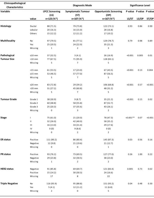
The main clinicopathological characteristics of the three groups are summarized in Table 2. The most commonly found subtype was ductal carcinoma, at similar proportions between study groups. Among all groups there were no statistically significant differences with regard to multifocality, with the majority of tumours emerging as a single node.
With respect to pathological tumour size 32.5% of tumours from LPCC screening presented 10mm or less, 53.5% less than 15mm and 72.8% 20mm or less. For tumour diameter of 10mm statistically significant differences were found between the three groups (P<0.001 LPCC screening versus symptomatic tumours (LS/ST); P=0.005 LPCC screening versus opportunistic screening (LS/OP); P=0.01 symptomatic tumours versus opportunistic screening (ST/OP)), wherein symptomatic and opportunistic tumours exhibit greater dimensions. For the remaining comparisons (15mm and 20mm) no statistically significant differences were found between LPCC screening tumours and opportunistic screening tumours and it was found that LPCC screening tumours were significantly smaller than symptomatic and opportunistic screening tumours (P<0.001 for LS/ST; P=0.004 and P<0.001 for ST/OP).
Regarding tumour grade, LPCC screening tumours were better differentiated with 30.9% of the tumours being well differentiated, 48.8% moderately differentiated and 20.3% poorly differentiated. Symptomatic tumours emerge significantly poorer differentiated being 35.6% poorly differentiated, 55.8% moderately differentiated and 8.7% well differentiated (P<0.001). Amongst symptomatic tumours and tumours from opportunistic screening the same tendency was found with symptomatic tumours being less differentiated (P=0.02). No statistically significant differences were found between LPCC and opportunistic screening tumours.
No tumour from LPCC and opportunistic screening showed distant metastasis at diagnosis, existing no differences between these groups. On the other hand 8.5% of symptomatic tumours exhibited distant metastasis at the date of diagnosis thus existing statistically differences between these and LPCC and opportunistic screening tumours (P=0.001 and P<0.001, respectively).
Concerning the stage at diagnosis, LPCC screening tumours were found in earlier stages with 61% of them being in stage I. Less proportion of tumours from opportunistic screening were in stage I (47.3%) however this difference wasn´t statistically significant. In comparison with symptomatic tumours both had significantly earlier stages (P<0.001 for both) with only 20% of symptomatic tumours in stage I.
Results
18 symptomatic tumours, however there were only statistically significant differences
between LPCC screening tumours and symptomatic tumours (P=0.03). For Progesterone Receptors the results were similar among the three groups in study.
A substantial number of missing values was found in the HER2 analysis in the three groups. Nevertheless, there was a significantly higher proportion of negatives in LPCC screening tumours (85.8%) and opportunistic tumours (83.4%) compared to symptomatic tumours (69.7%) (P=0.005 and P=0.02, respectively).
Triple negative tumours showed higher proportion in symptomatic tumours, being statistically significant the difference when compared to LPCC screening tumours (P=0.04).
Clinicopathological Characteristics
Variable LPCC Screening Symptomatic Tumour Opportunistic Screening P value P value P value (LS) (ST) (OP)
value n=123 (%*) n=107 (%*) n=167 (%*) LS/ST LS/OP ST/OP Histology Ductal 88 (71.5) 79 (73.8) 122 (73.1) 0.93 0.86 0.90 Lobular 20 (16.3) 16 (15.0) 28 (16.8) Others 15 (12.2) 12 (11.2) 17 (10.2) Multifocality No 97 (79.5) 81 (77.1) 129 (78.7) 0.79 0.98 0.89 Yes 25 (20.5) 24 (22.9) 35 (21.3) Missing 1 2 3 Pathological ≤10 mm 37 (32.5) 3 (4.1) 26 (16.9) <0.001 0.005 0.01 Tumour Size >10 mm 77 (67.5) 71 (95.9) 128 (83.1) Missing 5 7 5 <15 mm 61 (53.5) 17 (23.0) 67 (43.5) <0.001 0.13 0.004 ≥15 mm 53 (46.5) 57 (77.0) 87 (56.5) Missing 5 7 5 ≤20 mm 83 (72.8) 29 (39.2) 106 (68.8) <0.001 0.57 <0.001 >20 mm 31 (27.2) 45 (60.8) 48 (31.2) Missing 5 7 5
Tumour Grade Grade 1 38 (30.9) 9 (8.7) 35 (21.2) <0.001 0.15 0.02 Grade 2 60 (48.8) 58 (55.8) 87 (52.7) Grade 3 25 (20.3) 37 (35.6) 43 (26.1) Missing 0 3 2 Stage I 75 (61.0) 21 (20.0) 78 (47.3) <0.001** 0.07 <0.001 II 32 (26.0) 42 (40.0) 58 (35.2) III 16 (13.0) 33 (31.4) 29 (17.6) IV 0 (0) 9 (8.6) 0 (0) Missing 0 2 2 ER status Positive 111 (90.2) 86 (80.4) 145 (87.3) 0.03 0.56 0.16 Negative 12 (9.8) 21 (19.6) 21 (12.7) Missing 0 0 1 PR status Positive 93 (76.2) 73 (69.5) 127 (77.0) 0.26 1.00 0.22 Negative 29 (23.8) 32 (30.5) 38 (23.0) Missing 1 2 2
HER2 status Negative 91 (85.8) 69 (69.7) 121 (83.4) 0.005 0.73 0.02 Positive 15 (14.2) 30 (30.3) 24 (16.6) Missing 17 8 22 Triple Negative No 116 (95.9) 95 (88.8) 151 (93.2) 0.04 0.48 0.30 Yes 5 (4.1) 12 (11.2) 11 (6.8) Missing 2 0 5 Significance Level Diagnostic Mode
Table 2 - Clinicopathological characteristics of invasive breast cancers from LPCC screening, Opportunistic screening or Symptomatic tumours diagnosed in 2011 and treated exclusively at IPO.
* Percents were calculated excluding cancers with unknown values; ** 25.0% of cells expected a count lower than 5; LS/ST, LPCC screening tumours compared to Symptomatic tumours; LS/OP, LPCC screening tumours compared to Opportunistic screening tumours; ST/OP, Symptomatic tumours compared to Opportunistic screening tumours; ER, oestrogen receptors; PR, progesterone receptors; HER2, epidermal growth factor receptor 2.
Results
20
Variable LPCC Screening Symptomatic Tumour Opportunistic Screening P value P value P value (LS) (ST) (OP)
value n=123 (%*) n=107 (%*) n=167 (%*) LS/ST LS/OP ST/OP Type of treatment Surgery / Surgery + other 119 (96.7) 76 (71.0) 159 (95.2) <0.001 0.72 <0.001
QT / QT + other 4 (3.3) 31 (29.0) 8 (4.8)
Treatment schemes Surgery+others 64 (53.8) 9 (12.3) 47 (29.9) <0.001 <0.001 0.006 Surgery+QT+others 55 (46.2) 64 (87.7) 110 (70.1)
Treatments Diagnostic Mode Significance Level
Different therapeutic schemes were applied when treating women from the different groups (Table 3). Women from LPCC screening underwent a higher proportion of surgeries as first treatment (96.7%). The same occurred with women from opportunistic screening (95.2%). From women with symptoms at diagnosis only 71% attended surgery firstly, existing thus statistically significant differences between this group and the others (P<0.001). For women in which surgery was used as first treatment, LPCC screening women underwent significantly decreased use of chemotherapy as adjuvant therapy (46.2% compared to 87.7% for symptomatic tumours and 70.1% for opportunistic tumours). All groups showed statistically significant differences (P<0.001 LS/ST and LS/OP; P=0.006 ST/OP).
Table 3 - Treatments applied to women with invasive breast cancers diagnosed in 2011 and treated exclusively at IPO.
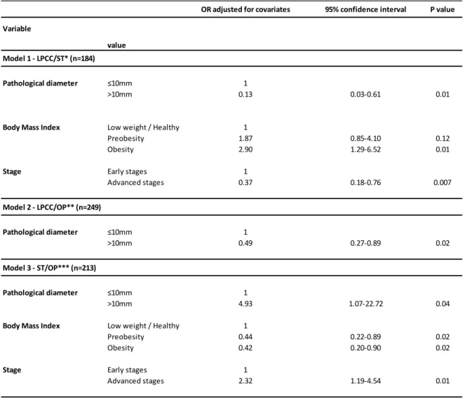
Through unconditional multivariate logistic regression the association between diagnostic modality and clinicopathological and personal characteristics of tumours adjusted for possible confounding factors was tested.
When testing LPCC screening tumours versus symptomatic tumours the final model included pathological diameter, stage at diagnosis and BMI. LPCC tumours were smaller, with earlier stages and women were fatter.
For LPCC screening tumours versus opportunistic screening tumours, pathological tumour size was the unique feature in the final model.
Regarding symptomatic tumours versus opportunistic screening tumours testing, the final model included pathological tumour size, stage at diagnosis and BMI. More advanced stages and higher pathological diameters presented higher chance of being
* Percents were calculated excluding cancers with unknown values; LS/ST, LPCC screening tumours compared to Symptomatic tumours; LS/OP, LPCC screening tumours compared to Opportunistic screening tumours; ST/OP, Symptomatic tumours compared to Opportunistic screening tumours.
part of the symptomatic tumours group and higher BMI with greater chance of coming from opportunistic screening.
Table 4 - Multivariate logistic regression for the association between clinicopathological characteristics of breast cancers and diagnostic modality. Three models comparing pairwise the three studied groups.
OR adjusted for covariates 95% confidence interval P value Variable
value Model 1 - LPCC/ST* (n=184)
Pathological diameter ≤10mm 1
>10mm 0.13 0.03-0.61 0.01
Body Mass Index Low weight / Healthy 1
Preobesity 1.87 0.85-4.10 0.12
Obesity 2.90 1.29-6.52 0.01
Stage Early stages 1
Advanced stages 0.37 0.18-0.76 0.007 Model 2 - LPCC/OP** (n=249) Pathological diameter ≤10mm 1 >10mm 0.49 0.27-0.89 0.02 Model 3 - ST/OP*** (n=213) Pathological diameter ≤10mm 1 >10mm 4.93 1.07-22.72 0.04
Body Mass Index Low weight / Healthy 1
Preobesity 0.44 0.22-0.89 0.02
Obesity 0.42 0.20-0.90 0.02
Stage Early stages 1
Advanced stages 2.32 1.19-4.54 0.01
OR, odds ratio; *LPCC/ST, LPCC screening tumours compared to Symptomatic tumours; **LPCC/OP, LPCC screening tumours compared to Opportunistic screening tumours; ***ST/OP, Symptomatic tumours compared to Opportunistic screening tumours.
Discussion
In this study a comparison of three groups of breast cancers (tumours arising from organized screening, tumours from opportunistic screening and symptomatic tumours) diagnosed in women during 2011 and treated in IPO was performed. This evaluation was carried out trying to draw conclusions about the potentially imposed differences by different diagnostic modalities. It was found that women from organized LPCC screening had significantly smaller tumours than the symptomatic group, better differentiated, at lower stages, with a higher percentage of positivity for oestrogen receptor and a larger amount of negativity for HER2 receptor and triple negative tumours. Compared to opportunistic screening tumours, significant differences were found for tumours with 10 mm or less, being LPCC screening tumours smaller.
Through the research of patients’ records it wasn’t possible to distinguish if cancers were detected in initial or in subsequent screenings or if the cases corresponded to interval cancers. The main limitation of this study comes from the fact that it is hospital based, therefore it doesn’t provide the complete scope of participation in breast cancer screening modality and its results. Furthermore, the reduced number of cases might have impaired some of the results.
A tumour is considered clinically palpable when it has one cm or more (42). In this study, over thirty per cent of LPCC tumours presented a size smaller than one cm making the tumour unreachable at palpation. For tumours from opportunistic screening this percentage was lower prowling seventeen per cent of the cases. As expected, almost all symptomatic tumours presented sufficient size to be felt through breast palpation. The remaining symptomatic tumours showed other symptoms as skin alterations, breast enlargement, nipple changes and pain, among others.
Considering the histological tumour type, the distribution of tumours was similar among the three groups of interest. According to the literature, earlier stages are an expected result of adherence to organized population-based screening programs (15, 38). In line with the predictable (38, 43), LPCC screening tumours and opportunistic screening tumours presented earlier stages comparing to symptomatic tumours.
Other variables related to early cancer detection like tumour size and grade were also analysed. According with the expected, LPCC screening tumours were smaller and better differentiated which was reflected in the treatments applied (4, 26, 27, 37, 38). This group of tumours required less aggressive treatments and has been subjected to less use of chemotherapy in association with other therapeutics which is in accordance with the
Discussion
24 Triple negative tumours, described in literature as having an inherently aggressive
disease phenotype (9, 11), were as expected more frequent in the symptomatic tumours group. For this type of tumours no specific treatment is known yet (9, 11).
HER2 positive tumours, more common in the symptomatic tumours group, are known to possess an aggressive disease phenotype as well but, in contrast with triple negative tumours, there have been introduced HER2 targeted therapies. With these therapies patients with HER2 positive tumours no longer have a worse prognosis than the negative ones (9, 11, 15).
Symptomatic tumours evaluated in this study were worse differentiated, with higher stages and having even metastasis at diagnosis, as expected (26, 38). These malignancies emerged mainly in women healthy or underweight.
In general, obesity is positively associated with risk of breast cancer development mainly in post-menopausal women and possibly in pre-menopausal ones considering breast density. This risk factor is also associated with worse prognosis (2). BMI has also been associated with the women’s participation in population screening, relating women with higher BMI usually as non-attenders of organized screening when compared with women with lower BMI. Hellmann et al. (44) found an association between obesity and non-participation in screening among Danish women. The same association was described previously in US, where opportunistic rates were higher in obese women (44) compared to organized screening rates. The results observed in our study are in disagreement with the previous findings, as the women that were in the LPCC screening group had a higher BMI than the women in the symptomatic group. Possible explanations for these results would require further BMI analysis of the population from which these women were originated; also, the socio-economic status of these women can have an impact on BMI.
Mammographic density, which might impair tumour detection, is according to literature, inversely associated with body mass index (44, 45). Applying this finding to the results obtained in this study this would mean that LPCC screening women, which had higher body mass index, would have less dense breasts.
In the aforementioned study in Danish women, the existence of diabetes was also reported as a barrier to screening, although this finding was not statistically significant due to the limited number of cases (44). In the present study no statistically significant differences were found when comparing the different groups in relation to diabetes with or without organ damage.
In general, therapeutic schemes applied were substantially different in terms of aggressiveness among the three study groups. LPCC screening tumours were mostly treated through the use of surgery alone or together with other treatment options like radiotherapy and hormone therapy (53.8%). Opportunistic screening tumours underwent more aggressive treatments; in 70.1% chemotherapy was added. Finally, symptomatic tumours were treated in 87.7% of the cases with surgery and chemotherapy combined or not with other treatment options. The fact that symptomatic tumours were subjected to more aggressive and mutilating treatments is in line with the expected as these cases were in more advanced stages, corroborating that screening contributes to the earlier detection of tumours, culminating in a less need for treatments (15).
Interestingly, although tumour stages evaluated as initial or advanced weren’t significantly different between organized and opportunistic screening, this last group of women underwent significantly more chemotherapy combined with surgery. Even after stratifying for the presence or absence of comorbidities, women from the opportunistic screening experienced chemotherapy more frequently.
The association between diagnostic modality and personal and clinicopathological characteristics of the cases was evaluated through unconditional multivariate regression adjusting for possible confounding factors. For LPCC screening versus opportunistic screening, pathological tumour size was the unique feature in the final model significantly different in these two groups. This tumour characteristic is possibly the main reason for the differences in treatments discussed above.
Through general analysis of the results of this study it can be accepted that opportunistic screening presents similar efficiency to LPCC screening in the early detection of cancer, however it implies disadvantages, mainly economics. The recurrent and simultaneous use of mammography and ultrasonography as well as reduced time intervals between exams carries unnecessary costs to the health system. According to ARS-N in 2007 and 2008 over 16% of women repeated screening mammography and 59% attended additional breast ultrasound, as a result of opportunistic screening (46).
Organized breast cancer screening programme allows a higher coverage of the population and a reduction in the number of exams performed, without compromising its quality, making it an intervention with higher cost-effectiveness. Tumours are detected mainly in early stages contributing to reduce aggressive and mutilating treatments. Lastly, with more initial stages and efficient treatments it is expected that breast cancer mortality will be significantly lower in the future.
Conclusion and Future Perspectives
The question “is screening having an impact?” was rhetoric. It cannot be answered in a valid and conclusive way with a cross-sectional study like the one presented here. The question just pointed a way. Specifically designed studies have to be performed. There are clear guidelines on what to do. In the end, answering the question will be done using critical appraisal, judging on the evidence available in several written reports and different types of published studies. Within that process, this study will be no doubt assessed. Data here collected and corresponding analysis is compatible with the hypothesis that the existing organized screening program is resulting in more favourable (for prognosis) breast cancers being diagnosed and less aggressive treatment strategies performed. Differences with symptomatic tumour cases and those diagnosed during opportunistic screening are clear. Findings are compatible with an effective screening program but cannot prove it.
Just increasing the sample size could be useful. Meanwhile, this study would be enriched with the analysis of some additional features such as the existence of previous breast benign or in situ tumours or familiar history of breast malignancies. Additionally, the evaluation of socio-economic status, obtained for example by education level information, could contribute to a better understanding of the influence of socio-economic factors in the screen modality choice. Using the subjects of this study in case-control could be one more step in the right direction.
Performing the recommended analytical epidemiology studies that can answer the question “is screening having an impact?” is the desirable future.
References
1. Zervoudis, S., Iatrakis, G., Tomara, E., Bothou, A., Papadopoulos, G., and Tsakiris, G. (2014). Main controversies in breast cancer. World J Clin Oncol 5, 359-373. 2. Ferrini, K., Ghelfi, F., Mannucci, R., and Titta, L. (2015). Lifestyle, nutrition and breast cancer: facts and presumptions for consideration. Ecancermedicalscience 9, 557. 3. Scoccianti, C., Key, T.J., Anderson, A.S., Armaroli, P., Berrino, F., Cecchini, M., Boutron-Ruault, M.C., Leitzmann, M., Norat, T., Powers, H., et al. (2015). European Code against Cancer 4th Edition: Breastfeeding and cancer. Cancer Epidemiol.
4. Soerjomataram, I., Louwman, M.W., Ribot, J.G., Roukema, J.A., and Coebergh, J.W. (2008). An overview of prognostic factors for long-term survivors of breast cancer. Breast Cancer Res Treat 107, 309-330.
5. GLOBOCAN (2012). Breast Cancer. Estimated Incidence, Mortality and Prevalence Worldwide in 2012. IARC.
6. Pinheiro, P.S., Tyczyński J.E., Bray F., Amado J., Matos E., Parkin D.M. (2003). Cancer incidence and mortality in Portugal. Eur J Cancer 39, 2507-20.
7. RORENO (2015). Registo Oncológico Regional do Norte 2009. Instituto Português de Oncologia do Porto.
8. Kumar V., Abbas A.K., Fausto N., Mitchell R.N. (2008). Robbins Patologia Básica. 8th ed. Elsevier.
9. Arpino, G., Milano, M., and De Placido, S. (2015). Features of aggressive breast cancer. Breast.
10. Prat, A., Pineda, E., Adamo, B., Galván, P., Fernández, A., Gaba, L., Díez, M., Viladot, M., Arance, A., and Muñoz, M. (2015). Clinical implications of the intrinsic molecular subtypes of breast cancer. Breast.
11. Li, J., Chen, Z., Su, K., and Zeng, J. (2015). Clinicopathological classification and traditional prognostic indicators of breast cancer. Int J Clin Exp Pathol 8, 8500-8505. 12. Lehmann, U. (2015). Lobular breast cancer - the most common special subtype or a most special common subtype? Breast Cancer Res 17, 99.
13. Senkus, E., Kyriakides, S., Penault-Llorca, F., Poortmans, P., Thompson, A., Zackrisson, S., Cardoso, F., and Group, E.G.W. (2013). Primary breast cancer: ESMO Clinical Practice Guidelines for diagnosis, treatment and follow-up. Ann Oncol 24 Suppl 6,
References
30 14. Edge, S., Byrd, D.R., Compton, C.C., Fritz, A.G., Greene, F.L., Trotti, A. (2010).
AJCC Cancer Staging Handbook. 7th ed. Springer.
15. Cianfrocca, M., and Goldstein, L.J. (2004). Prognostic and predictive factors in early-stage breast cancer. Oncologist 9, 606-616.
16. Bretthauer, M., and Kalager, M. (2013). Principles, effectiveness and caveats in screening for cancer. Br J Surg 100, 55-65.
17. Balmaña, J., Díez, O., Rubio, I.T., Cardoso, F., and Group, E.G.W. (2011). BRCA in breast cancer: ESMO Clinical Practice Guidelines. Ann Oncol 22 Suppl 6, vi31-34. 18. Pruthi, S., Heisey, R.E., and Bevers, T.B. (2015). Chemoprevention for Breast Cancer. Ann Surg Oncol 22, 3230-3235.
19. Nielsen, M., Vachon, C.M., Scott, C.G., Chernoff, K., Karemore, G., Karssemeijer, N., Lillholm, M., and Karsdal, M.A. (2014). Mammographic texture resemblance generalizes as an independent risk factor for breast cancer. Breast Cancer Res 16, R37. 20. Boyd, N.F., Li, Q., Melnichouk, O., Huszti, E., Martin, L.J., Gunasekara, A., Mawdsley, G., Yaffe, M.J., and Minkin, S. (2014). Evidence that breast tissue stiffness is associated with risk of breast cancer. PLoS One 9, e100937.
21. Boyd, N.F., Martin, L.J., Yaffe, M.J., and Minkin, S. (2011). Mammographic density and breast cancer risk: current understanding and future prospects. Breast Cancer Res 13, 223.
22. Boyd, N.F., Martin, L.J., Bronskill, M., Yaffe, M.J., Duric, N., and Minkin, S. (2010). Breast tissue composition and susceptibility to breast cancer. J Natl Cancer Inst 102, 1224-1237.
23. Harvie, M., Howell, A., and Evans, D.G. (2015). Can diet and lifestyle prevent breast cancer: what is the evidence? Am Soc Clin Oncol Educ Book 35, e66-73.
24. Marmot, M.G., Altman, D.G., Cameron, D.A., Dewar, J.A., Thompson, S.G., and Wilcox, M. (2013). The benefits and harms of breast cancer screening: an independent review. Br J Cancer 108, 2205-2240.
25. Løberg, M., Lousdal, M.L., Bretthauer, M., and Kalager, M. (2015). Benefits and harms of mammography screening. Breast Cancer Res 17, 63.
26. Allgood, P.C., Duffy, S.W., Kearins, O., O'Sullivan, E., Tappenden, N., Wallis, M.G., and Lawrence, G. (2011). Explaining the difference in prognosis between screen-detected and symptomatic breast cancers. Br J Cancer 104, 1680-1685.
27. Dawson, S.J., Duffy, S.W., Blows, F.M., Driver, K.E., Provenzano, E., LeQuesne, J., Greenberg, D.C., Pharoah, P., Caldas, C., and Wishart, G.C. (2009). Molecular characteristics of screen-detected vs symptomatic breast cancers and their impact on survival. Br J Cancer 101, 1338-1344.
28. Hofvind, S., Lee, C.I., and Elmore, J.G. (2012). Stage-specific breast cancer incidence rates among participants and non-participants of a population-based mammographic screening program. Breast Cancer Res Treat 135, 291-299.
29. Wilson, J., and Jungner, G. (1968). Principles and practices of screening for disease. Public Health Papers.
30. Dourado, F., Carreira, H., and Lunet, N. (2013). Mammography use for breast cancer screening in Portugal: results from the 2005/2006 National Health Survey. Eur J Public Health 23, 386-392.
31. Direção Geral de Saúde. (2014). Programa Nacional para as Doenças Oncológicas. Avaliação e Monitorização dos Rastreios Oncológicos Organizados de Base Populacional de Portugal Continental.
32. US Preventive Services Task Force (2009). Screening for breast cancer: U.S. Preventive Services Task Force recommendation statement. Ann Intern Med 151, 716-726, W-236.
33. Berry, D.A. (2013). Breast cancer screening: controversy of impact. Breast 22 Suppl 2, S73-76.
34. Drukteinis, J.S., Mooney, B.P., Flowers, C.I., and Gatenby, R.A. (2013). Beyond mammography: new frontiers in breast cancer screening. Am J Med 126, 472-479.
35. Perry, N., Broeders, M., de Wolf, C., Tornberg, S., Holland, R., and von Karsa, L. (2006). European guidelines for quality assurance in breast cancer screening and diagnosis (Luxembourg: Office for Official Publications of the European Communities). 36. Brodersen, J., and Siersma, V.D. (2013). Long-term psychosocial consequences of false-positive screening mammography. Ann Fam Med 11, 106-115.
37. Plotogea, A., Chiarelli, A.M., Mirea, L., Prummel, M.V., Chong, N., Shumak, R.S., O'Malley, F.P., Holloway, C.M., and Group, B.S.S. (2014). Clinical and prognostic factors associated with diagnostic wait times by breast cancer detection method. Springerplus 3, 125.
References
32 38. Nagtegaal, I.D., Allgood, P.C., Duffy, S.W., Kearins, O., Sullivan, E.O.,
Tappenden, N., Wallis, M., and Lawrence, G. (2011). Prognosis and pathology of screen-detected carcinomas: how different are they? Cancer 117, 1360-1368.
39. Meshkat, B., Prichard, R.S., Al-Hilli, Z., Bass, G.A., Quinn, C., O'Doherty, A., Rothwell, J., Geraghty, J., Evoy, D., and McDermott, E.W. (2015). A comparison of clinical-pathological characteristics between symptomatic and interval breast cancer. Breast 24, 278-282.
40. Elston, C.W., and Ellis, I.O. (2002). Pathological prognostic factors in breast cancer. I. The value of histological grade in breast cancer: experience from a large study with long-term follow-up. Histopathology 41, 154-161.
41. World Health Organization. Body Mass Index - BMI. [Access date: 08-05-2015]. Available from: http://www.euro.who.int/en/health-topics/disease-prevention/nutrition/a-healthy-lifestyle/body-mass-index-bmi.
42. Cooper, K., and Gosnell, K. (2015). Adult Health Nursing. 7th ed. Elsevier.
43. Pálka, I., Kelemen, G., Ormándi, K., Lázár, G., Nyári, T., Thurzó, L., and Kahán, Z. (2008). Tumor characteristics in screen-detected and symptomatic breast cancers. Pathol Oncol Res 14, 161-167.
44. Hellmann, S.S., Njor, S.H., Lynge, E., von Euler-Chelpin, M., Olsen, A., Tjønneland, A., Vejborg, I., and Andersen, Z.J. (2015). Body mass index and participation in organized mammographic screening: a prospective cohort study. BMC Cancer 15, 294. 45. Andersen, Z.J., Baker, J.L., Bihrmann, K., Vejborg, I., Sørensen, T.I., and Lynge, E. (2014). Birth weight, childhood body mass index, and height in relation to mammographic density and breast cancer: a register-based cohort study. Breast Cancer Res 16, R4.
46. Administração Regional de Saúde do Norte. (2011). Prescrição de Mamografia de rastreio em mulheres do grupo etário 45 a 69 anos, nas Unidades de Cuidados de Saúde Primários. Circular normativa nº 1.
Attachments
34
Attachments
AJCC´s Breast Cancer TNM Staging
Primary tumour Characteristics
TX Primary tumour cannot be assessed
T0 No evidence of primary tumour
Tis Carcinoma in situ
T1mi Tumour ≤ 1mm in greatest dimension
T1a Tumour > 1mm but ≤ 5mm in greatest dimension
T1b Tumour > 5mm but ≤ 10mm in greatest dimension
T1c Tumour > 10mm but ≤ 20mm in greatest dimension
T2 Tumour > 20mm but ≤ 50mm in greatest dimension T3 Tumour > 50mm in greatest dimension
T4a Extension to the chest wall, not including only pectoralis muscle adherence / invasion
T4b Ulceration and / or ipsilateral satellite nodules and / or edema (including peaud’orange) of the skin,
which do not meet the criteria for inflammatory carcinoma
T4c Both T4a and T4b
T4d Inflammatory carcinoma
cN Characteristics
cNX Regional lymph nodes cannot be assessed (for example, previously removed)
cN0 No regional lymph node metastases
cN1 Metastases to movable ipsilateral level I, II axillary lymph node(s)
cN2a Metastases in ipsilateral level I, II axillary lymph nodes fixed to one another (matted) or to other structures
cN2b Metastases only in clinically detected* ipsilateral internal mammary nodes and in the absence of
clinically evident level I, II axillary lymph node metastases
cN3a Metastases in ipsilateral infraclavicular lymph node(s)
cN3b Metastases in ipsilateral internal mammary lymph node(s) and axillary lymph node(s
pN Characteristics
pNX Regional lymph nodes cannot be assessed (for example, previously removed, or not removed for
pathologic study)
pN0 No regional lymph node metastasis identified histologically
pN0(i−) No regional lymph node metastases histologically, negative IHC
pN0(i+) Malignant cells in regional lymph node(s) no greater than 0.2 mm (detected by H&E or IHC including ITC)
pN0(mol−) No regional lymph node metastases histologically, negative molecular findings (RT-PCR)
pN0(mol+) Positive molecular findings (RT-PCR)**, but no regional lymph node metastases detected by histology
or IHC
pN1mi Micrometastases (greater than 0.2 mm and/or more than 200 cells, but none greater than 2.0 mm)
pN1a Metastases in 1–3 axillary lymph nodes, at least one metastasis greater than 2.0 mm
pN1b Metastases in internal mammary nodes with micrometastases or macrometastases detected by
sentinel lymph node biopsy but not clinically detected
pN1c Metastases in1–3 axillary lymph nodes and in internal mammary lymph nodes with micrometastases or
macrometastases detected by sentinel lymph node biopsy but not clinically detected
pN2a Metastases in 4–9 axillary lymph nodes (at least one tumour deposit greater than 2.0 mm)
pN2b Metastases in clinically detected internal mammary lymph nodes in the absence of axillary lymph node
metastases
pN3a Metastases in 10 or more axillary lymph nodes (at least one tumour deposit greater than 2.0 mm); or
metastases to the infraclavicular (level III axillary lymph) nodes
pN3b
Metastases in clinically detected ipsilateral internal mammary lymph nodes in the presence of one or more positive axillary lymph nodes; or in more than three axillary lymph nodes and in internal mammary lymph nodes with micrometastases or macrometastases detected by sentinel lymph node biopsy but not clinically detected
pN3c Metastases in ipsilateral supraclavicular lymph nodes
M Characteristics
M0 No clinical or radiographic evidence of distant metastases
cM0(i+)
No clinical or radiographic evidence of distant metastases, but deposits of molecularly or microscopically detected tumour cells in circulating blood, bone marrow, or other nonregional nodal tissue that are no larger than 0.2 mm in a patient without symptoms or signs of metastases
M1 Distant detectable metastases as determined by classic clinical and radiographic means and/or
Attachments
36
Clinicopathological characteristics of breast cancers treated in a hospital: is
screening having an impact?
Informação geral:
1.Número de formulário: ___________________________________________________________________ 2.Data de nascimento………..………..………..… |__|__|/|__|__|/|__|__|__|__| 3.Data de diagnóstico ………..………....… |__|__|/|__|__|/2011 4.Localização topográfica primária ………..………..……. C50.|__| 5.Morfologia ………..….………...….….. |__|__|__|__| 6.Comportamento (3 – maligno) ……….…..….3 7.Grau (1- Grau 1, bem diferenciado; 2- Grau 2, moderadamente diferenciado; 3- Grau 3, pouco diferenciado; 4- Grau 4, indiferenciado /anaplásico; 9- desconhecido) ………...……….………… |__| 8.Base de diagnóstico (1 - microscópico (histologia de um tumor primário, citologia, histologia de uma metástase)) ………..……….……….. 1 9.Tumor múltiplo, se aplicável (colocar o número a que corresponde o tumor em estudo) ……….…………..….. 1
Comorbilidades:
10.Índice de Charlson (entre 0 e 90) ……….………..…..… |__|__| 11.Enfarte do miocárdio (1-sim; 2-não; 9-desconhecido) ……….………..……… |__| 12.Insuficiência cardíaca congestiva (1-sim; 2-não; 9-desconhecido) ………...….……….………..… |__| 13.Doença vascular periférica (1-sim; 2-não; 9-desconhecido) ……….….……….….…..… |__| 14.Doença cerebrovascular (1-sim; 2-não; 9-desconhecido) ………..…….……….……….……..… |__| 15.Demência (1-sim; 2-não; 9-desconhecido) .……….……….…………..… |__| 16.Doença pulmonar obstrutiva crónica (1-sim; 2-não; 9-desconhecido) ………….………..… |__| 17.Doença do tecido conjuntivo/ doença reumatológica (1-sim; 2-não; 9-desconhecido) ………...… |__| 18.Úlcera péptica (1-sim; 2-não; 9-desconhecido) ……….……….……….………...… |__| 19.Doença hepática ligeira (1-sim; 2-não; 9-desconhecido) ……….………..………….…. |__| 20.Diabetes sem lesão nos órgãos (1-sim; 2-não; 9-desconhecido) ………….….………..……… |__| 21.Hemiplegia/Paraplegia (1-sim; 2-não; 9-desconhecido) ………….……….….………..………. |__| 22.Doença renal moderada ou severa (1-sim; 2-não; 9-desconhecido) …….………..……….………..………. |__| 23.Diabetes com lesão nos órgãos (1-sim; 2-não; 9-desconhecido) ……….………. |__| 24.Outro Tumor sólido maligno sem metástases (1-sim; 2-não; 9- desconhecido)…….………..… |__| 25.Linfoma (1-sim; 2-não; 9-desconhecido) ……….….……….………..… |__| 26.Leucemia (1-sim; 2-não; 9-desconhecido) ………..……….………..… |__| 27.Doença hepática moderada ou severa (1-sim; 2-não; 9-desconhecido) ……...……….…………..… |__| 28.Tumor sólido metastático (1-sim; 2-não; 9-desconhecido) ……….… |__| 29.SIDA/HIV (1-sim; 2-não; 9-desconhecido) ……….…..……… |__|
30.Fumador (1- sim, atualmente; 2- sim, anteriormente; 3- não, nunca; 9- desconhecido) ……….…|__| 31.IMC à data de diagnóstico ou à data do primeiro tratamento …….……….…………..… |__|__|,|__|
Nível de desempenho
32.Tipo de escala do nível de desempenho (1- ECOG; 9- não disponível) ……….………… |__| 33.Score do nível de desempenho ………..……….………..… |__|__|__|
Follow-up
34.Recidiva (0- sem recidiva; 1- recidiva local; 2- recidiva em nódulos regionais ou tecidos/órgãos adjacentes; 3- metástases à distância; 9- desconhecido) ………….……….………..… |__| 35.Data da recidiva ……..……….…..………..… |__|__|/|__|__|/|__|__|__|__| 36.Topografia do segundo tumor ………..………..… C|__|__|.|__| 37.Data de Incidência do segundo tumor……….………..… |__|__|/|__|__|/|__|__|__|__| 38.Estado vital à data do último contacto (1- vivo; 2- falecido) ..……….…….………..………..… |__| 39.Causa de morte ……….……….……..………____________________ 40.Data do último contacto……….….……….………...… |__|__|/|__|__|/|__|__|__|__|
Outras informações
41.Estado da menopausa à data de diagnóstico (1- pré-menopausa ou peri-menopausa; 2- pós menopausa; 9- desconhecido) ………..……….……….……….… |__| 42.Modalidade de diagnóstico (0- rastreio LPCC; 1- tumor sintomático; 2- rastreio oportunista) ………..………..………..….…..… |__| 43.Lateralidade (1- esquerda; 2- direita; 9- desconhecido)……….……….… |__| 44.Multifocalidade (1- sim; 2- não; 9- desconhecido) ………..………..……… |__|
Exames de diagnóstico (até 3 meses antes ou após o diagnóstico)
45.Mamografia (1- sim; 2- não; 9- desconhecido) ……….……….…………..……….……..… |__| 46.Data da mamografia ……….………..….|__|__|/|__|__|/|__|__|__|__| 47.Ecografia Mamária (1- realizada; 2- não realizada; 9- desconhecido) …………..……..….………….…………..…|__| 48.Biópsia (1- realizada; 2- não realizada; 9- desconhecido) ……….…….|__| 49.Ressonância magnética (1- realizada; 2- não realizada; 9- desconhecido)……….………..…....|__| 50.Extemporâneo (1- realizado; 2- não realizado; 9- desconhecido)………..…………...………..|__|
Imagiologia para deteção de metástases (até 3 meses antes ou após o diagnóstico)
51.No fígado (1- realizado; 2- não realizado; 9- desconhecido)………..……….………... |__| 52.No pulmão (1- realizado; 2- não realizado; 9- desconhecido) ……….…………..……….………..… |__|