Association of PKC
f
Expression with Clinicopathological
Characteristics of Breast Cancer
Jian Yin1,2,3*, Zhipei Liu4,5, Haixin Li6, Jingyan Sun1,2,3, Xinzhong Chang1,2,3, Jing Liu1,2,3, Shanshan He1,2,3, Binghui Li7
1Department of Breast Surgery, Tianjin Medical University Cancer Institute and Hospital, National Clinical Research Center of Cancer, Tianjin, China,2Key Laboratory of Cancer Prevention and Therapy, Tianjin, China,3Key Laboratory of Breast Cancer Prevention and Therapy, Tianjin Medical University, Ministry of Education, Tianjin, China,
4Production and R&D Center, Tianjin Binhai Union Gene Technology Co. LTD, Tianjin, China,5Gene Bank, Union Stem Cell & Gene Engineering Co. LTD, Tianjin, China,
6Tumor Tissue Banking Facility, Department of Epidemiology and Biostatistics, Tianjin Medical University Cancer Institute and Hospital, Tianjin, China,7Laboratory of Cancer Cell Biology, Tianjin Medical University Cancer Institute and Hospital, Tianjin, China
Abstract
The protein kinase C (PKC) family has been functionally linked to cancer. It has been suggested that atypical PKCs contribute to cell proliferation and cancer progression. With respect to breast cancer, PKCfhas been found to play a key role in intracellular transduction of mitogenic and apoptotic signals using mammary cell lines. However, little is known about its functionin vivo. Here we examined the correlation between PKCfprotein levels and important clinicopathologic factors in
breast cancer using patient samples. To conduct the study, 30 invasive ductal carcinoma cases and their paired normal tissues were used for tissue microarray analysis (TMA) and 16 were used for western blot analysis. In addition, the correlation between PKCfexpression levels and clinicopathologic characteristics was determined in 176 cases with relevant clinical data. Finally, the correlation between PKCfand epithelial growth factor receptor 2 (HER2) expressions was determined using three breast cancer cell lines by western blot analysis. Both TMA and western blot results showed that PKCfprotein was highly expressed in primary tumors but not in paired normal tissue. The correlation study indicated that high PKCflevels were associated with premenopausal patients (p = 0.019) and worse prognostic factors, such as advanced clinical stage, more lymph node involvement and larger tumor size. Both disease-free survival and overall survival rates were lower in the high PKCfgroup than those in the low PKCfgroup. No correlation was observed between PKCflevels and age, histological grade, or estrogen or progesterone receptor expression status. A positive correlation between PKCfand HER2 levels was observed in both tumor samples and cell lines. Our observations link PKCfexpression with factors pointing to worse prognosis, higher HER2 levels and a lower survival rate. This suggests that PKCfprotein levels may serve as a prognostic marker of breast cancer.
Citation:Yin J, Liu Z, Li H, Sun J, Chang X, et al. (2014) Association of PKCfExpression with Clinicopathological Characteristics of Breast Cancer. PLoS ONE 9(3): e90811. doi:10.1371/journal.pone.0090811
Editor:William B. Coleman, University of North Carolina School of Medicine, United States of America
ReceivedSeptember 10, 2013;AcceptedFebruary 4, 2014;PublishedMarch 6, 2014
Copyright:ß2014 Yin et al. This is an open-access article distributed under the terms of the Creative Commons Attribution License, which permits unrestricted use, distribution, and reproduction in any medium, provided the original author and source are credited.
Funding:The study has been supported by Chinese National Natural Science Foundation (No. 81202100). The funders had no role in study design, data collection and analysis, decision to publish, or preparation of the manuscript.
Competing Interests:Zhipei Liu is affiliated to Tianjin Binhai Union Gene Technology Co. LTD and Union Stem Cell & Gene Engineering Co. LTD. There are no patents, products in development or marketed products to declare. This does not alter our adherence to all the PLOS ONE policies on sharing data and materials, as detailed online in the guide for authors.
* E-mail: [email protected]
Introduction
Protein kinase C (PKC) displays key regulatory roles in a wide variety of fundamental cellular processes, including signal trans-duction, regulation of gene expression and cell cycle control. Abnormal expression, activation and/or localization of PKC can dramatically alter cell growth status, inducing proliferation or apoptosis, which may cause various diseases including cancer [1]. A functional link between PKC and cancer is suggested by the fact that PKCs are major cellular receptors for the tumor-promoting phorbol esters [2,3].
The PKC family is composed of 12 structurally related members, grouped into three subclasses: the classical (cPKC: a, bI,bII,c), the novel (nPKC:d,e,g,h) and the atypical (aPKC:f, l/i, m). The last subclass is structurally and functionally distinct
from the other PKC subclasses. They are not sensitive to diacylglycerol, calcium or phosphatidylserine, but are regulated
by 3-phosphoinositides and phosphoinositide-dependent kinase 1 (PDK1) phosphorylation [4–6]. The unique N-terminal regulatory domain on aPKCs interacts with partitioning-defective (PAR)-3 and PAR-6 proteins, which play roles in asymmetrical cell division and cell polarization processes [7]. During cancer progression, this interaction is involved in the epithelial-to-mesenchymal transition that is characteristic of the invasive phenotype associated with metastatic carcinomas [8–10].
suggests that PKCfmay be used as a prognostic marker for breast cancer.
Materials and Methods
Patients and clinical specimens
All specimens used in this study were taken from a tumor tissue bank of the Tianjin Medical University Cancer Institute and Hospital. Invasive ducal carcinoma specimens used in this study were randomly selected from those obtained from female patients who had undergone mastectomy in 1996–1997 in the department of Breast Surgery at Tianjin Cancer Hospital. Survival status of patients was followed for up to 132 months post-surgery.
During the sample collection procedure of the tissue bank, samples were routinely cut into two parts for different treatments. Those for frozen were snap-frozen in liquid nitrogen, and then transferred and preserved in280uC freezers. Those for fixation were rinsed in 10% formalin and then embedded in paraffin wax. Embedded samples were stored at room temperature. Specimens for protein isolation were snap-frozen and stored at280uC, and those for immunohistochemistry (IHC) were fixed with 10% formalin and embedded in paraffin wax. Information on patient age, menopausal status, stage, tumor size at operation, lymph node status, histologic grade, estrogen receptor (ER), progesterone receptor (PR), and human epithelial growth factor receptor 2 (HER2) status, and relapse and survival times was retrieved from patient charts.
Tissue microarray
The tissue microarray (TMA) was constructed from paraffin-embedded blocks of 30 paired invasive ducal carcinoma cases using a Beecher tissue arrayer (Beecher Instruments Inc., Sun Prairie, WI, USA). In brief, donor blocks were prepared after a thorough evaluation of hematoxylin and eosin-stained slides. One representative section of tumor or normal tissue for each cancer case was selected after the identification of representative tumor areas in each whole-mount slide. A single core (0.6 mm in diameter) was punched from the selected part of each donor block, using a specific orientation and placed into a pre-molded recipient paraffin wax block. Consecutive 4-mm-thick sections were cut from the recipient blocks and placed onto adhesive-coated slides for IHC analysis.
Immunohistochemistry and image analysis
IHC was performed using VECTASTAINH ABC Kit as described by the manufacturer (Vector, Burlingame, CA, USA).
Figure 1. PKCfis expressed strongly in invasive tissue, but weakly/not in normal tissue.A) IHC images of TMA analysis from a paired invasive ductal carcinoma case. Arrows point to areas of carcinoma (C) or normal (N) tissue. B) Positive ratio of PKCfstaining in TMA, 56.7% (17 of 30 cases) in invasive samples and 10% (3 of 30 cases) in normal samples. **, p,0.001 by Fisher’s exact test. C) Western blots of PKCfand actin from four paired cases; carcinoma (C) and normal (N) samples. D) Relative PKCflevels in western blots. PKCflevels were normalized by ß-actin levels as loading control. PKCflevels were much higher in cancer than in normal specimens (5.0964.42 vs 0.5260.39). **, p,0.01 by t-test.
doi:10.1371/journal.pone.0090811.g001
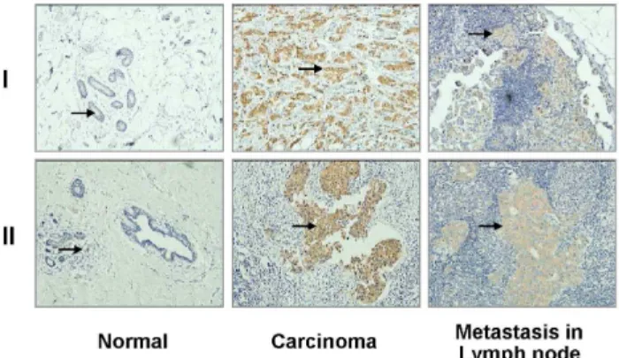
Figure 2. PKCf expressed strongly in carcinoma tissue, but weakly/not in normal tissue and in lymph node metastasis.
Samples of IHC images were from two independent individuals (I and II). Arrows point to PKCfsignals.
In brief, after dewaxing and rehydration, array sections and tissue slices were pretreated with EDTA and H2O2for antigen retrieval
and for quenching endogenous peroxidase activity. Then the sample slides were blocked with 2% bovine serum albumin (BSA), and incubated with a primary antibody against PKCf(1:50 at 4uC overnight; Santa Cruz Biotechnology, Dallas, TX, USA) and secondary antibody successively. Slides were visualized using 3, 3-diaminobenzidine (DAB) and lightly counterstained with hema-toxylin. Negative controls were obtained by omitting the primary antibody.
Images were graded according to the percentage of tumor cells stained and the intensity of the staining. The percentage of cells stained was scored from 0 to 4+ by calculating the percent of positive to total epithelial cells in an area covering 25% of the tumor. In brief, 0 for 0% positive, 1+ for 25% and less, 2+for more than 25% but less than or equal to 50%, 3+for more than 50% but less than or equal to 75% and 4+for more than 75%. The intensity of immunostaining was scored semiquantitatively as: 0 for no obvious yellow particles in epithelial cell plasma membrane or cytoplasm; 1 for weak (light yellow particles); 2 for
moderate (moderately yellow particles); 3+for strong (deep yellow particles). Two pathologists independently determined scores for all samples. In cases where the difference in score exceeded two, the slides were re-examined until the pathologists reached an agreement.
The scores of percentage of positive cells and intensity of staining from the same specimen were added. When the sum was greater than or equal to 3, the staining of the specimen was taken as positive.
Protein isolation and western blot
Protein was isolated from cell lines or tumor and their paired normal tissue samples using lysis buffer containing 1% SDS, 10 mmol/L Tris-Cl (pH 7.6), 150 mmol/L NaCl, 20 g/L apro-tinin, 20 g/L leupeptin, and 1 mmol/L phenylmethanesulfonyl fluoride. Protein lysates (30mg/lane) were separated on 10% SDS-PAGE under denatured conditions, and transferred to PVDF membrane. Membranes were blocked with 1% BSA. Immobilized proteins were probed using a primary antibody against PKCf Table 1.Correlation between PKCfexpression and clinicopathologic characteristics of human breast carcinomas.
Clinicopathological parameters PKCfexpression t or x2 P value
Low (+/2) High (++/+++)
81 95
Age (y) 52.369.7 50.9610.2 0.739 0.814
Menopause
before 36 (44.4%) 59 (62.1%) 5.490 0.019
after 45 (55.6%) 36 (37.9%)
Clinical stage
I 20 (24.7%) 8 (8.4%) 23.112 ,0.001
II 43 (53.1%) 34 (35.8%)
III 14 (17.3%) 46 (48.4%)
IV 4 (4.9%) 7 (7.4%)
Lymph node metastasis
Negative 70 (86.4%) 39 (41.1%) 38.1683 ,0.0001
Positive$5 11 (13.6%) 56 (58.9%)
Histologic grade
1 8 (9.9%) 3 (3.2%) 5.344 0.691
2 62 (76.5%) 70 (73.7%)
3 11 (13.6%) 22 (23.2%)
Tumor size 27.2610.7 40.9620.3 24.612 ,0.001
ER
Negative 43 (53.1%) 57 (60.0%) 0.852 0.356
Positive 38 (46.9%) 38 (40.0%)
PR
Negative 47 (58.0%) 53 (55.8%) 0.089 0.765
Positive 34 (42.0%) 42 (44.2%)
HER2
Negative 60 (74.1%) 60 (63.2%) 12.784@ 0.004
1+ 6 (7.4%) 1 (1.1%)
2+ 9 (11.1%) 11 (11.6%)
3+ 6 (7.4%) 23 (24.2%)
@:Fisher’s exact test.
doi:10.1371/journal.pone.0090811.t001
(sc-216, Santa Cruz Biotechnology), followed by an HRP conjugated secondary antibody. Immunoreactive proteins were visualized with an enhanced chemiluminescence detection system (Amersham-Pharmacia Biotech, Piscataway, NJ, USA). The optical densities of western blot bands were analyzed with Image J software and statistically analyzed by t-test.
Statistics
Statistical analysis was performed using the Statistical Package for the Social Sciences software (SPSS, Chicago, IL, USA). The Chi-square test and Fisher’s exact test were used to examine the association between PKCf expression and various clinicopatho-logical parameters.
Ethical statements
All experiments and information collected were approved by the research ethics committee of Tianjin Medical University. All specimens were taken from the tumor tissue bank of the Tianjin Medical University Cancer Institute and Hospital. Data were analyzed anonymously.
Results
PKCfwas expressed highly in human breast carcinomas
To investigate PKCf expression in breast cancer, TMA and western blots were performed using paired samples from patients with invasive ductal carcinoma (Fig. 1). Compared with normal tissue, carcinoma tissue displayed much higher signals for PKCf by both methods. Thirty paired samples were analyzed using TMA, and the number of invasive samples that scored positive for PKCf (17/30, 56.7%) was much higher than that for normal samples (3/30, 10%) (p,0.01). Sixteen paired samples were analyzed by western blot. Only one single band around 80 kDa was detected by the antibody against PKCf. Similarly, relative PKCflevels (normalized by ß-actin) were much higher than those in normal smples (5.0964.42 vs 0.5260.39, p,0.01 by t-test).
Relationship between PKCfexpression levels and pathological characteristics of breast carcinomas
To analyze the relationship between PKCf expression levels and pathological characteristics of breast cancer, samples from 176 patients with invasive ductal carcinoma were investigated. PKCf levels were determined in tumor samples using IHC (Fig. 2). Patients were divided into two groups by PKCf level, and statistically analyzed according to each clinicopathological param-eter (Table 1). Between the low and high PKCfexpression groups, distribution of age, histological grade of tumor, and expression status of ER and PR showed no differences (p.0.05). Based on menopausal status, up to 62% of patients in the high PKCfgroup were premenopausal, while 44.4% were in the low PKCfgroup (p = 0.019). Of note, high PKCf levels displayed significant correlation with factors indicating worse prognosis. Compared with the patients in the low PKCfgroup, those in the high PKCf group were in advanced clinical stages (55.7 vs 22.2% in stage III/ IV, p,0.001), with more lymph node metastases (71.6 vs 46.1% positive, p = 0.0009) and larger tumor size (40.9620.3 mm vs 27.2610.7 mm, p,0.001). The correlation was further confirmed in the survival analysis. Both disease-free and overall survival rates were lower in the high PKCfexpression group (Fig. 3).
Positive correlation between PKCfand HER2 levels
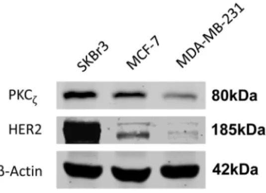
Interestingly, our data suggested that high PKCf levels were related to high HER2 levels. More patients in the high PKCf group were HER2 positive (36.8 vs 25.9%, p = 0.004) (Table 1). The difference was further enlarged (24.2 vs 7.4%) when compared with the HER2 (3+) subgroup. To confirm our observation, we investigated PKCf levels and HER2 levels in three breast cancer cell lines by western blot (Fig. 4.). The cell line data also indicated a positive correlation between PKCf and HER2.
Discussion
During our investigation, greater than 50% of breast cancer specimens overexpressed PKCf, and PKCf that were related to
pathological and prognostic characteristics, including clinical stage, lymph node metastasis status, tumor size, HER2 status and survival rate. Our results were different from those studies in which transcript levels were determined. For example, Awadelk-arim et al.found that PKCfwas overexpressed in only 9.6% of tumor specimens by qRT-PCR. Neither breast cancer molecular subtype, according to hormone receptor and HER2 status, nor relapse-free survival (RFS) was found to be related to PKCf transcript levels [11]. One of the possible reasons might be that the antibody against PKCftheoretically detected signals from other PKC forms, PKCl and PKCi. However, molecular weights of those PKC isoforms were different. As it was indicated in the datasheets from manufacturer, PKCfsignals were around 80kDa, while signals for PKCl and PKCi were around 74 kDa and 65 kDa, respectively. The specificity of the antibody against PKCf has been conformed by several studies [12,13]. Since we detected and analyzed only one single band around 80kDa, the merge signals from other PKC isoforms were unlikely the major cause. Another reason could be due to differences between PKCfmRNA and protein levels, whereby elevated proteins levels may result from regulation of PKCfexpression downstream of transcription, such as protein translation, stability and solubility. Drosophila deficient in INAD (inactivation no afterpotential D) protein, which
is required for the stability of PKC, caused a reduction in PKC protein [14]. Fas-associated protein with death domain (FADD) has been found to regulate cPKC phosphorylation and the subsequent change in stability and solubility of cPKC in human and mouse cells [15]. It has been reported that modification of proteins by phosphorylation, acetylation and redox reactions, altered not only the activity but also the stability of some proteins. The tumor suppressor p53 is a well-known example [16]. Further investigation is required to characterize PKCf protein status in vivo.
Prognosis of breast cancer is affected critically by metastasis, in which cell motility and chemotaxis are involved [17]. Early studies linked PKCfto chemokine-triggered cell migration, not only in multiple types of cancer cells [18–20], but also in metastasis-related macrophages [21]. Investigations with specific inhibitors against PKCfalso suggested that PKCfis associated with cancer with respect to both chemotaxis and immune responses [22,23]. Therefore, PKCf may serve as a potential target for cancer therapy and as a prognostic biomarker. To date, several groups have shown that PKCf regulates proliferation, invasion and metastasis of breast cancer cellsin vitro [19,24,25]. There is also some evidence linking PKCfwith prognosis of several tumor types in patients, including non-gastrointestinal stromal tumor soft tissue sarcomas (non-GIST STSs) [26], renal cell carcinoma [27] and ovarian cancer [28]. Though overexpression of PKCf has been observed in breast tumor tissue rather than in adjacent breast normal tissue, no consistent conclusion has been reported in breast cancer yet [29]. Our observation linked overexpression of PKCf with advanced clinical stages, greater lymph node metastasis and increased HER2 expression, suggesting that PKCfprotein levels may be related to prognosis of breast cancer in patients. Since PKCfhas been reported to be a mitogenic downstream mediator of EGFR in cell signal transduction [30], inhibiting PKCfcould target HER2 or EGFR and potentially enhance breast cancer chemotherapy.
Author Contributions
Conceived and designed the experiments: JY ZL BL. Performed the experiments: JY ZL. Analyzed the data: JY HL. Contributed reagents/ materials/analysis tools: JS XC JL SH. Wrote the paper: JY ZL.
References
1. Fields AP, Gustafson WC (2003) Protein kinase C in disease: cancer. Methods Mol Biol 233: 519–537.
2. Castagna M, Takai Y, Kaibuchi K, Sano K, Kikkawa U, et al. (1982) Direct activation of calcium-activated, phospholipid-dependent protein kinase by tumor-promoting phorbol esters. J Biol Chem 257: 7847–7851.
3. Nishizuka Y (1995) Protein kinase C and lipid signaling for sustained cellular responses. FASEB J 9: 484–496.
4. Nakanishi H, Brewer KA, Exton JH (1993) Activation of the zeta isozyme of protein kinase C by phosphatidylinositol 3,4,5-trisphosphate. J Biol Chem 268: 13–16.
5. Dong LQ, Zhang RB, Langlais P, He H, Clark M, et al. (1999) Primary structure, tissue distribution, and expression of mouse phosphoinositide-dependent protein kinase-1, a protein kinase that phosphorylates and activates protein kinase Czeta. J Biol Chem 274: 8117–8122.
6. Le Good JA, Ziegler WH, Parekh DB, Alessi DR, Cohen P, et al. (1998) Protein kinase C isotypes controlled by phosphoinositide 3-kinase through the protein kinase PDK1. Science 281: 2042–2045.
7. Liu XF, Ishida H, Raziuddin R, Miki T (2004) Nucleotide exchange factor ECT2 interacts with the polarity protein complex Par6/Par3/protein kinase Czeta (PKCzeta) and regulates PKCzeta activity. Mol Cell Biol 24: 6665–6675. 8. Joberty G, Petersen C, Gao L, Macara IG (2000) The cell-polarity protein Par6 links Par3 and atypical protein kinase C to Cdc42. Nat Cell Biol 2: 531–539. 9. Etienne-Manneville S, Hall A (2003) Cell polarity: Par6, aPKC and cytoskeletal
crosstalk. Curr Opin Cell Biol 15: 67–72.
10. Wilson MI, Gill DJ, Perisic O, Quinn MT, Williams RL (2003) PB1 domain-mediated heterodimerization in NADPH oxidase and signaling complexes of atypical protein kinase C with Par6 and p62. Mol Cell 12: 39–50.
11. Awadelkarim KD, Callens C, Rosse C, Susini A, Vacher S, et al. (2012) Quantification of PKC family genes in sporadic breast cancer by qRT-PCR: Evidence that PKCiota/lambda overexpression is an independent prognostic factor. Int J Cancer 131(12): 2852–2862.
12. Iakoubov R, Ahmed A, Lauffer LM, Bazinet RP, Brubaker PL (2011). ‘‘Essential role for protein kinase Czeta in oleic acid-induced glucagon-like peptide-1 secretion in vivo in the rat.’’ Endocrinology 152(4): 1244–1252.
13. Zhang F, Zhang X, Li M, Chen P, Zhang B, et al. (2010). ‘‘mTOR complex component Rictor interacts with PKCzeta and regulates cancer cell metastasis.’’ Cancer Res 70(22): 9360–9370.
14. Venkatachalam K, Wasserman D, Wang X, Li R, Mills E, et al. (2010) Dependence on a retinophilin/myosin complex for stability of PKC and INAD and termination of phototransduction. J Neurosci 30: 11337–11345. 15. Cheng W, Wang L, Zhang R, Du P, Yang B, et al. (2012) Regulation of Protein
Kinase C Inactivation by Fas-associated Protein with Death Domain. J Biol Chem 287: 26126–26135.
16. Hollstein M, Hainaut P (2010) Massively regulated genes: the example of TP53. J Pathol 220: 164–173.
17. Page DL (1991) Prognosis and breast cancer. Recognition of lethal and favorable prognostic types. Am J Surg Pathol 15: 334–349.
18. Guo H, Gu F, Li W, Zhang B, Niu R, et al. (2009) Reduction of protein kinase C zeta inhibits migration and invasion of human glioblastoma cells. J Neurochem 109: 203–213.
19. Sun R, Gao P, Chen L, Ma D, Wang J, et al. (2005) Protein kinase C zeta is required for epidermal growth factor-induced chemotaxis of human breast cancer cells. Cancer Res 65: 1433–1441.
Figure 4. Positive correlation between PKCfand HER2 levels in breast cancer cell lines. Expression of PKCf and HER2 were determined using western blots. ß-actin served as a loading control. doi:10.1371/journal.pone.0090811.g004
20. Liu Y, Wang B, Wang J, Wan W, Sun R, et al. (2009) Down-regulation of PKCzeta expression inhibits chemotaxis signal transduction in human lung cancer cells. Lung Cancer 63: 210–218.
21. Guo H, Ma Y, Zhang B, Sun B, Niu R, et al. (2009) Pivotal Advance: PKCzeta is required for migration of macrophages. J Leukoc Biol 85: 911–918. 22. Li H, Wu J, Ying G, Chen L, Lai L, et al. (2012) J-4: a novel and typical
preclinical anticancer drug targeting protein kinase C zeta. Anticancer Drugs 23: 691–697.
23. Wu J, Zhang B, Wu M, Li H, Niu R, et al. (2010) Screening of a PKC zeta-specific kinase inhibitor PKCzI257.3 which inhibits EGF-induced breast cancer cell chemotaxis. Invest New Drugs 28: 268–275.
24. Castoria G, Migliaccio A, Di Domenico M, Lombardi M, de Falco A, et al. (2004) Role of atypical protein kinase C in estradiol-triggered G1/S progression of MCF-7 cells. Mol Cell Biol 24: 7643–7653.
25. Liu Y, Wang J, Wu M, Wan W, Sun R, et al. (2009) Down-regulation of 3-phosphoinositide-dependent protein kinase-1 levels inhibits migration and experimental metastasis of human breast cancer cells. Mol Cancer Res 7: 944– 954.
26. Valkov A, Sorbye SW, Kilvaer TK, Donnem T, Smeland E, et al. (2011) The prognostic impact of TGF-beta1, fascin, NF-kappaB and PKC-zeta expression in soft tissue sarcomas. PLoS One 6: e17507.
27. Pu YS, Huang CY, Chen JY, Kang WY, Lin YC, et al. (2012) Down-regulation of PKCzeta in renal cell carcinoma and its clinicopathological implications. J Biomed Sci 19: 39.
28. Nazarenko I, Jenny M, Keil J, Gieseler C, Weisshaupt K, et al. (2010) Atypical protein kinase C zeta exhibits a proapoptotic function in ovarian cancer. Mol Cancer Res 8: 919–934.
29. Lin YM, Su CC, Su WW, Hwang JM, Hsu HH, et al. (2012) Expression of protein kinase C isoforms in cancerous breast tissue and adjacent normal breast tissue. Chin J Physiol 55: 55–61.