Doutoramento em Medicina
Especialidade de Cirurgia Geral
Universidade de Lisboa
Faculdade de Medicina
The Role of Systemic Metabolism in
Breast Cancer Progression
Catarina Sofia Rodrigues dos Santos Granja da Fonseca
Todas
As opiniões expressas nesta publicação são da exclusiva responsabilidade
do seu autor.
Universidade de Lisboa
Faculdade de Medicina
The Role of Systemic Metabolism in
Breast Cancer Progression
Catarina Sofia Rodrigues dos Santos Granja da Fonseca
Orientadores
Professor Doutor Sérgio Dias
Professor Doutor José Crespo Mendes de Almeida
Doutoramento em Medicina
Especialidade de Cirurgia Geral
A impressão desta dissertação foi aprovada pelo Conselho Científico da Faculdade de Medicina de Lisboa em reunião de 28 de Outubro de 2014.
O projecto de doutoramento foi desenvolvido no âmbito do Programa de Formação Médica Avançada, 2ª edição, 2009.
Agradecimentos
“Depois de escalar uma grande colina, apenas descobrimos que há muitas mais colinas para escalar”. Nelson Mandela Esta frase tem estado bem presente para mim. Aplica-se a cada momento em que trabalhei neste projecto. O trabalho não fica terminado quando concluímos uma tarefa. Isso apenas nos indica que há muitos mais caminhos.
Mas aplica-se também a outras dimensões da vida. Nos últimos tempos, em que me preparava para terminar a especialidade de cirurgia geral e a tese de doutoramento, tive a sensação de estar a subir a colina mais difícil, a mais árdua de sempre, ou talvez duas colinas ao mesmo tempo. Hoje chego ao ponto de onde vejo bem que a escalada está apenas no início e que muitas e maiores colinas existem.
O medo de deixar atrás algum lugar não explorado ou de não encontrar o caminho acompanhou-me desde o primeiro momento. Por vezes foi mesmo o maior peso que carreguei. Mas, nesta caminhada tive a sorte de ter ao meu lado aqueles que amo e que hão-de perpetuar esse amor, aqueles que me ensinaram a escolher o caminho, aqueles que me confortaram em cada chegada, e em cada nova partida, aqueles me ofereceram a sua amizade e até aqueles que me levaram a mudar de direção.
Não poderia chegar aqui sem que muitas pessoas tivessem contribuído para isso. Com a certeza de que vou pecar por omissão, quero deixar uma palavra de agradecimento escrita para
a Rosinda, pela MÃE que és,
o Fernando, pelo amor incondicional,
o Guilherme e a Maria Sofia, por darem sentido à minha vida, por todas as palavras que não vos ouvi dizer, por todos os primeiros passos a que não assisti, por todos os dentes que não vi cair, peço desculpa;
a Teresa, a Bia e a Matilde, por todo o tempo em conjunto que vos roubei, a Laurentina e ao Manuel Fernando por me fazerem sentir uma verdadeira filha, a todos os meus amigos, pela paciente aceitação das minhas ausências,
o Professor Doutor José Fernandes e Fernandes pela confiança que sempre depositou no meu percurso académico;
o Professor Doutor Mendes de Almeida, por todo o suporte e pelo exemplo inspirador que é para mim;
o Professor Doutor Sérgio Dias, por me ter recebido no seu laboratório, orientado o meu projecto e por todo o empenho e motivação que colocou no meu trabalho;
a Professora Doutora Isabel Fonseca pela disponibilidade para a revisão, sempre atenta, dos meus resultados;
a Professora Doutora Jacinta Serpa, pela genrosidade com que reviu criticamente a minha tese;
os colegas de laboratório, a quem agradeço a ajuda, os ensinamentos e a genuína amizade. Em especial à Tânia Carvalho e à Germana Domingos pelo apoio em tarefas laboratoriais cruciais do meu projecto;
a Professora Doutora Leonor Parreira, pela oportunidade de integrar o Programa Doutoral da Gulbenkian que foi uma experiência única de aprendizagem científica e humana; aos Amigos do meu Programa Doutoral da Gulbenkian (Alexandra B., Alexandra S.; Inês; Nuno; Susana; Jaime; Diogo; Sandra e Claúdia) pelos momentos irrepetíveis que partilhámos.
Lisboa, 16 de Novembro de 2014 Catarina Rodrigues dos Santos
7
Resumo
Em cada dez novos casos de cancro diagnosticados, no mundo, por ano, um é cancro de mama. Apesar do aumento da sobrevivência nos países mais desenvolvidos (e com alta incidência), relacionado com a existência de rastreio, diagnóstico rápido e melhoria no tratamento, o cancro da mama continua a ser a principal causa de morte por cancro em mulheres.
A investigação das últimas décadas tem-se centrado fundamentalmente no tumor e na sua genómina considerando-o como uma entidade independente do organismo. Mesmo sendo a terapêutica individualizada um dos objectivos major desta pesquisa, as características do individuo têm sido poucas poucas vezes valorizadas.
A hipótese principal deste trabalho foi a de que o tumor é um microsistema a evoluir num macrosistema hospedeiro devendo existir uma interdependência entre estes. O conceito da influência do macroambiente na evolução tumoral é consistente com a teoria Darwiniana da evolução que postula que a seleção é uma propriedade essencial dos sistemas biológicos. Isto é, assim como as forças ambientais poderão explicar os padrões de incidência do cancro da mama na população mundial, também os factores do hospedeiro poderão selecionar os fenótipos do tumor. De facto, dados recentes indicam que alguns factores de risco conhecidos para o cancro da mama são condições sistémicas como a obesidade ou a diabetes. A prevalência epidémica da obesidade e das suas comorbilidades têm sido investigadas como possíveis razões para o aumento da incidência do cancro nas sociedades ocidentais. No entanto, apesar de as alterações do perfil lipídico, nomeadamente a hipercolesterolémia, serem também prevalentes nestas regiões o papel do metabolismo lipídico sistémico no cancro da mama permanece por esclarecer.
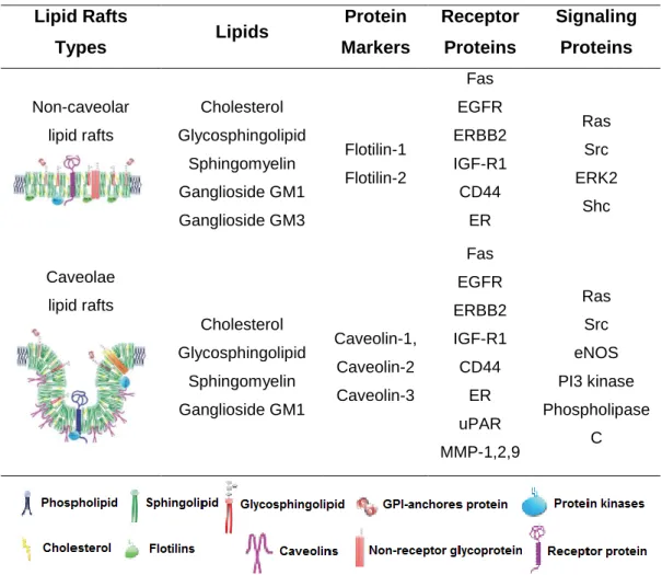
Os lípidos são fundamentais para as células, onde actuam quer como constituintes celulares quer como moléculas de sinalização. O colesterol, especificamente, é um componente essencial das membranas celulares onde se concentra nos denominados “lipid rafts”, microdomínios da membrana envolvidos na polaridade da célula, migração, proliferação, sobrevivência e angiogénese. O colesterol é também o precursor obrigatório das hormonas esteróides como o estrogénio e progesterona, sendo a vasta maioria dos tumores de mama hormono-sensíveis.
8
Até agora,a maioria dos estudos publicados sobre relação entre o colesterol e o cancro da mama tentaram demonstrar causalidade entre os níveis séricos e a incidência de cancro. Raros estudos exploraram a eventual associação com a agressividade tumoral ou com a sua progressão. Portanto, a importância do colesterol plasmático na progressão do cancro da mama é desconhecida. Esta Tese visou estudar o papel do colesterol
sistémico na progressão do cancro da mama e os mecanismos moleculares subjacentes
Para tal foi desenhado um estudo observacional prospectivo que seguiu um coorte de mulheres com cancro de mama (estadios I-III), sem tratamento prévio. Os resultados mostraram que o nível de LDL-C no momento do diagnóstico se correlaciona positivamente com o tamanho e estadio do tumor. Aos 2 anos de seguimento, mulheres com níveis de LDL-C mais elevados no diagnóstico tiveram menor tempo livre de doença. Estes dados apontam o LDL-C como um biomarcador de agressividade tumoral (factor de prognóstico) e um possível alvo terapêutico (factor preditivo).
Para provar a relação causal do ambiente enriquecido em colesterol na progressão do tumor foram usados modelos bem estabelecidos in vitro e in vivo que permitiram demonstrar que a sinalização mediada pelo LDL-C induz fenótipos tumorais, semelhantes aos observados nos doentes com cancro de mama e caracterizados por maior proliferação e capacidade invasora. As células expostas a concentrações maiores de LDL-C apresentam características genotípicas distintas, compatíveis com um fenótipo mais agressivo, como confirmado por análise de expressão génica. Experimentalmente foi demonstrado que a expressão tumoral do ABCA1 (principal exportador celular de cholesterol) num ambiente enriquecido em colesterol é um marcador do fenótipo induzido pelo LDL-C.
Em conjunto, os resultados desta tese ilustram a influência do metabolismo sistémico na progressão do cancro da mama e sugerem que o perfil lipídico deve ser avaliado em todos os doentes com este diagnóstico. Sendo a hipercolesterolémia tão prevalente nas sociedades ocidentais com uma distribuição global paralela à incidência do cancro da mama, é de esperar que a modulação do perfil lipídico nestes doentes tenha um impacto elevado como estratégia de prevenção (secundária). Isto parece particularmente relevante para os doentes com tumores não hormono-sensíveis já que não existem outras formas de quimioprevenção.
9
Abstract
One in ten of all new cancers diagnosed worldwide, each year, is a breast cancer. Despite the more favorable survival of breast cancer patients in (high-incidence) developed regions due to screening, faster diagnosis and improved treatment, it remains the most frequent cause of cancer-related death in women.
The research of the last decades focused primarily on tumor genomics considering the tumor as an entity independent of the organism. Although the personalized therapy has been the ultimate goal of this aproach, host characteristics were seldom valued.
Our working hypothesis states that the tumor is a microsystem evolving within a host macrosystem and an- interdependence between them must exist. The concept of the influence of the macroenvironment in tumor development is consistent with the Darwinian theory of evolution that postulates selection as a fundamental property of biological systems. As environmental forces may explain breast cancer incidence patterns in the world population, host environmental factors may drive breast cancer phenotypes. Recent data indicate that some known risk factors for breast cancer are indeed systemic conditions such as obesity or diabetes. The epidemic prevalence of obesity and its associated comorbidities have been investigated as possible reasons for the increased incidence of cancer in Western societies. Although altered lipid profiles, including hypercholesterolemia, are extremely prevalent in these areas the role of systemic lipid metabolism in breast cancer is poorly understood.
Lipids are fundamental to cells, where they act either as constituents of the cell or as cell signaling molecules. Cholesterol, in particular, is an essential component of cell membrane bilayer, where it concentrates on the so-called lipid rafts-membrane microdomains which are involved in cell polarity, migration, proliferation, survival and angiogenesis. Cholesterol is also an obligatory precursor of steroid hormones, such as estrogen and progesterone, being the vast majority of breast tumors hormone responsive. To date, most studies have seek to find a causal relation between cancer incidence and cholesterol plasma levels, however, fewer studies adressed a possible link in tumor aggressiveness or on its progression. Thus, for now, the importance of plasma cholesterol in breast cancer progression is largely unknown.
This Thesis aimed to study the role of systemic cholesterol in breast cancer progression and the underlying molecular mechanisms.
10
To do that, an observational prospective study was designed to follow a cohort of women with breast cancer (stages I-III), without previous treatment. Results show that plasma LDL-C at diagnosis positively correlates with tumor size and stage. At 2 years of follow up, higher levels of LDL-C were associated with reduced disease-free survival. These data indicate LDL-C as a biomarker of tumor aggressiveness (prognostic factor) and a possible therapeutic target (predictive factor).
To demonstrate a causal implication of cholesterol-enriched environment in tumor progression, well established in vitro and in vivo models were used. Results revealed that LDL-C signaling induces a tumor phenotype, also observed in breast cancer patients characterized by increased cell proliferation and invasion. Cells exposed to higher LDL-C concentrations have distinct genetic expression, supporting an aggressive phenotype, as confirmed by gene expression analysis. It was also experimentally shown that tumor ABCA1 (main cellular membrane cholesterol exporter) expression in cholesterol-enriched environment is a marker of LDL-C induced phenotype.
Together, the results of this thesis illustrate the influence of systemic metabolism in breast cancer progression and suggest that lipid profile must be assessed in all breast cancer patients. Being hypercholesterolemia so prevalent in Western societies with a global incidence pattern superimposing that of breast cancer, the modulation of cholesterol plasma levels are expected to have a major impact in (secondary) prevention. This seems particularly relevant for patients with hormone unresponsive tumors, for which no chemopreventive strategy exists.
11
Abbreviations
°C: degrees Celsius
µl: microliter µM: micromolar
ABCA1: ATP-binding cassette protein A1
ABCG1: ATP-binding cassette protein G1
ACAT-1: acetyl-CoA acetyltransferase 1
AJCC: American Joint Committee on Cancer
Akt: Akt protein kinase
AMPK: mitogen activated protein kinase ANOVA: analysis of variance
ATP: adenosine 5′-triphosphate
BMDC: bone marrow derived cells
BMI: body mass index
CDC: Centers for Disease Control and Prevention
cDNA: complementary deoxyribonucleic acid CI: confidence interval
c-Myc v-myc avian myelocytomatosis viral oncogene homolog protein
c-myc-: v-myc avian myelocytomatosis viral oncogene homolog
CO2: carbon dioxide
COX-2: cyclooxygenase-2
CXCR4: C-X-C chemokine receptor type 4
DAPI: 4',6-diamidino-2-phenylindole
DCIS: ductal carcinoma in situ
DFS: disease-free survival
DHCR7: 7-dehydrocholesterol reductase DMEM: Dulbecco’s modified Eagle’s medium
DMSO: dimethyl sulfoxide DNA: deoxyribonucleic acid
12 ECM: extracellular matrix
EDTA: ethylenediaminetetraacetic acid
EGF: epidermal growth factor
EGFR: epidermal growth factor receptor
EMT: epithelial mesenchymal transition
ER: estrogen receptor
ERBB: epidermal growth factor B
ERBB2 (=HER2; =Her2-neu receptor): human epidermal growth factor receptor-2 or v-erb-b2 avian erythroblastic leukemia viral oncogene homolog 2
ERK: extracellular-signal-regulated kinases
FAK: focal adhesion kinase
Fas: CD95 or apoptosis antigen 1 (APO-1)
FBS: fetal bovine serum
FBSLF: fetal bovine serum lipoprotein free FDG-PET: F-18 fluoro-2-deoxyglucose PET
FU: follow-up
g: gram
GFP: green fluorescent protein
GLY: glyburide
Gy: gray
H&E: hematoxylin and eosin
h: hours
HD: hypercholesterolemic diet
HDL (=HDL-C): high-density lipoprotein
HIF-1: hypoxia-inducible factor 1
HMGR: hidroxi-3-methyl-glutaril-CoA reductase HR: hazard ratio;
IDL: intermediate-density lipoprotein
IGF-1R: insulin-like growth factor 1 receptor
IHC: immunohistochemistry
13 JNK: jun amino-terminal kinase
Ki-67: antigen identified by monoclonal antibody Ki-67
LDL (=LDL-C): low-density lipoprotein
LDLR: low-density lipoprotein receptor
LRP1: low density lipoprotein receptor-related protein 1
LVI: lymphovascular invasion
LXR: liver X receptors α (LXRα) and β (LXRβ)
MAPK: mitogen activated protein kinase mg: milligram
microRNA (=miRNA): micro ribonucleic acid
mL: milliliter
mm: millimeter
mM: millimolar
MMP: matrix metalloproteinase mRNA: messenger ribonucleic acid
mTOR: mammalian target of rapamycin
NCCN: National Comprehensive Cancer Network
ND: normal diet
NFĸB: nuclear factor kappa-light-chain-enhancer of activated B
NOS: not otherwise specified
NSAID’s: non-steroid anti-inflammatory drugs
OLR1: ox-LDL receptor
OR: odds ratio
OS: overall survival
P: p-value
p53: tumor supressor protein p53
PAGE: polyacrylamide gel electrophoresis PAI-1: plasminogen activator inhibitor type 1
PCR: polymerase chain reaction
14 PI3K: phosphoinositide 3-kinase
PLC: phospholipase C
PR: progesterone receptor
PTEN: phosphatase and tensin homolog
PUFA: polyunsaturated fatty acids
RB: retinoblastoma-associated gene
Rb: retinoblastoma-associated protein
RE: endoplasmasmic reticulum RIPA: radioimmunoprecipitation assay
RNA: ribonucleic acid
ROS/RNS: reactive oxygen species/reactive nitrogen species
RR: relative risk;
RTC: randomized control trials
SCAP: sterol regulatory element-binding protein cleavage activating protein SDF-1: stromal cell-derived factor 1
SDS: sodium dodecyl sulfate
SEM: standard error of the mean
SFK: Src family of kinases
Shh Sonic hedgehog protein
siRNA: small interfering ribonucleic acid SOS: son of sevenless
SR-A1/2: scavenger receptors class A
SR-BI: scavenger receptors class B
SREBP: sterol regulatory element-binding protein
TC: total cholesterol
TGFα: transforming growth factor alfa
TGF-β: transforming growth factor beta
Tie-2: angiopoietin receptor 2
TNF: tumor necrosis factor
15 uPA: urokinase-type plasminogen activator
uPAR: urokinase-type plasminogen activator receptor
VEGF-A: vascular endothelial growth factor-A
VLDL: very low-density lipoprotein
WHO: World Health Organization
WNT: wingless-type MMTV integration site family
17
Contents
Agradecimentos ... 5 Resumo ... 7 Abstract ... 9 Abbreviations ...11 Contents ...17 List of Figures ...21 List of Tables ...22 List of Graphics ...22 General Introduction ...23I. Breast cancer–The problem ...23
Incidence and mortality ...23
Etiopathogenesis ...24
Phenotypes ...28
Tumor biology ...31
Tumor environment ...40
Tumor dissemination ...42
II. Dyslipidemia-The problem ...45
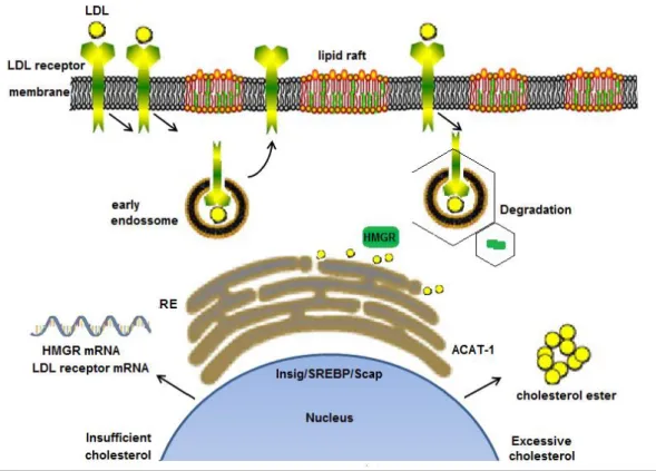
Cholesterol and cholesterol metabolism ...47
Cholesterol metabolism ...48
Cholesterol biological functions ...53
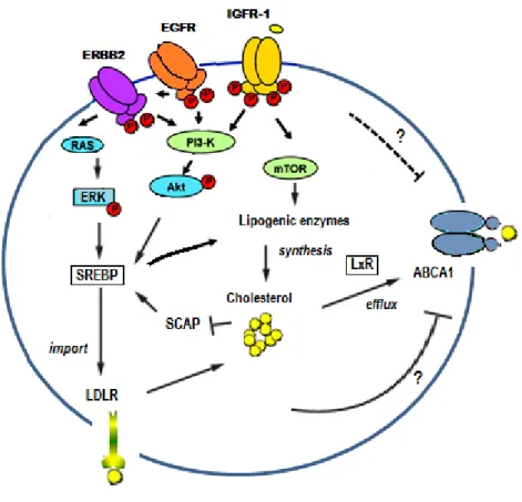
Cholesterol and cholesterol metabolism in (breast) cancer cells ...56
Cholesterol and breast cancer ...62
Lipid profile and breast cancer risk...62
Lipid profile in breast cancer patients ...63
Insights from experimental studies ...66
Statins use and breast cancer ...67
Hypothesis and Aims ...73
18
Plasma Level of LDL-Cholesterol is a Predictive Factor of Breast Tumor
Aggressiveness ...75
Introduction ...75
Methods ...77
Study population and data collection ...77
Biospecimen collection and plasma lipid and lipoproteins assays ...77
Statistical analysis ...78
Results ...78
Spearman correlations ...80
Univariate associations ...82
Multivariate logistic regression ...83
Discussion ...84
Conclusion ...87
Chapter 2 ...89
LDL-cholesterol signaling induces breast cancer proliferation and invasion ...89
Introduction ...89
Material and Methods ...90
Cell lines and reagents ...90
Cell proliferation assay ...90
Migration assay...90
Adhesion assay ...91
RNA extraction and microarray analysis ...91
Protein extraction and western blotting analysis...91
Statistical analysis ...91
In vivo models ...91
Statistical analysis ...93
Results ...93
LDL-cholesterol stimulation induces breast cancer cell lines proliferation, migration and reduces cell adhesion ...93
19
High LDL-cholesterol promotes breast cancer growth in animal models ...96
Statins treatment does not reduced systemic LDL level in mice trial ...98
Discussion ...98
Chapter 3 ... 103
ABCA1 silencing reduces breast cancer aggressive phenotype induced by LDL-cholesterol ... 103
Introduction ... 103
Material and Methods ... 104
Cell lines and reagents ... 104
Human samples analysis ... 105
Cell proliferation assay ... 105
Apoptosis and cell death ... 105
Migration assay... 105
Cholesterol measurements ... 106
Quantification of mRNA levels ... 106
Western blotting analysis ... 106
Small interfering RNA ... 107
Immunofluorescence ... 107
In vivo models ... 107
Statistical analysis of experimental data ... 108
Results ... 108
Exogenous LDL-cholesterol induces ABCA1 expression ... 108
Systemic level of LDL-C correlates with ABCA1 expression ... 110
ABCA1 inhibition reduces LDL-cholesterol-induced proliferation and migration and increases apoptosis of breast cancer cells ... 111
siRNA ABCA1 inhibition prevents LDL-C-induced phenotype of breast cancer cells ... 115
ABCA1 inhibition increases cellular cholesterol content ... 117
Glyburide ABCA1 inhibition reduces ERK protein phosphorylation ... 119
20
Discussion ... 121
Conclusion ... 124
Chapter 4 ... 125
Plasma level of LDL-cholesterol, at diagnosis is a breast cancer prognostic factor .... 125
Introduction ... 125
Methods ... 126
Study population and data collection ... 126
Biospecimen collection. Pathological and Immunohistochemistry assays ... 127
Statistical analysis ... 127
Results ... 128
Discussion ... 130
Conclusion ... 131
Final Discussion ... 133
Importance of the findings and future perspectives ... 141
Bibliography ... 143 Additional Tables ... 165 Additional Figures ... 169 Publications ... 171 Presentations ... 171 Awards ... 172 Papers ... 173
21
List of Figures
Figure 1: Major pathways regulating proliferation of breast cancer cells...33 Figure 2: Glucose catabolism in normal and tumor cells ...37 Figure 3: Determinants of the tumor metabolic phenotype. ...39 Figure 4: Primary tumor microenvironment ...40 Figure 5: Selective pressures and steps from primary tumor growth to metastasis ...41 Figure 6: Breast Cancer Incidence, Worldwide in 2008 ...46 Figure 7: Mean blood cholesterol, ages 25+, age standardized, females 2008 ...46 Figure 8: Cholesterol metabolism products. ...47 Figure 9: Overview of lipoprotein metabolism...49 Figure 10: Cholesterol biosynthesis pathway ...50 Figure 11: Schematic representation of the intracellular cholesterol homeostasis. ...51 Figure 12: Regulation of cholesterol homeostasis and cancer signaling pathways. ...57 Figure 13: Study Fluxogram ...77 Figure 14: Tumor characteristics-LDL tertiles ...83 Figure 15: Phenotype of breast tumors exposed to high levels of cholesterol-Proposed Model. ...86 Figure 16: Proliferation, migration and loss of adhesion induced by LDL-C in breast cancer cell lines. ...94 Figure 17: Activated cellular networks, at 48h, in LDL treated breast cancer cell line. ...95 Figure 18: LDL-C induces ERK and Akt protein phosphorylation ...96 Figure 19: Hypercholesterolemic diet induces a breast cancer phenotype characterized by large and more proliferative tumors. ...97 Figure 20: Tumor size of hypercholesterolemic diet fed mice treated with statins show no significant differences between hypercholesterolemic diet fed and control mice. ...98 Figure 21: Phenotype of breast tumors exposed to high levels of cholesterol-Proposed Model. ... 101 Figure 22: Exogenous LDL-cholesterol induces ABCA1 expression. ... 109 Figure 23: ABCA1 expression in human breast tumors ... 110 Figure 24: ABCA1 inhibition reduces LDL-induced proliferation and migration of breast cancer cell lines. ... 112 Figure 25: Apoptosis and cell death. ... 113 Figure 26: Glyburide decreases ABCA1 transcription and expression. ... 114 Figure 27: Down regulation of ABCA1 by siRNA specifically reduces LDL induced proliferation and migration of MDA MB 231 cells. ... 116 Figure 28: ABCA1 inhibition increases cellular cholesterol content. ... 117
22
Figure 29:ABCA1 inhibition decreases LDLR and HMGR expression. ... 118 Figure 30: ABCA1 inhibition by glyburide reduces ERK protein phosphorylation. ... 119 Figure 31: Glyburide reduces tumor growth and lung metastasis incidence. ... 120 Figure 32: ABCA1 as a marker of LDL-C induced phenotype-Proposed Model... 124 Figure 34: Study Fluxogram ... 126 Figure 35: Overall and disease-free survival in LDL-C tertiles groups. Kaplan–Meier curves. ... 128 Figure 36: Overall and disease-free survival in LDL-C tertiles groups adjusted to BMI ... 129
Additional figure 1: LDLR expresion in human breast tumors ... 169 Additional figure 2: Inhibition of LDLR does not prevent the accumulation of intracellular cholesterol and LDL-C induced phenotype ... 170
List of Tables
Table 1: Risk factors for breast cancer and approximate strength of association ...27 Table 2: Lipid and protein composition of caveolae and non-caveolae lipid rafts ...53 Table 3: Alterations in lipid profile of breast cancer patients ...64 Table 4:Statins ...67 Table 5: Epidemiologic studies on the association of statins use and breast cancer risk. .70 Table 6: Clinical and tumor-related characteristics of the study population (N=244) ...79 Table 7: Lipid profile in tumor stage1 and in prognostic groups2 ...81 Table 8: Patient characteristics in LDL-C levels tertiles ...82 Table 9: Univariate and multivariate logistic regression to the risk of tumor size ≥20mm. 83 Table 10: Cox multivariate regression model for disease-free survival ... 130
Additional Table 1: Univariate logistic regression to the risk of ... 165 Additional Table 2: Breast Cancer Treatment ... 166 Additional Table 3: Gene expression of LDL-C treated MDA MB 231 ... 167 Additional Table 4: Lipid Profile in Mice Trails ... 168
List of Graphics
Graphic 1: Estimated number of new cases from cancer by site in Europe in 2012. ...23 Graphic 2: Percentage of new cases of breast cancer, by age. ...24
23
General Introduction
The general introduction of this Thesis is organized in two parts. The first one reviews the actual knowledge of breast cancer ethiopathogenesis and highligths some gaps in breast cancer research. The second part, briefly describes cholesterol functions and metabolism exploring published data on the relationship between cholesterol and breast cancer.
I. Breast cancer–The problem
Incidence and mortality
Cancer and cardiovascular diseases are the leading causes of death in the West1,2 and are dramatically rising in Asian countries3,4.
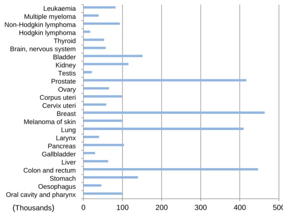
During 2012, there were over 3,4 million new cases of cancer, in Europe (excluding skin non-melanoma cancers) (Graphic 1). The most common cancer site was breast (464,000 cases, 13,5% of all cancer cases) and among women, breast cancer was the leading cause of death (131,000, 16,8% of all cancer)5
Graphic 1: Estimated number of new cases from cancer by site in Europe in 2012. ( Data from J. Ferlay et al, ,20136.)
Oral cavity and pharynx Oesophagus Stomach Colon and rectum Liver Gallbladder Pancreas Larynx Lung Melanoma of skin Breast Cervix uteri Corpus uteri Ovary Prostate Testis Kidney Bladder Brain, nervous system Thyroid Hodgkin lymphoma Non-Hodgkin lymphoma Multiple myeloma Leukaemia 0 100 200 300 400 500 (Thousands)
24
In Portugal, breast cancer is the most frequent cancer in women, and despite being the cancer specific site (excluding skin cancers) responsible for the largest volume of oncologic treatments, including oncologic surgery, it still is the first cause of cancer-related death7.
Faster diagnosis and progresses in systemic treatment have improved survival rates in the last three decades8,9,10, but advanced cases (stage IV) have 5-year survival rate of 25%11. Identifying cases that will progress and that should be treated aggressively remains a major problem.
Etiopathogenesis
Breast cancer has been attributed to a combination of genetic susceptibility and other patient factors including age, reproductive, hormonal and lifestyle features.
Age
Breast cancer is extremely rare among women younger than 20 years old and is uncommon among women younger than 30 years old. Incidence increases sharply with age, being two thirds of invasive breast cancers found in women older than 55 years11(Graphic 2).
Graphic 2: Percentage of new cases of breast cancer, by age.
Percentage of new cases in females of all races, 2007-2011. Data from Surveillance, Epidemiology and End Results Program (www.seer.cancer.gov)11.
0,0% 1,8% 9,3% 22,0% 25,5% 21,3% 14,4% 5,7% <20 20-34 35-44 45-54 55-64 65-74 75-84 >84 P er ce n tage o f new ca se s Age
25
Genetic factors
By 1980, a significant body of evidence supported the presence of inherited factors responsible for familial clustering of breast cancer12, and scientific efforts shifted to determine its inheritance pattern. Thirty years later, high-penetrance cancer susceptibility genes appear directly responsible for only 5-10% of all breast cancers. BRCA1 and BRCA2 gene mutations are the most common and well-studied genes associated with inherited breast cancer and account for almost all these cases13. Mutations in p53 (Li-Fraumeni syndrome14), PTEN (Cowden syndrome15), CHEK216, STKI/LKB1 (Peutz-Jeghers syndrome17), ATM18, MSH2/MLH1 (Muir-Torre syndrome19), PALB220, BRIP121, were also described in breast cancer families but are less common.
The search for low penetrance alleles, either by candidate gene approach or genome wide association studies, did not produce major clinical impact. Apart from the identification of some loci strongly associated with breast cancer risk22,23,24, the understanding how this knowledge can be applied clinically, is still missing. On the other hand, the identification of small interfering ribonucleic acids (siRNA) uncovers a new level of regulation of gene expression. Ribonucleic acids (RNAs) are the direct products of genes, and these small RNAs, also called micro RNAs (miRNAs) can bind to other specific messenger ribonucleic acid (mRNA) molecules and either increase or decrease their activity, resulting in gene silencing via translational repression or target degradation, thus, preventing protein expression25. It is now believed that as much as 92% of gene expression is regulated by siRNAs26. In breast cancer miRNAs show differential expression across molecular subtypes, and show both oncogenic and tumor-suppressive roles dependent on the context27.
Non-genetic factors
Reproductive factors, hormonal factors and nutritional/lifestyle are considered non-genetic breast cancer risk factors. Whereas the causal effect of some of them is accepted, their individual strength and how they correlated with each other is more difficult to define.
Reproductive factors
Early age menarche28, late menopause29 and nuliparity28, have all been consistently associated with increased risk of breast cancer28. Most studies point the early onset of ovulatory menstrual cycles and the greater lifetime exposure to endogenous hormones to be the reason of such association30. First pregnancy leads to proliferation of breast cells, which results in differentiation into mature cells prepared to lactation. After differentiation, epithelial cells have a longer cell cycle and spend more time in the G1 phase allowing for DNA repair31. In the same trend, lactation may be a protective factor because
breast-26
feeding results in further terminal differentiation of the breast epithelium and delays the return of ovulatory menstrual cycles after pregnancy.
Hormonal factors
Several lines of evidence have long suggested that sex hormones play a central role in the etiology of breast cancer. In animals, estrogens, progesterone, and prolactin all promote mammary tumors. Data from randomized controlled trials (RTC) have confirmed the epidemiologic relations of hormone therapy to increased risk of breast cancer and estrogen plus progestin therapy is now classified as a human carcinogen32,33,34. Conversely, hormonal manipulations, such as anti-estrogens and aromatase inhibitors are useful in the treatment of breast cancer and in reducing breast cancer incidence in high risk women35,36,37..
Nutrition/ lifestyle factors
Around the world, nutritional factors have been prominent among the hypothesized environmental determinants of breast cancer incidence and in the large increase in rates of migrant populations from low-incidence to high-incidence countries38,39. The dominant hypothesis has been that high-fat intake increases the risk of breast cancer. In agreement, overweight and obesity were clearly associated with increased incidence of breast cancer in postmenopausal women and a higher mortality rates40,41,42,43. Studies specifically addressing high-fat diet in breast cancer risk, showed increased occurrence of mammary tumors in rodents44,45 although some of these studies loses the strong association when adjusted to energy intake46. Human ecological studies evidences that fat consumption per
capita is highly correlated with breast cancer mortality rates47; and intervention studies
show that the implementation of weight loss and low fat intake programs reduce breast cancer risk in 9% between interventional and control groups48.
Other dietary elements possibly associated to breast cancer, include a positive association with alcohol consume49 and a negative association with vitamin D50,51,52 intake. Physical exercise is suggested as protective53, through body mass index (BMI) reduction, menarche delay, sex hormones and insulin-like growth factors lowering and by improving immune function54, 55.
Other factors
Other variables searched for a relation with breast cancer risk include; proliferative breast diseases, ionizing radiations, active and passive smoking, silicone breast implants, diabetes mellitus, thyroid cancer, non-steroid anti-inflammatory drugs (NSAID’s), statins and antidepressants use. As for comproved risk factors, the relative weight of each factor
27
is hard to predict in the individual case (Table 1) and for some of them the literature is very scarce.
Proliferative breast diseases without atypia slightly increases the risk of breast cancer while atypical hyperplasia represents a moderate increased risk of breast cancer applying for both glands even when it is only unilateral56.
Ionizing radiation to the chest, in cumulative doses at young age, substantially increases breast cancer risk. Evidence on this topic comes from atomic bomb survivors57, therapeutic58 and occupational59 radiation use studies. The regular diagnostic radiation use, such as the mammographic screening, shows no significant increased risk of breast cancer60.
Recent studies have shown the association of type 2 diabetes with the incidence of breast cancer61,62,63 and cancer-specific mortality. This association was most pronounced in postmenopausal women and ER-positive disease63. This effect is attributed to insulin, which acts as a breast cancer cell growth factor64,65. But because many other conditions lead to hyperinsulinemia, further studies on the relationship between breast cancer and insulin resistance are warranted. Nevertheless, clinical trials with oral anti-diabetics drugs are in course66.
Table 1: Risk factors for breast cancer and approximate strength of association
Reproductive Factors Hormonal Factors Nutritional/Lifestyle Factors Others Factors Early age at menarche + Oral contraceptives use (current vs none) + Obesity (BMI>30vs>25) Premenopausal – Postmenopausal + Family history (mother and sister)
+++ Age at first birth
(>35vs<20) ++
Estrogen replacement (10 years vs none) +
Adult weight gain (postmenopausal) ++ Family history (1st– degree relative) ++ No. of births (0 vs 1 child) + Hormone replacement (>5 years vs none) ++
Alcohol (one or more drink vs none) + Jewish heritage (yes vs no) + Age at menopause (5-years increment) + ↑ blood estr./andr (post menopause) +++ Physical activity (>3hours/week) - Ionizing radiation (yes vs no) + Breast-feeding (>1year vs none) -
High blood prolactin ++
Monounsaturated fat (vs saturated fat) -
Benign breast disease ++ - no risk; +low risk; ++moderate risk; +++high risk
28
Although the relative strength of known or suspected breast cancer risk factors are modest in magnitude (RR are usually in the range of 1.3 to 1,8) the impact of risk factors control could be very large. When considering (primary) prevention, it is important to remember that even small changes at individual level can produce substantial changes in population rates of disease67. A recent study estimates that up to 27% of breast cancer-related deaths would be avoided with control of key behavioral and environmental risk factors such as alcohol use, overweight and obesity and physical inactivity68.
Nevertheless, only about 30% of breast cancers are estimated to be explained by known risk factors69, leading to the hypothesis that other environmental factors could play a major role. On the other hand, mechanisms linking known and suspected risk factors to the initiation and development of breast cancer are poorly understood. Therefore, there may still be a large amount of unknown risk factors to breast cancer etiopathogenesis.
Phenotypes
Invasive breast cancers are a heterogeneous group of lesions differing with regard to their clinical presentation, imagiological appearance, pathological features, gene expression profiling and biological behavior.
The phenotype is usually based on the histological characteristics of the tumor, enclosing a range of genotypes and biological behaviors. In clinical practice, the phenotype is used to define therapy and prognosis.
Based on World Health Organization (WHO)70, the most widely used histological classification system of breast cancers, the most common histological type is invasive (infiltrating) ductal carcinoma (53-70%), recently denominated invasive carcinoma, not otherwise specified (NOS). The other types comprises a group of invasive breast cancers with specific or special histological features: invasive lobular carcinoma (5-16%), medullar (3-9%), tubular (1-3%), mucinous (1-2%), and other rare types71,72,73. Non-epithelial breast cancers such as sarcomas or lymphomas are even rarer.
The routine pathologic examination of invasive breast cancers defines the histological type and the grade. Histological grade is based on the degree of differentiation of the tumor tissue. For breast cancer the most reproduced system is the Nottingham (Elston-Ellis) grade system74. It refers to the semi-quantitative evaluation of three morphological characteristics: degree of tubule or gland formation, nuclear pleomorphism and mitotic count. The final score shows a very strong correlation with prognosis; patients with grade I tumors have a significantly better survival than those with grade II and III tumors75.
29
Additionally, three immunohistochemical markers (estrogen receptor (ER), progesterone receptor (PR) and human epidermal growth factor receptor 2 (ERBB2 or Her-2/neu receptor or HER2)) are generally used to identify biological properties. And in the last years, proliferative markers such as Ki67 are also used to determine tumor index proliferation rate, since it was demonstrated to have correlation with prognosis76.
The tumor subgroups identified by immunohistochemistry closely resemble the molecular subtypes, defined by Sorlie et al, based on gene expression profiling studies: Luminal A, Luminal B, HER2 type and basal-like77.
Luminal A and luminal B cancers generally have a good prognosis and show high expression of hormone receptors and associated genes. Together, these two subtypes account for approximately 70% of all breast cancers. The luminal B cancers tend to be higher grade than the luminal A and some of them may overexpress HER2. Both luminal A and luminal B cancers usually respond to hormone therapy, with luminal A cancers showing the improved response. Response of the luminal cancers to chemotherapy is variable, with the luminal B cancers generally showing better response.
The HER2 type cancers show high expression of HER2 and low expression of ER and associated genes. They account for approximately 15% of all breast cancers and are generally ER or PR negative. HER2 cancers are more likely to be high grade and have positive lymph nodes. These cancers show the best response to trastuzumab and to anthracycline-based chemotherapy but, overall have a poor survival prognosis.
The basal-like breast cancers show high expression of basal epithelial genes and basal cytokeratins, low expression of ER and ER associated genes as well as low expression of HER2. They constitute approximately 15% of all breast cancers and are often referred to as triple negative cancers, because they are invariably ER, PR, and HER2 negative. The basal-like tumor phenotype is especially common in African-American women and is also the most common phenotype of BRCA1-associated breast cancers. Basal-like cancers have a poor prognosis and are not amenable to treatment with either hormonal or biological therapy.
.
Prognostic and predictive markers
Despite usefulness of pathological characteristics, in which clinical practice and therapeutic decision are based, they do not linearly correlate with tumor biology and
30
behavior. In order to define prognosis and predict therapeutic response, clinicians classify a newly diagnosed breast cancer through stage and a panel of associated factors.
The American Joint Committee on Cancer (AJCC) system is the most used to stage tumors. It is both a clinical and pathological staging and is based on the TNM system, in which T refers to tumor, N to regional lymph nodes, and M to distant metastasis78
At present, the prognostic and predictive factors of primary breast cancers used in clinical practice are79:
1. Axillary lymph node status, including micrometastasis (Nmic) 2. Tumor size
3. Histological subtype 4. Histological grade
5. Proliferation indices, including mitotic index
6. Estrogen and progesterone receptor status (mainly as predictive markers of response to hormonal therapies)
7. HER2 amplification or overexpression (mainly as a predictive marker of response to trastuzumab and possibly as predictive of benefit from anthracyclines)
8. Multiparameter-based markers (mainly as prognostic indicator of recurrence risk). The identification of a single biomarker to predict prognosis and response to therapy would be a major step forward in oncology. However, one of the hallmarks of cancer is the redundancy of cellular pathways, leading this single biomarker difficult to obtain.
In the last decades technology for individualizing therapy on the basis of gene arrays for tumor characteristics has bloomed and was heralded to dictate the future individualized therapy. For instance, gene arrays, also termed gene expression arrays or DNA microarrays, are a method to simultaneously determine the expression levels of up to 25,000 human genes in a tumor or normal tissue simultaneously. Among several gene expression based prognosticators80,81,82 only few was validated, but they do not replace traditional prognostic factors. Even the most validated assays have been studied only in relatively small datasets or as subsets of larger clinical trials and many of them have not been validated at all on independent test sets. Moreover, the field of breast cancer therapy is rapidly changing and the natural history of breast cancer can be very long. In
31
these circumstances the evidence on prognosis and efficacy of a given approach can be obsolete during development of validation studies.
On opposition relation of tumor with other host pathological or physiological pathways has barely been explored. For example, the effectiveness of certain drugs, such as tamoxifen and the chemotherapy irinotecan, is mediated by metabolism of cytochrome P450 enzymes, mainly in the liver. Based on genetic polymorphisms the activity of such enzyme complex have considerable inter-individual variability83. This aspect may contribute to the observed variability in the response to hormone therapy. This underlines the dependency of the host and how systemic characteristics need to be considered for tumor biology interpretation and treatment strategy.
The future of prognostication and prediction may rely on the integration of classic biomarkers, such as ER status and stage, with genomic biomarkers and individual characteristics.
Tumor biology
Normal cells became neoplastic and lead to the onset of cancer by progressively acquiring hallmark capabilities such as: sustained proliferative signaling, cell death resistance, evasion to growth suppressors, replicative immortality and angiogenic and invasion (metastastatic) potential. Reprogramming energy metabolism and evasion from immune destruction are now being considered as emerging hallmarks84.
Breast cancer proliferation genetics and signaling pathways
Proliferation signals are conveyed in large part by growth factors that bind cell-surface receptors, typically containing intracellular tyrosine kinase domains. These emit signals via branched intracellular signaling pathways that regulate cell cycle, progression and proliferation. Often, these signals influence other cell-biological properties, such as cell survival and energy metabolism.
Cancer cells may produce growth factor ligands themselves, resulting in autocrine proliferative stimulation or, alternatively send signals to stimulate normal cells within the supporting tumor-associated stroma, which supply cancer cells with various growth factors85,86. Receptor signaling can also be deregulated by elevating the levels of receptor proteins at the cancer cell surface, rendering such cells hyperresponsive. Growth factor independence may also derive from the structural alterations in the receptor molecules or constitutive activation of elements of signaling pathways operating downstream of these
32
receptors. Given that a number of distinct downstream signaling pathways radiate from a ligand-stimulated receptor, the activation of one or another of these downstream pathways may only recapitulate a subset of the regulatory instructions transmitted by an activated receptor. In breast cancer, some of these mechanisms have been demonstrated. Estrogen and progesterone induce proliferation and differentiation of normal breast epithelium. Their effects are mediated trough ER and PR, respectively. ER is highly elevated in nearly all precursors87,88 and drugs targeting this receptor (e.g. tamoxifen) reduce breast cancer by 50%36,89. Nuclear/genomic ER can activate growth factor pathways by increasing the expression of ligands (transforming growth factor α-TGFα, amphiregulin), receptors (insulin-like growth factor 1 receptor -IGF-1R), or other signaling intermediate molecules (insulin receptor substrate-1) which are estrogen regulated and important for growth factor activity90. In addition to the genomic activity, ER has a non-genomic action by membrane initiated steroid signaling91. Membrane ER may exist as a cytoplasmic entity tethered to the inner face of the plasma membrane bilayer through binding to proteins of lipid rafts, scaffold or adaptor proteins92,93, or possibly associating with other membrane receptors, such as IGF-1R94,95 epidermal growth factor receptor (EGFR)96, or ERBB292,97.
ERBB2 (also known as neu oncogene or HER2) amplification, with resultant ERBB2 protein overexpression, has been shown to play a role in sustaining multiple cancer pathways, including self-sufficiency in growth signals, sustained angiogenesis, increased cell division, and enhanced invasion98,99,100. ERBB2 receptor (amplifications or overexpression)101 are present in 15-20% of breast cancers and inhibition of ERBB2 membrane signaling in these cancer cells through administration of humanized anti-ERBB2 antibodies (trastuzumab) or administration of small molecule inhibitors of anti-ERBB2 tyrosine kinase activity (lapatinib) is associated with improved patient outcomes for women with both primary and metastatic disease102,103,104.
Constitutive activation of signaling circuits usually triggered by activated growth factor receptors is also a cancer cell mechanism to proliferate. Mutations in the phosphoinositide 3-kinase (PI3K) pathway are frequent in breast cancer, causing resistance to ERBB2-targeting agents and, possibly, to hormonal agents as well. Multiple PI3K inhibitors are currently under development, including pure PI3K inhibitors, compounds that block both PI3K and mammalian target of rapamycin receptor (mTOR) (dual inhibitors), pure catalytic mTOR inhibitors, and inhibitors that block Akt105.
Disruptions of negative-feedback mechanisms that attenuate proliferative signaling are another way of tumor cell perpetuates proliferation. Defects in ras GTPase, phosphatase and tensin homolog (PTEN) phosphatase and mTOR kinase are examples105 (Figure 1).
33
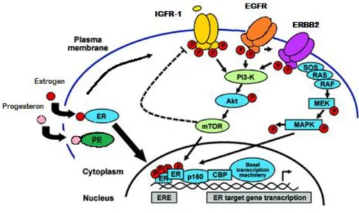
Figure 1: Major pathways regulating proliferation of breast cancer cells.
Abbreviations: CBP:CREB binding protein; ER:estrogen receptor; ERE:estrogen-responsive element; HER2: human epidermal growth factor receptor 2; MAPK:mitogen-activated protein kinase; MEK:mitogen-activated protein kinase/extracellular signal–related kinase kinase; mTOR:mammalian target of rapamycin; PI3K:phosphoinositide-3kinase; SOS:son of sevenless. (Adaptated from Di Cosimo S et al, Management of breast cancer with targeted agents: Importance of heterogenicity,2010106)
In addition, evading growth suppressors will also promote proliferation. The two prototypical tumor suppressors encode retinoblastoma-associated protein (Rb) and tumor suppressor protein p53 (p53). TP53 (p53 gene) mutations are common in precursors lesions and breast cancer and are supposed to be the cause of 1% of hereditary cases13,107.
Growth factor–dependent and hormone-dependent signaling are not the only important signal transduction pathways that derive from the cell surface. Two important classes of adhesion-dependent signals also play roles in cell growth, differentiation, and survival, as well as in motility and invasion of breast cancer cells: integrin and cadherin adhesion molecules108,109.
Integrins carry signals from the extracellular matrix into the cell. Integrins signal cells through many pathways, but one that has received considerable current interest is the focal adhesion kinase (Fak)-Src pathway. Activation of these two tyrosine-kinase– encoding oncoproteins activates the PI3K and Akt candidate oncoproteins, leading to multiple aspects of malignant behavior108,109. Of considerable interest, the tumor-suppressive phosphatase PTEN acts on Fak, PI3K, and Akt as its substrates to suppress
34
survival and induce apoptosis. However, mutations of PTEN do not appear to be particularly common in sporadic breast cancer110,111.
One of the functions of E-cadherin is to restrict cell motility, but it also diminishes the cytoplasmic pool of the important proliferation-modulating βcatenin transcription factor. The tumor-suppressive E-cadherin gene is commonly silenced by DNA methylation or mutation in breast cancer, releasing βcatenin to potentially up-regulate expression of the c-myc protooncogene (v-myc avian myelocytomatosis viral oncogene homolog)112,113. The βcatenin pathway may also be up-regulated by growth factor pathways, such as epidermal growth factor (EGF), hyperglycemic-glycogenolytic factor, and WNT (wingless) signaling114.
Cell death mechanisms
By 2000, it was widely accepted that cancer cells require unlimited replicative potential in order to generate macroscopic tumors. However, a balance between replication and death is required and involved in cancer progression. Cell death can occur by several mechanism including apoptosis or oncosis.
The concept that programmed cell death by apoptosis serves as a natural barrier to cancer development has been established by compelling functional studies 115,116,117. The apoptotic machinery is composed of both upstream regulators and downstream effector components115. The regulators, in turn, are divided into two major circuits, one receiving and processing extracellular death-inducing signals (the extrinsic apoptotic program, involving for example the Fas ligand/Fas receptor), and the other sensing and integrating a variety of signals of intracellular origin (the intrinsic program). Each one culminates in the activation of a normally latent proteases (caspases 8 and 9), that proceed to initiate a cascade of proteolysis involving effector caspases responsible for the execution phase of apoptosis, in which the cell is progressively disassembled and then consumed, both by its neighbors and by professional phagocytic cells. The ‘‘apoptotic trigger’’ that conveys signals between the regulators and effectors is controlled by counterbalancing pro- and anti-apoptotic members of the Bcl-2 family of regulatory proteins115. Although the cellular conditions that trigger apoptosis remain to be fully enumerated, several abnormality sensors that play key roles in tumor development have been identified115,116. Most notable is DNA damage sensor that functions via the p53 tumor suppressor protein118. As referred previously, mutations in p53 leads tumor cells to evade growth suppression.
Trigger apoptosis signaling circuit is the mechanism of action of some anticancer therapy but is also a response to various physiological stresses that cancer cells experience
35
during the course of tumorigenesis and is substantially elevated in proliferative lesion such as higher grade DCIS119,120. This exposes that much more remains to elucidate about this process.
In contrast to apoptosis, cells dying by oncosis (commonly denominated necrosis, which refers just to the pathological aspect of cell death) become bloated and explode, releasing their contents into the local tissue microenvironment. Although oncosis has historically been viewed much like organismic death, as a form of system-wide exhaustion and breakdown, the conceptual landscape is changing: cell death by necrosis is clearly under genetic control in some circumstances, rather than being a random and undirected process121,122. Perhaps more important, necrotic cell death releases pro-inflammatory signals into the surrounding tissue microenvironment, in contrast to apoptosis and autophagy, which do not. Consequently, necrotic cells can recruit inflammatory cells of the immune system. Multiple lines of evidence indicate that immune inflammatory cells can promote tumor progression, given that such cells are capable of fostering angiogenesis, cancer cell proliferation, and invasiveness123,121.
Autophagy is a process by which cells clear damaged or superfluous proteins and organelles. The recycling of these intracellular constituents also serves as an alternative energy source during periods of metabolic stress to maintain homeostasis and viability. Although not a cell death process, recent evidence suggests that autophagy provides a protective function to limit tumor necrosis and inflammation, and to mitigate genome damage in response to metabolic stress and defects in apoptosis124. The mechanism behind this has not been totally determined but is expected to be an anticancer target in near future125.
Tumor neo-vascularization
Like normal tissues, tumors require sustenance in the form of nutrients and oxygen as well as an ability to evacuate metabolic wastes and carbon dioxide. The tumor-associated neovasculature, generated by the process of angiogenesis, addresses these needs. During embryogenesis, the development of the vasculature involves the generation of new endothelial cells and their assembly into tubes (vasculogenesis) in addition to the sprouting (angiogenesis) of new vessels from existing ones. Following this morphogenesis, the normal vasculature becomes largely quiescent. In the adult, in physiological processes such as wound healing and female reproductive cycling, angiogenesis is turned on, but only transiently. In contrast, during tumor progression, an ‘‘angiogenic switch’’ is almost always activated and remains on, causing normally quiescent vasculature to continually sprout new vessels that help to sustain expanding
36
neoplastic growth126. A compelling body of evidence indicates that the angiogenic switch is governed by factors that either induce or oppose angiogenesis127,128. The well-known prototypes of angiogenesis inducers and inhibitors are vascular endothelial growth factor-A (VEGF-factor-A) and thrombospondin-1, respectively129. Histological studies of premalignant, noninvasive lesions, including dysplasias and in situ carcinomas arising in a variety of organs, have revealed the early triggering of the angiogenic switch126. Although angiogenesis is well documented in breast cancer progression anti-angiogenic drugs failed to show the expected clinical success130.
Cancer metabolism
The chronic and often uncontrolled cell proliferation that represents the essence of neoplastic disease involves not only deregulated control of cell proliferation but also corresponding adjustments of energy metabolism in order to fuel cell growth and division. The existence of this metabolic switch in cancer cells was recognized since the pioneering work of Otto Warburg in the first half of the twentieth century131. Despite this feature has received little or no attention for decades, it has been the object of major research effort in recent years132,133.
Cancer cells exhibit increased nutrient uptake
Cancer cells increase their glucose uptake, but instead of oxidizing most of this glucose to efficiently generate adenosine 5′-triphosphate (ATP) by oxidative phosphorylation, they ferment the excess glucose to lactate. This phenomenon is observed even in the presence of oxygen, and is referred as the Warburg effect or aerobic glycolysis134,135(for review of molecular mechanism136)(Figure 2). Previously, aerobic glycolysis was suggested to be a consequence of mitochondrial damage131 or an adaptive response to tumor hypoxia137. However, mitochondria remain functional in most tumors, and aerobic glycolysis is observed in cancer cells even in normoxia134,135.
Aerobic glycolysis, may allow individual cancer cells to increase uptake and incorporation of nutrients into the biomass (nucleotides, amino acids, and lipids) and facilitate the construction of new cells. According; 1) several signaling pathways implicated in cell proliferation also regulate metabolic pathways that incorporate nutrients into biomass; and 2) certain cancer-associated mutations enable cancer cells to metabolize nutrients in a manner conducive to proliferation rather than efficient ATP production. In support of this idea, aerobic glycolysis is a characteristic of many rapidly proliferating normal tissues and microorganisms134. Satisfying the metabolic needs of proliferation and redox control beyond ATP production may be advantages of aerobic glycolysis138. Moreover, studies have shown that lactate, a metabolite of aerobic glycolysis is also mediating the malignant
37
transformation and selection of surrounding cells leading to tumor progression and invasion139,140.
Although glucose catabolism through aerobic glycolysis has in large part been recognized a hallmark of cancer, it alone cannot explain all the metabolic changes necessary to support the requirements of cell growth141. Many normal mammalian tissues use nutrients other than glucose, and consumption of alternative fuel sources is observed in some cancer cells. Glutamine is the most abundant amino acid in both serum and cell culture medium, and glutamine is an important source of nitrogen for cells138,142. The carbon skeleton of glutamine can be oxidized to generate ATP and replenish citrate cycle intermediates. Finally, in some contexts reductive glutamine metabolism can provide carbon for lipid synthesis143,144. Indeed, after glucose, glutamine is the nutrient most highly consumed by cancer cells in tissue culture142,145. Emerging evidence suggests that other nutrients, including fatty acids and other amino acids, can also play key roles in some contexts 145,146,147.
Increased nutrient uptake is exploited in the clinic as a way to image tumors. F-18 fluoro-2-deoxyglucose PET (FDG-PET) is used to visualize tumors activity. This technique serves as a measure of glucose uptake in patient tissues by coupling positron-emitting 18F to an analog of glucose that is taken up and trapped in cells by phosphorylation but is not subject to further metabolism148. FDG-PET is most useful clinically as a staging tool and can also be used to monitor therapy response149. PET scanning to monitor uptake of other nutrients, such as glutamine and glutamate analogs has also been described in research settings149.
Cancer cells use different metabolic programs
While cancer
metabolism is often considered as a property that differs from normal cell metabolism, there is evidence that tumor cells exhibit a diversity of metabolic phenotypes150,145,151,15
38
2,153. Heterogeneous expression of metabolic genes is observed across tissue types, and
the metabolic network of an individual tumor more closely resembles that of the normal tissue from which the tumor arose than it does with other tumors that develop in different organ sites150. This expression pattern may reflect the propensity of cancer cells to adapt the pre-existing metabolic network to support their needs.
Indeed, the metabolic phenotype of tumors must be a function of both the genetic lesion driving tumorigenesis and the tissue from which the cancer arose152. Expression of oncogenes promotes increased consumption of glucose, glutamine, and proteins and can reprogram metabolism to support cell growth and proliferation146,154,155,156. Increasing evidence indicates that tumor suppressor genes function in part through effects on metabolism157, and the combination of genetic mutations in specific tissues facilitates altered metabolic regulation to support abnormal tissue growth.
Tumor cell metabolism is influenced by external factors
Altered tumor metabolism is not simply the final outcome of some combination of cell genetic modifications. Instead, a non-genetic component in the form of the tumor microenvironment must additionally be considered as part of the equation that influences metabolic changes in cancer cells158(Figure 3). Solid tumors are poorly vascularized, and therefore their surrounding environment can expose distinct regions of the tumor to spatial and temporal gradients of oxygenation, pH, and nutrient availability159. For example, fluctuating oxygen gradients across the microenvironment can drive sporadic hypoxia, the stabilization of hypoxia-inducible factor 1 (HIF1), and a corresponding induction of the HIF1-induced transcriptional program160. Regardless of whether HIF1-induced transcriptional effects are promoted through inappropriate genetic regulation or in response to hypoxic stress, one of its downstream consequences remains the conversion of a large percentage of glycolytic pyruvate to secreted lactate. The secreted lactate in turn triggers additional metabolic responses as a result of local acidification within the tumor microenvironment. It has also been suggested that this lactate-driven acidification can promote both tumor invasion and immune evasion161,162, which are among the other denoted hallmarks of cancer. Moreover, lactate secretion may have a functional role within a larger system of metabolic cooperation and symbiosis between cells in the microenvironment. Described as a “2-compartment” model of tumor metabolism, the symbiosis is characterized as the potential for anabolic malignant cells to extract high-energy metabolites (lactate, glutamine, and fatty acids) from adjacent catabolic cells (within the tumor or neighboring stromal cells) through a network of nutrient sharing that can stimulate tumor proliferation and metastasis163,164.
39
Studies reporting 2-compartment tumor metabolism have recently emerged in the context of breast cancer cells and their neighboring fibroblasts165,166,167, as well as for ovarian cancer cells and their neighboring adipocytes168. However, the complex interplay between genetics, microenvironment, and tissue heterogeneity is poorly understood. In addition, whole body metabolic regulation can affect tumor tissue metabolism, and patients with cancer often have perturbations in whole body metabolism169. Altered organismal metabolism can affect cancer outcomes as evidenced by the relationships between cachexia170,171, obesity40 or diabetes61 and poor patient survival.
Powerful homeostatic mechanisms exist at the organismal level to maintain a relatively constant supply of nutrients available to both normal and malignant tissues. This complex system cannot be understood from cell cultures studies or simple models and remains a challenge for the field (Figure 3).
Figure 3: Determinants of the tumor metabolic phenotype.
The metabolic phenotype of tumor cells is controlled by intrinsic genetic mutations and external responses to the tumor environment. Oncogenic signaling pathways controlling growth and survival are often activated by the loss of tumor suppressors (such as p53) or the activation of oncoproteins (such as PI3K). The resulting altered signaling modifies cellular metabolism to match the requirements of the cell division. Abnormal microenvironment conditions such as hypoxia, low pH and or nutrient deprivation elicit responses from tumor cells, including autophagy, which further affect metabolic activity. These adaptations optimize tumor metabolism for proliferation by providing appropriate levels of energy in the form of ATP, biosynthetic capacity and the maintenance of balanced redox status. The influence of the systemic metabolism on this microsystem is not known. AMPK: AMP-activated protein kinase; HIF-1: hypoxia-inducible factor 1.